Review
. 2023 Feb 2;5(1):20220026.
doi: 10.1259/bjro.20220026. eCollection 2023.

Imaging in patients with acute dyspnea when cardiac or pulmonary origin is suspected

Affiliations
Review

Imaging in patients with acute dyspnea when cardiac or pulmonary origin is suspected

Ruxandra-Iulia Milos et al. BJR Open. .

Abstract

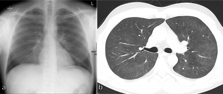
A wide spectrum of conditions, from life-threatening to non-urgent, can manifest with acute dyspnea, thus presenting major challenges for the treating physician when establishing the diagnosis and severity of the underlying disease. Imaging plays a decisive role in the assessment of acute dyspnea of cardiac and/or pulmonary origin. This article presents an overview of the current imaging modalities used to narrow the differential diagnosis in the assessment of acute dyspnea of cardiac or pulmonary origin. The current indications, findings, accuracy, and limits of each imaging modality are reported. Chest radiography is usually the primary imaging modality applied. There is a low radiation dose associated with this method, and it can assess the presence of fluid in the lung or pleura, consolidations, hyperinflation, pneumothorax, as well as heart enlargement. However, its low sensitivity limits the ability of the chest radiograph to accurately identify the causes of acute dyspnea. CT provides more detailed imaging of the cardiorespiratory system, and therefore, better sensitivity and specificity results, but it is accompanied by higher radiation exposure. Ultrasonography has the advantage of using no radiation, and is fast and feasible as a bedside test and appropriate for the assessment of unstable patients. However, patient-specific factors, such as body habitus, may limit its image quality and interpretability. Advances in knowledge This review provides guidance to the appropriate choice of imaging modalities in the diagnosis of patients with dyspnea of cardiac or pulmonary origin.


Conflict of interest statement

Competing interests: Ruxandra-Iulia Milos, Carmen Bartha, Benedikt H. Heidinger, Florian Prayer, Lucian Beer, Christian Wassipaul, Daria Kifjak, Martin Watzenböck – no conflict of interests to declare; Sebastian Röhrich – consulting activities contextflow GmbH; Helmut Prosch – Speaker honoria: AstraZeneca, BMS, Boehringer Ingelheim, Siemens Healthcare, Takeda. Research grants: Boehringer Ingelheim

Figures

Figure 1.
Figure 1.
Diagnostic CXRs in dyspneic patients: (a) 55-year-old male patient with posterior wall infarct, followed by ventricular fibrillation and resuscitation with signs of pulmonary edema of cardiac origin; (b) 76-year-old female patient with known metastasized breast cancer and malignant pleural effusion on the left side; (c) 34-year-old male patient with a spontaneous tension pneumothorax on the right side; (d) 78-year-old male patient with COVID-19 pneumonia showing scattered air space opacities and areas of interstitial thickening in both lungs. No pleural effusion. CXR, chest X-ray.
Figure 2.
Figure 2.
(a) Supine CXR of an 80-year-old male dyspneic patient with pneumothorax and deep sulcus sign (arrow); (b) 72-year-old male dyspneic patient with acute asthma exacerbation; supine CXR showing a small area of increased radiolucency in the para-aortic region (arrow) and the appearance of sharp edges of the descending aorta; (c) 49-year-old female patient 11 years after kidney transplant with dyspnea; bedside CXR shows signs of pleural effusion with thickening of the horizontal fissure, veil-like increased density of the hemithorax on the right side, and an enlarged heart silhouette.
Figure 3.
Figure 3.
65-year-old male patient with COPD IV with massive aggravated dyspnea and tachypnea over the 3 days prior to admission: (a) CXR presenting a newly developed non-specific small opacity and gas-fluid level above the right diaphragm (circle), compared to an older image (c), while chest CT (b) depicts a newly demarcated consolidation in the dependent areas of the bullous parenchymal destruction of the middle lobe, as an inflammatory superinfection. (d) CT examination taken previously. COPD, chronic obstructive pulmonary disease; CXR, chest X-ray.
Figure 4.
Figure 4.
43-year-old male patient with renal insufficiency, hypertension, and signs of AHF: enlarged heart, bilateral interlobular thickening, bilateral pleural effusion, and bilateral ground-glass opacification. AHF, acute heart failure.
Figure 5.
Figure 5.
40-year-old male dyspneic patient after kidney transplant and with B-cell lymphoma under chemotherapy and an unremarkable CXR. On CT, diffuse ground-glass opacities as a radiological manifestation of Pneumocystis jirovecii pneumonia. CXR, chest X-ray.
Figure 6.
Figure 6.
79-year-old female patient with dyspnea due to bilateral and centrally located acute pulmonary embolism with signs of right ventricle dysfunction and contrast reflux to the inferior vena cava (note the acute angle of the embolus with the vessel, arrow), as well as peripheral infarcted lung parenchyma (circle).
Figure 7.
Figure 7.
Diagnostic chest CT in dyspneic patients: (a) 48-year-old female patient with inhalation trauma and extensive ground-glass opacities as signs of permeability (non-cardiogenic) edema; (b) 52-year-old female patient with ill-defined ground-glass centrilobular nodules indicative of hypersensitivity pneumonitis; (c) 36-year-old male patient with endocarditis after tricuspid valve repair, Staphylococcus aureus sepsis and septic emboli; (d) 61-year-old female patient with lung hemorrhage as a result of systemic sclerosis vasculitis.
Figure 8.
Figure 8.
63-year-old male dyspneic patient who was referred for CTPA with suspicion of PE. The CT revealed, in addition to the segmental PE in the left lower lobe (arrowhead), a mass in the right upper lobe with pathological mediastinal lymphadenopathy (a). The patient also underwent an ultra-low-dose chest CT, which showed a good correlation with the standard CT in depicting an infarcted area of lung parenchyma in the left lower lobe (circle) and an atelectasis in the right lower lobe (arrow) (b) standard CT and (c) ultra-low-dose CT. CTPA, CT pulmonary angiography; PE, pulmonary embolism.
Figure 9.
Figure 9.
Example of a quantitative pattern analysis report regarding the percentage of lung anomalies (consolidation and ground-glass opacities) detected on a chest CT scan of a 42-year-old-patient with COVID-19 pneumonia.
Figure 10.
Figure 10.
(a, b) Lung ultrasound of a 61-year-old male dyspneic patient depicting B-lines indicative of cardiogenic pulmonary edema; (c) 17-old-year male patient with complex parapneumonic pleural effusion showing septa.
Figure 11.
Figure 11.
(a) CXR of a 9-year-old boy with dyspnea and pneumonia and (b) corresponding ultrasound image of the pneumonia, depicting the consolidation of the lung parenchyma with punctiform hyperechogenic spots that correspond to the air bronchograms. CXR, chest X-ray.

References

    1. Cairns C, Kang K, L S. National Hospital Ambulatory Medical Care Survey: 2018 emergency department summary tables. Available from: https://www.cdc.gov/nchs/data/nhamcs/web_tables/2018_ed_web_tables-508.pdf
    1. Kelly AM, Keijzers G, Klim S, Graham CA, Craig S, Kuan WS, et al. An observational study of dyspnea in emergency departments: the Asia, Australia, and New Zealand dyspnea in emergency departments study (AANZDEM). Acad Emerg Med 2017; 24: 328–36. doi: 10.1111/acem.13118 - DOI - PubMed
    1. Parshall MB, Schwartzstein RM, Adams L, Banzett RB, Manning HL, Bourbeau J, et al. An official American thoracic Society statement: update on the mechanisms, assessment, and management of dyspnea. Am J Respir Crit Care Med 2012; 185: 435–52. doi: 10.1164/rccm.201111-2042ST - DOI - PMC - PubMed
    1. Laribi S, Keijzers G, van Meer O, Klim S, Motiejunaite J, Kuan WS, et al. Epidemiology of patients presenting with dyspnea to emergency departments in Europe and the Asia-Pacific region. Eur J Emerg Med 2019; 26: 345–49. doi: 10.1097/MEJ.0000000000000571 - DOI - PubMed
    1. Ray P, Birolleau S, Lefort Y, Becquemin M-H, Beigelman C, Isnard R, et al. Acute respiratory failure in the elderly: etiology, emergency diagnosis and prognosis. Crit Care 2006; 10: R82. doi: 10.1186/cc4926 - DOI - PMC - PubMed