. 2023 Apr 10;8(7):e167306.
doi: 10.1172/jci.insight.167306.

Single-epitope T cell-based vaccine protects against SARS-CoV-2 infection in a preclinical animal model

Single-epitope T cell-based vaccine protects against SARS-CoV-2 infection in a preclinical animal model

Takuya Tada et al. JCI Insight. .

Abstract

Currently authorized COVID-19 vaccines induce humoral and cellular responses to epitopes in the SARS-CoV-2 spike protein, though the relative roles of antibodies and T cells in protection are not well understood. To understand the role of vaccine-elicited T cell responses in protection, we established a T cell-only vaccine using a DC-targeted lentiviral vector expressing single CD8+ T cell epitopes of the viral nucleocapsid, spike, and ORF1. Immunization of angiotensin-converting enzyme 2-transgenic mice with ex vivo lentiviral vector-transduced DCs or by direct injection of the vector induced the proliferation of functional antigen-specific CD8+ T cells, resulting in a 3-log decrease in virus load upon live virus challenge that was effective against the ancestral virus and Omicron variants. The Pfizer/BNT162b2 vaccine was also protective in mice, but the antibodies elicited did not cross-react on the Omicron variants, suggesting that the protection was mediated by T cells. The studies suggest that the T cell response plays an important role in vaccine protection. The findings suggest that the incorporation of additional T cell epitopes into current vaccines would increase their effectiveness and broaden protection.

Keywords: COVID-19; Dendritic cells; Immunology; T cells.


Conflict of interest statement

Conflict of interest: The authors have declared that no conflict of interest exists.

Figures

Figure 1
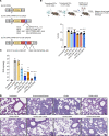
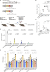
Figure 1. DC vaccine protects mice against SARS-CoV-2 infection.
(A) Schematic of lentiviral vectors expressing murine CD40L and CD8+ T cell epitopes N219-227, N105-113, ORF11637-1646, S539-546, and N219-227-S539-546. (B) The experimental scheme for testing vaccine protection is diagrammed. hACE2-KI mice were immunized by 2 IV injections of 1 × 10⁶ transduced DCs, 1 week apart (n = 6). The mice were challenged 1 week after the second immunization with 2 × 10⁴ PFU SARS-CoV-2 WA1/2020. SAMHD1-KO BMDCs were transduced with lentiviral vectors at MOI = 5, and CD40L expression was analyzed 3 dpi by flow cytometry (bottom). The experiment was done twice with similar results. hACE2-KI, human angiotensin-converting enzyme 2–knockin; dpi, days postinfection. (C) Viral subgenomic RNA in the lungs of the immunized mice was measured by RT-qPCR 3 days postchallenge. The y axis of the histograms shows the viral RNA copy numbers/g lung tissue. Statistical significance was determined by Kruskal-Wallis test with post hoc Dunn’s test. Confidence intervals are shown as the mean ± SD. (**P ≤ 0.01, ****P ≤ 0.0001.) The experiment was done 3 times with similar results. (D) Hematoxylin and eosin staining of lung sections from unimmunized and lentiviral vector–immunized mice (original magnification, 20×; scale bars: 50 μm). Each image is representative of 6 mice.
Figure 2
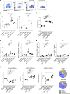
Figure 2. T cell response induced from lentivirus-based DC vaccine.
(A) The gating scheme used in the analysis of the antigen-specific CD8+ T cells induced by vaccination is shown for a representative sample from a single mouse. Splenocytes from mice immunized with 2 injections of transduced DCs and then challenged with SARS-CoV-2 WA1/2020 or splenocytes from unimmunized mice and then challenged with SARS-CoV-2 WA1/2020 were stained with anti-CD3, anti-CD8, and an MHC class I tetramer/N219-227 peptide complex. Infected, unimmunized and infected, immunized controls are shown. FSC, forward scatter. (B) At 3 dpi, the fraction of antigen-specific (TCR+) CD8+ T cells was quantified by flow cytometry as shown by the scheme in A using tetramers for the N219-227 and S539-546 epitopes. The immunizing vectors are labeled below. (C) The weight of the spleens of immunized and challenged mice was determined 14 days postimmunization. (D) The fraction of antigen-specific CD8+ T cells in the immunized mice that expressed IFN-γ and perforin was quantified by flow cytometry. (E) The fraction of total CD8+ T cells in the immunized mice that expressed CD107a and IL-2 was quantified by flow cytometry. (F) Naive, effector, and central memory T cells in the immunized mice were quantified. The results are summarized in the pie charts on the right. The numbers in the control pie chart (top) show the percentage of naive, effector, and central memory cells from uninfected, unimmunized and CD40L-immunized mice. The numbers in the immunized pie chart (bottom) show the percentage of naive, effector, and central memory T cells from CD40L-N105-113–, CD40L-N219-227–, CD40L-ORF11637-1646–, CD40L-S539-546–, and CD40L-N219-227- S539-546–immunized mice. Statistical significance was determined by Kruskal-Wallis test with post hoc Dunn’s test. Confidence intervals are shown as the mean ± SD. (*P ≤ 0.05, **P ≤ 0.01, ***P ≤ 0.001, ****P ≤ 0.001.) The experiment was done 3 times with similar results.
Figure 3
Figure 3. Enhancement of the antiviral response by CD40L.
(A) The schematic diagram of lentiviral vectors expressing nonfunctional CD40L. The red asterisk indicates the T146N mutation in CD40L. (B) SAMHD1-KO BMDCs were transduced (MOI = 5) with lentiviral vectors expressing CD40L, mutated CD40L, CD40L-N219-227, and CD40L.T146N-N219-227 virus. After 3 days, the cells were analyzed for CD83 and CD86 by flow cytometry. (C) Mice were injected twice with transduced BMDCs and challenged with SARS-CoV-2 WA1/2020. After 3 days, virus load in the lung was measured by RT-qPCR. (D) Antigen-specific CD8+ T cells, IFN-γ in TCR+CD8+ T cells, and CD69+ cells in the immunized and challenged mice were quantified by flow cytometry. Statistical significance was determined by Kruskal-Wallis test with post hoc Dunn’s test. Confidence intervals are shown as the mean ± SD. (*P ≤ 0.05, **P ≤ 0.01, ***P ≤ 0.001.) The experiment was done twice with similar results.
Figure 4
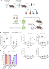
Figure 4. Vaccine-induced protection from infection is mediated by CD8+ T cells.
(A) The cytolytic and antiviral activity of different cell types induced by vaccination was analyzed by the diagrammed experimental scheme. Mice were immunized twice with 1 × 10⁶ DCs transduced with CD40L or CD40L-N219-227 vector. At 7 days after second immunization, splenic CD8+ T cell, CD4+ T cell, DC, and B cell populations were isolated on magnetic beads. A portion of the cells was analyzed in an in vitro cytolytic assay in which the cells were mixed with the CFSE-labeled SARS-CoV-2–infected naive lung cells of hACE2-KI mice and the number of lysed cells was quantified by flow cytometry. A portion of each population was reinfused into recipient mice (n = 5), which were challenged 5 days later with SARS-CoV-2 WA1/2020. (B) Seven days after the second immunization, the fraction of antigen-specific CD8+ and CD4+ T cells in splenocytes was quantified by flow cytometry using tetramers for the N219-227 epitopes. (C) Cytolytic activity of the CD4+ T cells, CD8+ T cells, DCs, and B cells of control CD40L alone and CD40L-N219-227 vector–immunized mice was analyzed with the assay diagrammed in A using SARS-CoV-2–infected primary mouse lung epithelial cell targets. (D) Splenic CD8+ T cell, CD4+ T cell, DC, and B cell populations from immunized mice were reinfused into recipient mice (n = 5). At 5 days postinjection, mice were challenged with SARS-CoV-2 WA1/2020. Subgenomic viral RNA in the lungs of the mice was quantified 3 dpi. Statistical significance was determined by Kruskal-Wallis test with post hoc Dunn’s test. Confidence intervals are shown as the mean ± SD. (*P ≤ 0.05, **P ≤ 0.01, ***P ≤ 0.001, ****P ≤ 0.0001.) The experiment was done twice with similar results.
Figure 5
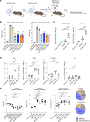
Figure 5. Direct lentivirus injection protects mice from SARS-CoV-2 infection.
(A) Schematic of direct lentivirus immunization. A total of 5 × 10⁶ IU lentiviral vector encoding CD40L and T cell epitopes N219-227, N105-113, ORF11637-1646, S539-546, and N219-227-S539-546. Lentiviral vectors were injected into hACE2-KI mice IV (n = 6). One week after the first immunization, the mice were re-immunized. One week or 30 days following the second immunization, the mice were challenged with 2 × 10⁴ PFU SARS-CoV-2 WA1/2020. (B) One week (left) or 30 days (right) following the second immunization, SARS-CoV-2 subgenomic viral RNA in the lung was quantified 3 dpi with SARS-CoV-2 WA1/2020. (C) Splenocytes were analyzed for the CD3+, CD8+, CD4+, and SARS-CoV-2–specific TCR+CD8+ T cells by flow cytometry. (D) IFN-γ, TNF-α, perforin, and IL-2 levels in TCR+CD8+ T cells were quantified by flow cytometry. (E) Naive, effector, and central memory T cells were distinguished by CD62L and CD44, then determined the population of naive (CD62LhiCD44lo), effector (CD62LloCD44hi), and central memory cells (CD62LhiCD44hi). The results are summarized in the pie charts on the right. The percentage of naive, effector, and central memory cells of uninfected, unimmunized, and CD40L-immunized mice is shown (top). The percentage of naive, effector, and central memory T cells from CD40L-N105-113–, CD40L-N219-227–, CD40L-ORF11637-1646–, CD40L-S539-546–, and CD40L-N219-227- S539-546–immunized mice (*P ≤ 0.05, **P ≤ 0.01, ***P ≤ 0.001, ****P ≤ 0.0001) is shown (bottom). Statistical significance was determined by Kruskal-Wallis test with post hoc Dunn’s test. Confidence intervals are shown as the mean ± SD. The experiment was done twice with similar results.
Figure 6
Figure 6. Comparison of protective efficacy of BNT162b2, lentivirus-based DC, and direct lentivirus vaccine against SARS-CoV-2 variants.
(A) The experimental scheme is diagrammed. hACE2-KI mice were immunized with mRNA vaccine BNT162b2 (5 μg), with N219-227 or S539-546 vector–transduced DCs (1 × 10⁶), or by direct lentivirus injection of the CD40L-N219-227 or CD40L-S539-546 lentiviral vectors (n = 5). Uninfected and unimmunized/infected mice served as controls. All mice were boosted 7 days after the first immunization. After another 7 days, the mice were challenged by infection with SARS-CoV-2 WA1/2020 or Omicron variants. (B) Antigen-specific CD8+ T cells of the immunized and challenged mice were stained with the corresponding N219-227 and S539-546 tetramers and analyzed by flow cytometry. (C) The tetramer+IFN-γ+CD8+ T cells of the immunized mice were quantified by flow cytometry. (D) Sera from the immunized mice was collected 7 days postboost. Neutralizing antibody titers against viruses with D614, BA.1, BA.2, and BA.5 S were measured. The IC50 of the serum from each mouse is shown. (E) Immunized mice were challenged with SARS-CoV-2 WA1/2020 or BA.1, BA.2, BA.5. At 3 dpi (SARS-CoV-2 WA1/2020) or 2 dpi (BA.1, BA.2, and BA.5), virus load in the lungs was determined. Statistical significance was determined by Kruskal-Wallis test with post hoc Dunn’s test with confidence intervals shown as the mean ± SD. (*P ≤ 0.05, **P ≤ 0.01, ***P ≤ 0.001.) The experiment was done twice with similar results.

References

    1. Scobie HM, et al. Monitoring incidence of COVID-19 cases, hospitalizations, and deaths, by vaccination status - 13 U.S. jurisdictions, April 4-July 17, 2021. MMWR Morb Mortal Wkly Rep. 2021;70(37):1284–1290. doi: 10.15585/mmwr.mm7037e1. - DOI - PMC - PubMed
    1. Hall V, et al. Protection against SARS-CoV-2 after covid-19 vaccination and previous infection. N Engl J Med. 2022;386(13):1207–1220. doi: 10.1056/NEJMoa2118691. - DOI - PMC - PubMed
    1. Harvey WT, et al. SARS-CoV-2 variants, spike mutations and immune escape. Nat Rev Microbiol. 2021;19(7):409–424. doi: 10.1038/s41579-021-00573-0. - DOI - PMC - PubMed
    1. Neidleman J, et al. mRNA vaccine-induced T cells respond identically to SARS-CoV-2 variants of concern but differ in longevity and homing properties depending on prior infection status. Elife. 2021;10:e72619. doi: 10.7554/eLife.72619. - DOI - PMC - PubMed
    1. Hurme A, et al. Long-lasting T cell responses in BNT162b2 COVID-19 mRNA vaccinees and COVID-19 convalescent patients. Front Immunol. 2022;13:869990. doi: 10.3389/fimmu.2022.869990. - DOI - PMC - PubMed

Publication types

Supplementary concepts