. 2023 Apr 10;8(7):e167422.
doi: 10.1172/jci.insight.167422.

Identification and tracking of HTLV-1-infected T cell clones in virus-associated neurologic disease

Affiliations

Identification and tracking of HTLV-1-infected T cell clones in virus-associated neurologic disease

Satoshi Nozuma et al. JCI Insight. .

Abstract

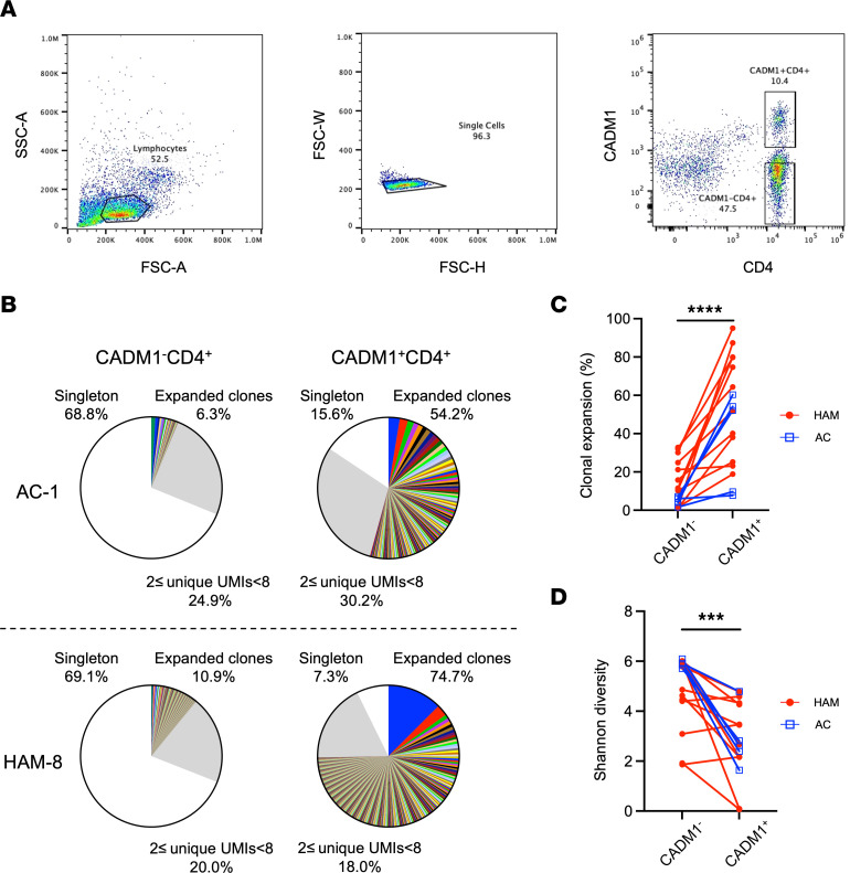
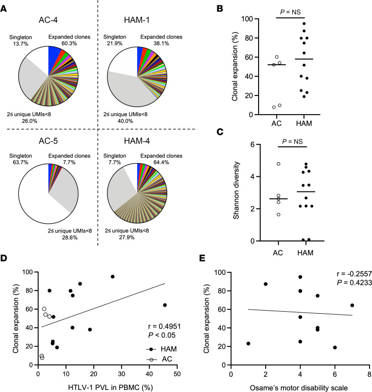
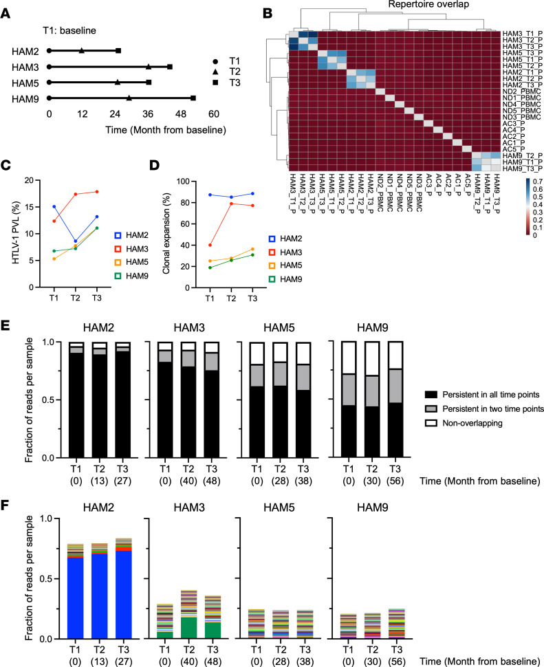
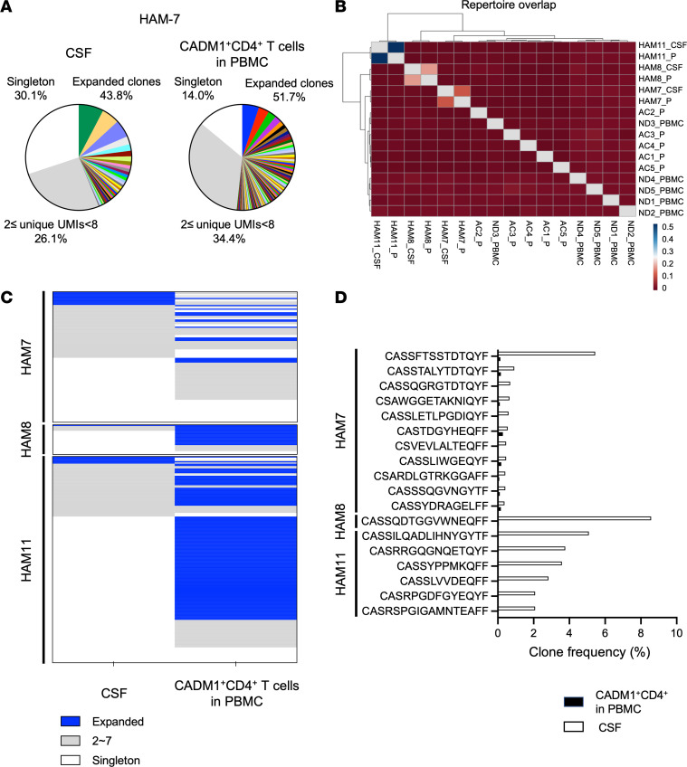
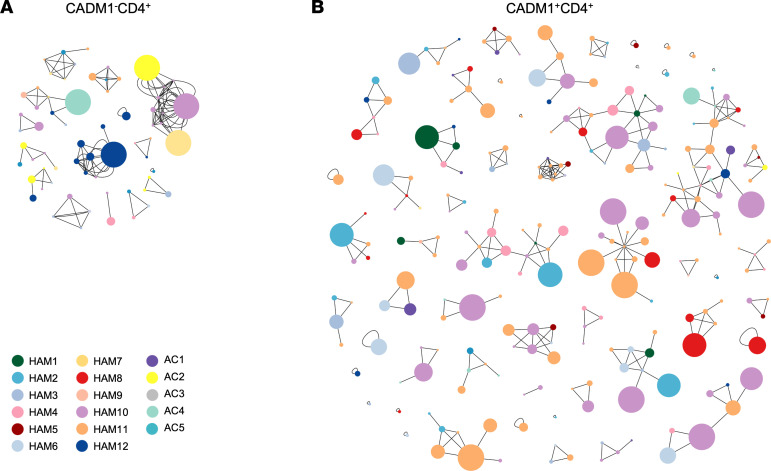
Human T lymphotropic virus type 1-assoicated (HTLV-1-associated) myelopathy/tropical spastic paraparesis (HAM/TSP) is a neuroinflammatory disease caused by the persistent proliferation of HTLV-1-infected T cells. Here, we performed a T cell receptor (TCR) repertoire analysis focused on HTLV-1-infected cells to identify and track the infected T cell clones that are preserved in patients with HAM/TSP and migrate to the CNS. TCRβ repertoire analysis revealed higher clonal expansion in HTLV-1-infected cells compared with noninfected cells from patients with HAM/TSP and asymptomatic carriers (ACs). TCR clonality in HTLV-1-infected cells was similar in patients with HAM/TSP and ACs. Longitudinal analysis showed that the TCR repertoire signature in HTLV-1-infected cells remained stable, and highly expanded infected clones were preserved within each patient with HAM/TSP over years. Expanded HTLV-1-infected clones revealed different distributions between cerebrospinal fluid (CSF) and peripheral blood and were enriched in the CSF of patients with HAM/TSP. Cluster analysis showed similarity in TCRβ sequences in HTLV-1-infected cells, suggesting that they proliferate after common antigen stimulation. Our results indicate that exploring TCR repertoires of HTLV-1-infected cells can elucidate individual clonal dynamics and identify potential pathogenic clones expanded in the CNS.

Keywords: Inflammation; Neurological disorders; T cell receptor; T cells; Virology.


Conflict of interest statement

Conflict of interest: The authors have declared that no conflict of interest exists.

Figures

Figure 1
Figure 1. Clonal expansion of TCRβ repertoire in HTLV-1–infected cells of patients with HAM/TSP and ACs.
(A) Gating strategy for flow cytometry sorting of CADM1+CD4+ and CADM1CD4+ T cells from PBMCs samples. (B) Representative examples of T cell clonal expansion in CADM1CD4+ and CADM1+CD4+ T cells of an AC (AC-1) and a HAM/TSP patient (HAM8). Clonal expansion of TCRβ repertoire is classified by the frequencies of clones ≥ 8 UMIs (color wedges), clones with 2 ≤ UMIs < 8 (gray), and singletons (white). In the group of expanded clones, each wedge exhibits a unique clonotype with a defined CDR3 sequence, and the identical clones shared by CADM1CD4+ and CADM1+CD4+ T cells in each individual are depicted in the same color. (C and D) Comparison of T cell clonal expansion by frequency of clones ≥ 8 UMIs (C) and Shannon diversity (D) between CADM1CD4+ and CADM1+CD4+ T cells from ACs (n = 5) and patients with HAM/TSP (n = 12) using Wilcoxon signed-rank test. ***P < 0.001, ****P < 0.0001.
Figure 2
Figure 2. Comparison of TCRβ clonal repertoire analysis of CADM1+CD4+ T cells from patients with HAM/TSP and ACs.
(A) Representative analysis of the clonal expansion of TCR repertoire in CADM1+CD4+ cells from ACs and patients with HAM/TSP. (B) Comparison of T cell clonal expansion between ACs (n = 5) and patients with HAM/TSP (n = 12) using Wilcoxon signed-rank test. The horizontal bar indicates the median. (C) Comparison of Shannon diversity between ACs (n = 5) and patients with HAM/TSP (n = 12) using Wilcoxon signed-rank test. The horizontal bar indicates the median. (D) Correlation of clonal expansion in CADM1+CD4+ T cells with HTLV-1 PVL in PBMCs from ACs (opened shapes; n = 5) and patients with HAM/TSP (closed shapes; n = 12) using Spearman’s rank correlation test. (E) Correlation of clonal expansion in CADM1+CD4+ T cells with OMDS of patients with HAM/TSP (n = 12) using Spearman’s rank correlation test.
Figure 3
Figure 3. Longitudinal analysis of the TCRβ repertoire in HTLV-1–infected T cells from patients with HAM/TSP.
(A) Scheme of longitudinal sampling. The duration from T1 to T3 is more than 2 years, and each time point represents a period of at least 8 months. (B) A heatmap analysis represents the repertoire overlap of TCRβ clonotypes shared among 3 longitudinal samples from 4 patients with HAM/TSP, 5ACs, and 5 normal donors using the overlap coefficient, a normalized measure of overlap similarity. (C and D) Longitudinal HTLV-1 PVL in PBMCs (C) and clonal expansion of HTLV-1–infected T cells (D) from 4 patients with HAM/TSP across 3 time points. (E) Dynamics of TCRβ clones in HTLV-1–infected cells. Persistent clones were investigated by the frequencies of the sequences persistent across 3 time points (black), at least 2 time points (gray), and nonoverlapping (white). (F) Tracking the 100 most dominant clonotypes shared across all time points in each HAM/TSP patient. The identical clones shared across all time points in each individual are depicted in the same color.
Figure 4
Figure 4. Differential distribution and enrichment of expanded HTLV-1–infected clones in the CSF compared with the peripheral blood of patients with HAM/TSP.
(A) Pie chart representations of T cell clonal expansion in CSF cells and CADM1+CD4+ cells of a HAM/TSP patient (HAM7). (B) A heatmap analysis exhibiting an overlap of TCRβ clonotypes among 3 paired CSF and CADM1+CD4+ cells from 3 patients with HAM/TSP, 5 ACs, and 5 normal donors using the overlap coefficient. (C) Heatmap of shared clonotypes in CSF cells and CADM1+CD4+ cells in PBMCs from 3 patients with HAM/TSP. Each row slice represents the shared TCRβ clonotypes between CSF cells and CADM1+CD4+ cells in PBMCs of each HAM/TSP patient. Expanded clonotypes with ≥ 8 UMIs are shown in the blue area, while clonotypes with 2 ≤ UMIs < 8 or singletons are visualized in the gray area and in the white area, respectively. For example, of 11 expanded clones shown in blue slices in the CSF of HAM7, only 1 clonotype (9.1%) was shared with the expanded clones in the peripheral CADM+CD4+ T cells and 10 clonotypes (90.9%) were shared with singleton (white) or 2 ≤ UMIs < 8 (gray). Conversely, of 16 expanded clones shown in blue slices in peripheral CADM1+CD4+ T cells of HAM7, only 1 clonotype (6.3%) was shared with the expanded clones in CSF cells and 15 clonotypes (93.7%) were shared with singleton or 2 ≤UMIs < 8. (D) Frequencies of 18 TCRβ expanded clonotypes in the CSF compared with peripheral CADM1+CD4+ cells in 3 patients with HAM/TSP.
Figure 5
Figure 5. Clustering analysis of similarity in TCRβ sequences in expanded HTLV-1–infected cells of patients with HAM/TSP and ACs.
(A and B) Network plot showing clusters of TCRβ in expanded CADM1CD4+ T cells (A) and CADM1+CD4+ T cells (B) in PBMCs of patients with HAM/TSP (n = 12) and ACs (n = 5) using GLIPH2 software. Each node represents a unique CDR3 sequence with node colors based on subjects and node size according to read counts.

Similar articles

Cited by

References

    1. Uchiyama T, et al. Adult T-cell leukemia: clinical and hematologic features of 16 cases. Blood. 1977;50(3):481–492. doi: 10.1182/blood.V50.3.481.481. - DOI - PubMed
    1. Osame M, et al. HTLV-I associated myelopathy, a new clinical entity. Lancet. 1986;1(8488):1031–1032. - PubMed
    1. Gessain A, et al. Antibodies to human T-lymphotropic virus type-I in patients with tropical spastic paraparesis. Lancet. 1985;2(8452):407–410. - PubMed
    1. Nagai M, et al. Analysis of HTLV-I proviral load in 202 HAM/TSP patients and 243 asymptomatic HTLV-I carriers: high proviral load strongly predisposes to HAM/TSP. J Neurovirol. 1998;4(6):586–593. doi: 10.3109/13550289809114225. - DOI - PubMed
    1. Nagai M, et al. Increased HTLV-I proviral load and preferential expansion of HTLV-I Tax-specific CD8+ T cells in cerebrospinal fluid from patients with HAM/TSP. Ann Neurol. 2001;50(6):807–812. doi: 10.1002/ana.10065. - DOI - PubMed

Publication types

Substances