. 2024 Jun 1;153(6):1273-1288.
doi: 10.1097/PRS.0000000000010556. Epub 2023 Apr 11.

The Deep Fascia of the Head and Neck Revisited: Relationship with the Facial Nerve and Implications for Rhytidectomy

Affiliations

The Deep Fascia of the Head and Neck Revisited: Relationship with the Facial Nerve and Implications for Rhytidectomy

Lennert Minelli et al. Plast Reconstr Surg. .

Abstract

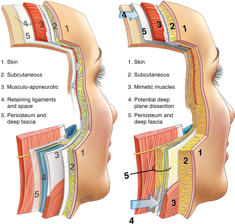
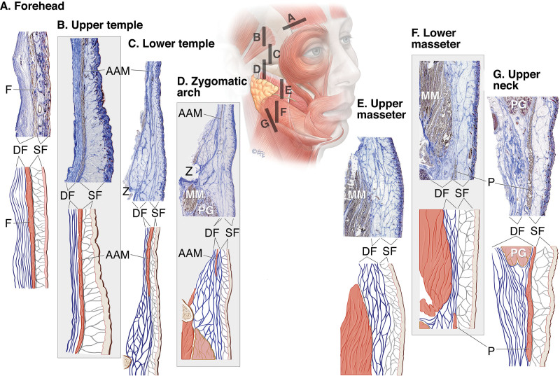
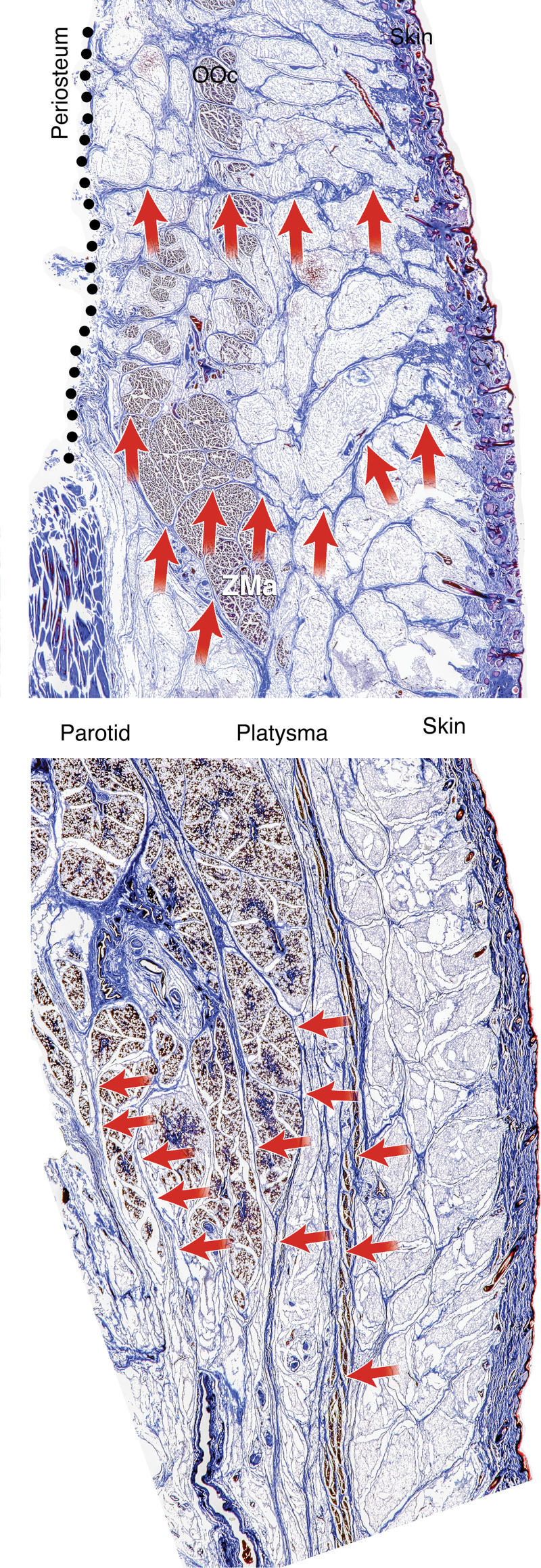
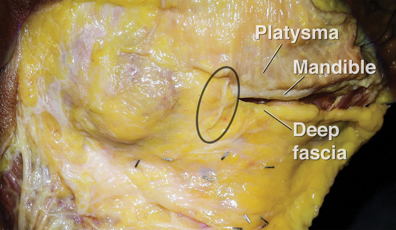
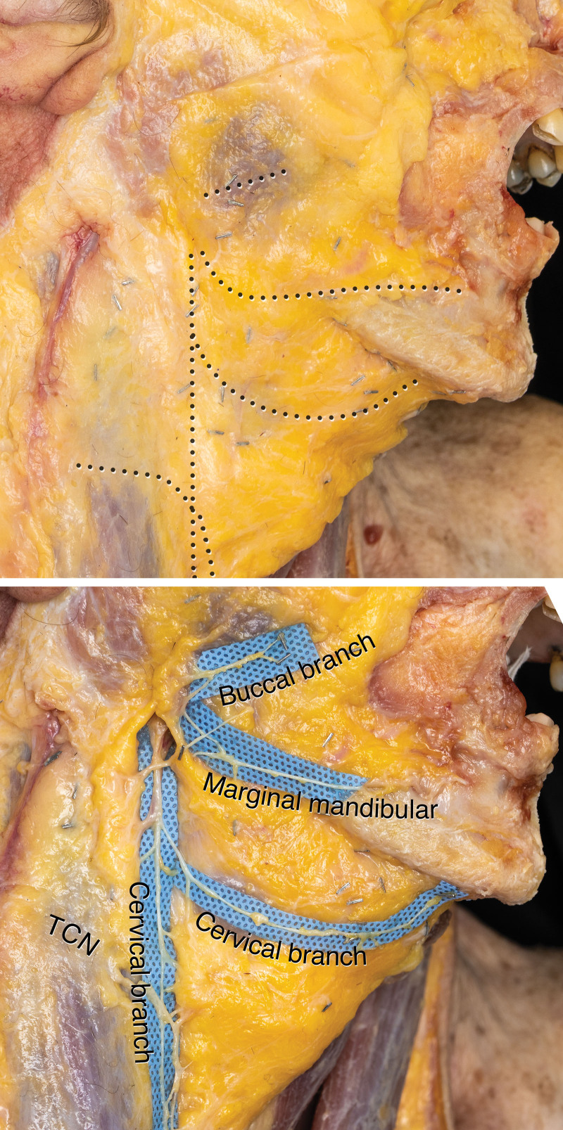
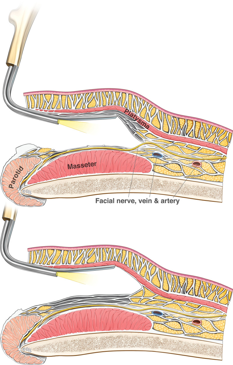
Background: The deep fascia is important in face-lift surgery, as it is relied on for protection of the facial nerve during surgical dissection. Lack of consistency with the term may be attributable to the term deep fascia having two different meanings. It is a specific type of connective tissue, classically thin, fibrous, and flat, whereas in the description of the layers of the face and neck, the deep fascia layer includes all the connective tissue deep to the superficial fascia layer. This cadaver study was undertaken to clarify the layered anatomy of the face and neck and its relationship with the facial nerve branches.

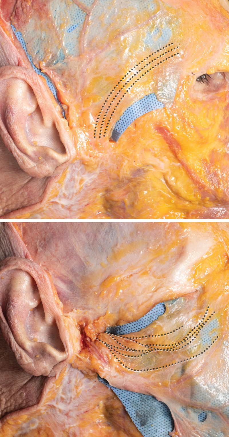
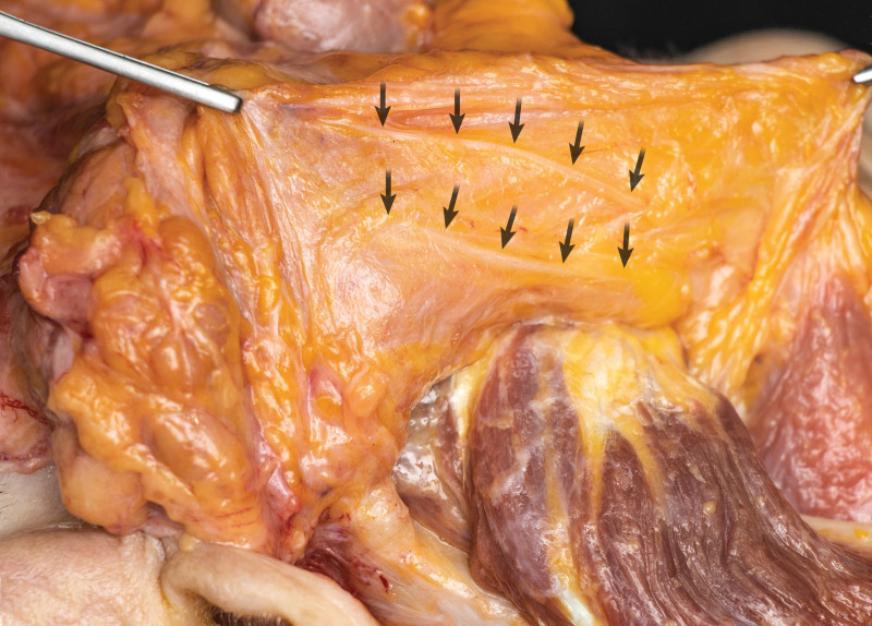
Methods: Preliminary dissections and macrosectioning, followed by a conclusive series of standardized layered dissections, histologic analysis, and sheet plastination, were performed on 50 cadaver heads.

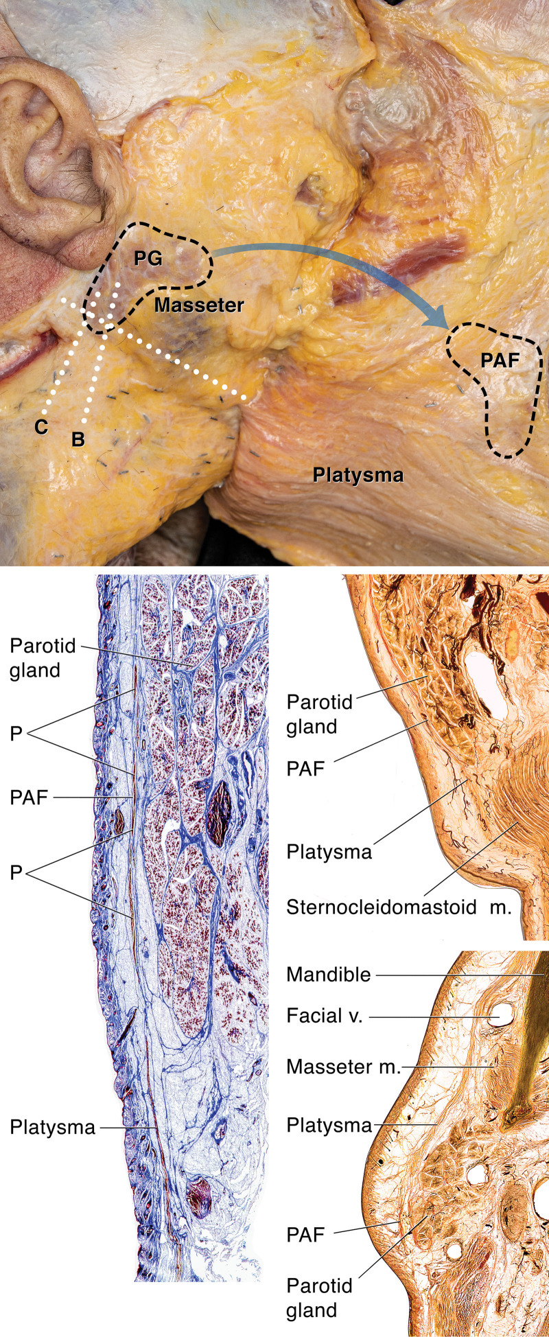
Results: The deep fascia is thin in convex areas of the face and neck, and thicker in concave areas; it is interspersed with deep fat. The facial nerve branches, after emerging from the parotid gland, are embedded within the deep fascia, not deep to it. They transition from deep within the deep fascia at specific locations to course in the most superficial part of the deep fascia where they underlie their target superficial fascia muscles and are at risk from deep plane face-lift dissection.

Conclusions: The deep fascia layer is a multilamellar fibrofatty layer of variable thickness, which includes the deep fat in which the facial nerve branches are embedded. In deep plane face-lift surgery, dissection must be performed in the most superficial level of this deep fascia layer.


Conflict of interest statement

The authors have no financial interests to disclose. No funding was received for this article.

Figures

Fig. 1.
Fig. 1.
Traditional textbook description of the layered anatomy of the face (left) and our current understanding (right). Traditionally, the concentric layers were as follows: layer 1, skin; layer 2, subcutaneous fat; layer 3, SMAS-platysma; layer 4, deep plane with spaces and ligaments; and layer 5, deep fascia. It is currently understood that the SMAS is not a complete anatomical layer and is only present in the form of the mimetic muscles but not the aponeurosis between the muscles. The deep fascia is not only a thin fibrous fascia, but a three-dimensional layer with fibrofatty and areolar tissue areas (spaces). The deep plane dissection (layer 4) is not a separate anatomical layer, but a potential dissection in the most superficial part of the deep fascia. Illustrations published with permission from Levent Efe. Copyright © 2022 Levent Efe.
Fig. 2.
Fig. 2.
Demonstration of the continuity of the platysma with the PAF. (Above) Right hemiface of a fresh cadaver. The strong PAF is lifted with the platysma from an incision in front of the ear and turned over 180 degrees to demonstrate its deep aspect. The PAF is the evolutionary aponeurotic remnant of the platysma, which overlies the parotid in humans. Lifting this SMAS-platysma (layer 3) exposes the parotid parenchyma, which is now covered only by its thin capsula propria (parotid capsule). (Below, left) Histologic image of the lower face and neck. Note the dispersed muscle fibers (P) inside the PAF demonstrating the evolutionary remnant of the platysma involuted to a fibromuscular structure. Sheet plastination sections of the parotid-platysma in a coronal plane (center, right), showing the continuation of platysma over the sternocleidomastoid muscle with the PAF over the parotid, and in an axial plane (below, right), showing the continuation of platysma over the mandible with the PAF over the parotid.
Fig. 3.
Fig. 3.
Dissection images of the deep fascia right side of a fresh cadaver. (Left) The deep fascia in situ. The deep fascia is thin over the cranium, upper temple, lower masseter, sternocleidomastoid, parotid gland, and submandibular gland, and it is thicker in the lower temple to the upper masseter, the anterior midcheek, and the anterior cervical triangle. (Center) After removal of the deep fascia. The buccal fat pad (BFP) is a separate entity altogether, and its temporal extension is seen. (Above, right) Close-up image of the same area in another cadaver. The deep fascia is reflected to show the underlying buccal fat pad and parotid duct (PD). Whereas the deep fascia features abundant connective tissue, the buccal fat pad lacks this and is more amorphous and pliable in contrast to the deep medial cheek fat (DMCF). (Below, right) Midline sagittal view of the deep cervical fascia after removal of the covering platysma muscle and removal of the right side of the deep fascia so that a cross-sectional view at the cut-end of the deep fascia is provided. The deep fascia is thin in the convex area over the thyroid and cricoid cartilages, but thick in the concave area overlying the hyoid bone. No separate deep fat layer could be distinguished from the deep fascia; they are one and the same layer.
Fig. 4.
Fig. 4.
Demonstration of the thickness of the deep fascia (in blue) filling the space between the platysma and the deeper structures. (Left) Axial macrosection through the neck at the level of the hyoid bone (C3) of a fresh cadaver. The deep fascia is a thin aponeurosis over the SCM but a thick fibrofatty layer that includes the deep fat of the neck in the other areas. (Center) Axial sheet plastination section through the neck at the level of the thyroid cartilage (C4). The deep fascia has a dense matrix of connective tissue. Not one but multiple sheets of connective tissue are visualized in the deep fascia. Up to 18 fascial sheets can be counted in the pretracheal area in this specimen (inset). Note how the connective tissue sheets of the deep fascia run parallel to the skin and SMAS-platysma and the connective tissue septa of the superficial fascia run perpendicular to the skin. (Right) Sagittal sheet plastination section through the masseter, mandible, submandibular gland, hyoid, and strap muscles. The deep fascia fills the space between the platysma and the deep muscles and extends continuously from the neck into the face, not stopping at the mandible as is traditionally described.
Fig. 5.
Fig. 5.
Full-thickness histologic specimens demonstrating the deep fascia of the different regions in the face. The area between the dotted lines is the deep fascia according to the classic understanding of deep fascia. The cranium and temporalis muscle were removed and are therefore not shown. Below each histologic image, an illustration depicts the general anatomy shown on histologic analysis. The deep fascia is depicted in blue. (Left) In the forehead between the frontalis (F) and the cranium, the deep fascia is thin. It is a multilaminated structure with up to numerous layers of connective tissue sheets (up to 41 in this specimen). Anatomical dissection could separate this deep fascia into separate layers to subcategorize them as “posterior frontalis fascia” for the superficial layers, “innominate fascia” for the middle layers, and the “cranial periosteum” for the deepest layers. However, these three entities are clearly parts of the same connective tissue entity known as the deep fascia. (Second from left) In the upper temple between the auricularis anterior muscle (AAM) and the temporalis muscle (removed), the deep fascia is also thin and similarly subcategorized as in the forehead, but instead of periosteum, the deepest layer is the deep temporal fascia. (Third from left) In the lower temple between the superficial fascia and the temporalis muscle (removed), the deep fascia gradually becomes thicker, with some of the fascial sheets splitting apart to give rise to the structures commonly known as the deep and superficial layer of the deep temporal fascia. However, even over the superficial layer of deep temporal fascia there is still a layer of deep fascia present in which the temporoparietal branches run. (Center) At the zygomatic arch between the superficial fascia and the zygomatic periosteum, the deep fascia is thick and fibrofatty. There is not simply zygomatic periosteum and the SMAS in this region. (Third from right) At the upper masseter between the superficial fascia and the masseter, the deep fascia is thick but becomes thinner toward the lower masseter. (Second from right) At the lower masseter between the superficial fascia and the masseter, the deep fascia is thin and multilaminated like the innominate fascia in the temple, allowing for an effortless dissection of the area (lower premasseter space). Usually, there is platysma muscle separating the superficial fascia from the deep fascia, but in this cadaver, the platysma did not come this high up. (Right) In the upper neck between the platysma and the strap muscles, the deep fascia is thick and multilaminated. Illustrations published with permission from Levent Efe. Copyright © 2022 Levent Efe.
Fig. 6.
Fig. 6.
Histologic specimens of the continuation of fascia. (Above) Superficial retinacula cutis (superficial fascia) extending into the orbicularis oculi and zygomatic major muscles to subdivide it in fascicles and continuing into the deep fascia as deep retinacula cutis. (Below) The deep retinacula cutis (deep fascia) extending into the parotid gland to subdivide it in lobules and extending into the platysma to continue as superficial retinacula cutis.
Fig. 7.
Fig. 7.
Histologic images demonstrating the relative depth and gradual transition of the frontotemporal branches (I), zygomatic branches (II), buccal branches (III), marginal mandibular branches (IV), and cervical branches (V) within the deep fascia. Yellow circles mark the facial nerve branches. Green circles mark the parotid duct. Frontotemporal branches gradually transition from deep within the deep fascia (I.A) to midway in the deep fascia over the zygomatic arch (I.B and I.C), to finally lie directly underneath the auricularis anterior muscle, orbicularis oculi muscle, and frontalis muscle (I.D and I.E). Zygomatic branches exit the parotid gland at its anterosuperior border deep within the deep fascia (II.A and II.B), run over the masseter while splitting into multiple smaller branches, some of which will go over (II.C) and some of which will go under (II.D) the zygomatic major muscle. In nine of 12 cadaver dissections and seven of eight histologic cadaver investigations, at least one small zygomatic branch ran over the zygomatic major muscle. Finally, they transition to the suborbicularis oculi fat along the zygomatic ligaments (II.E and II.F). closely passing the zygomatic minor muscle. Buccal branches exit the parotid gland deep within the deep fascia proximally (III.A), but progressively transition to a more superficial position along their trajectory over the masseter (III.B and III.C). The buccal branches that innervate the upper lip levator muscles (eg, zygomatic minor and major muscles, levator labii superioris, levator labii superioris alaeque nasi) from their deep aspect, run over the maxilla within the most superficial part of the deep fascia (III.D), which in this region is the deep medial cheek fat (DMCF). The buccal branches that innervate the buccinator and levator anguli oris from its superficial aspect run essentially in the epimysium of the buccinator, at the deepest aspect of the subcutaneous fat layer (III.E). Marginal mandibular branches exit the parotid gland at its anteroinferior border, with some leaving the gland over the angle of the mandible whereas some leave below it (IV.A). The branches that overlie the masseter run deep within the deep fascia (IV.B), starting to transition superficially only where the deep fascia becomes thicker at the anterior border of the masseter (IV.C). Whereas most of the branches cross the facial vessels superficially, in two of 12 dissections and one of eight histologic investigations, a small additional branch was seen deep to the facial vessels (IV.D). Anterior to the facial vessels, the main terminal branch becomes exposed in the most superficial aspect of the deep fascia where it curves over the mandible and around the mandibular ligament to continue forward deep to the depressor labii inferioris and depressor anguli oris toward the mentalis muscle (IV.E). The main cervical trunk exits the parotid gland deep within the deep fascia (V.A, V.B, and V.C), to run inferiorly deep within the deep fascia along the anterior border of the SCM. Smaller terminal branches coming off the trunk transition more superficially to run forward in the most superficial layer of the deep fascia at the deep surface of the platysma, sending terminal branches innervating the platysma muscle along its entire deep surface (V.D). No branches were visualized running within the platysma muscle itself. AAM, auricularis anterior muscle (temporoparietal muscle); BFP, buccal fat pad; BM, buccinator muscle; CBFN, cervical branch of facial nerve; DAO, depressor anguli oris; DLI, depressor labii inferioris; F, frontalis muscle; MM, masseter muscle; OOc, orbicularis oculi muscle; P, platysma; PDM, posterior digastric muscle; PG, parotid gland; SMG, submandibular gland; TCN, transverse cervical nerve; TM, temporalis muscle; Z, zygomatic arch; ZCL, zygomatic ligament; ZMa, zygomatic major muscle; ZMi, zygomatic minor muscle. Illustrations published with permission from Levent Efe. Copyright © 2022 Levent Efe.
Fig. 8.
Fig. 8.
Right temple of a fresh cadaver demonstrating the surgical deep temple dissection after removal of the dermis and subcutaneous fat, hereby isolating the superficial temporal fascia. (Above) The pathway of the frontotemporal branches is demonstrated. At the level of the inferior temporal septum, three related changes occur: (1) the frontotemporal branches now run within the most superficial part of the deep fascia (innominate fascia) at the underside of the temporoparietal muscle (at this location: auricularis anterior muscle); (2) the deep fascia becomes less fatty and more a collection of fascial connective tissue sheets (ie, mille-feuille pastry organization of the deep fascia known as the innominate fascia); (3) a deep temple dissection occurs within this innominate fascia and lifts the most superficial part of the innominate fascia together with the superficial temporal fascia. This means that the nerve branches, which lie within the most superficial part of the innominate fascia, will be seen in the roof of a temple space dissection. (Below) When the superficial temporal fascia is turned over, it becomes clear that the most superficial part of the deep fascia with the frontotemporal branches are present at the deep surface of the flap. A deep plane dissection of the temple occurs within the natural plane of the deep fascia’s areolar tissue, mille-feuille pastry like organization (ie, the innominate fascia). Thus, the superficial part of this innominate fascia together with the frontotemporal branches will be seen in the roof of the dissection as part of the “superficial temporal fascia,” whereas it in fact is still part of the “deep” innominate fascia. This explains how the surgical understanding of the fascial layers is slightly different from the true anatomical in vivo situation.
Fig. 9.
Fig. 9.
Right masseter fascia from an obese cadaver (body mass index, 27 kg/m2) lifted off the masseter muscle. Note the nerve branches (arrows) traveling deep within the masseter fascia but not deep to it.
Fig. 10.
Fig. 10.
Right side of the mandible with the platysma elevated to demonstrate the deep fascia beneath it. The main marginal mandibular nerve is seen emerging from within the deep fascia to run in the most superficial part of the deep fascia, separated from the platysma only by a thin membrane of deep fascia. It then crosses over the mandible posterior to the mandibular ligament (ie, the mandibular attachment of the platysma, depressor labii inferioris, and depressor anguli oris), passing around it to continue deep to the depressor labii inferioris toward the mentalis muscle.
Fig. 11.
Fig. 11.
Right side of the lower face and neck. (Above) Before removal of a layer of deep fascia. (Below) After removal of a layer of deep fascia. Note how most of the deep fascia still underlies the nerves, indicating that the nerves are within the deep fascia. TCN, transverse cervical nerve.
Fig. 12.
Fig. 12.
Artist illustration of the general architecture, variable thickness, and connectedness of the deep fascia. Note the variation of thickness of the deep fascia from over the masseter (MM) and SCM regions to more anteriorly. In the area of the lower masseter, this creates the so-called lower premasseter space, an area that is easy to dissect. However, in the area over the SCM, this results in the so-called cervical retaining ligaments, an adhesion zone between the muscle fascia of the platysma and the SCM. Illustration published with permission from Levent Efe. Copyright © 2022 Levent Efe.
Fig. 13.
Fig. 13.
Dissection in the deep fascia can be performed superficial on the underside of the platysma or slightly deeper, with implications for where the dissection plane would cross the facial nerve branches. The more superficial dissection crosses the facial nerve more distally and is therefore safer. It is therefore advised to maintain visualization of the platysma in the roof of the dissection to safely dissect up to the level of the facial vessels. Illustrations published with permission from Levent Efe. Copyright © 2022 Levent Efe.

Similar articles

Cited by

References

    1. Burns A. Observations on the Surgical Anatomy of the Head and Neck. Edinburgh: Thomas Bryce & Co; 1811.
    1. Wendell-Smith CP. Fascia: an illustrative problem in international terminology. Surg Radiol Anat. 1997;19:273–277. - PubMed
    1. Mendelson BC, Wong CH. Anatomy of the aging face. In: Neligan PC, ed. Plastic Surgery. Philadelphia: Elsevier; 2013:78–92.
    1. Cotofana S, Fratila A, Schenck T, Redka-Swoboda W, Zilinsky I, Pavicic T. The anatomy of the aging face: a review. Facial Plast Surg. 2016;32:253–260. - PubMed
    1. Last RJ, McMinn RMH. Last’s Anatomy: Regional and Applied. McMinn RMH, ed. New York: Churchill Livingstone; 1992.