

Computed Tomography Imaging for Monitoring of Marburg Virus Disease: a Nonhuman Primate Proof-Of-Concept Study
- PMID: 37036346
- PMCID: PMC10269526
- DOI: 10.1128/spectrum.03494-22
Computed Tomography Imaging for Monitoring of Marburg Virus Disease: a Nonhuman Primate Proof-Of-Concept Study
Abstract
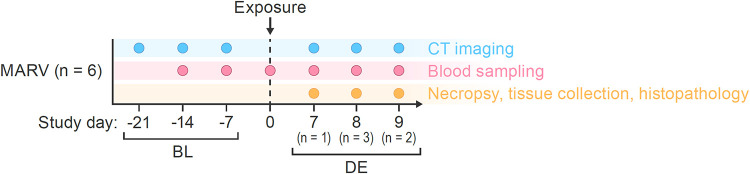
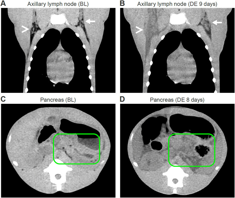
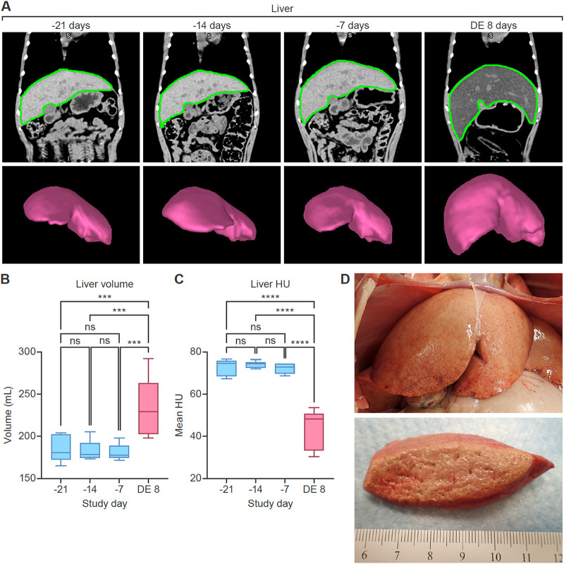
Marburg virus (MARV) is a highly virulent zoonotic filovirid that causes Marburg virus disease (MVD) in humans. The pathogenesis of MVD remains poorly understood, partially due to the low number of cases that can be studied, the absence of state-of-the-art medical equipment in areas where cases are reported, and limitations on the number of animals that can be safely used in experimental studies under maximum containment animal biosafety level 4 conditions. Medical imaging modalities, such as whole-body computed tomography (CT), may help to describe disease progression in vivo, potentially replacing ethically contentious and logistically challenging serial euthanasia studies. Towards this vision, we performed a pilot study, during which we acquired whole-body CT images of 6 rhesus monkeys before and 7 to 9 days after intramuscular MARV exposure. We identified imaging abnormalities in the liver, spleen, and axillary lymph nodes that corresponded to clinical, virological, and gross pathological hallmarks of MVD in this animal model. Quantitative image analysis indicated hepatomegaly with a significant reduction in organ density (indicating fatty infiltration of the liver), splenomegaly, and edema that corresponded with gross pathological and histopathological findings. Our results indicated that CT imaging could be used to verify and quantify typical MVD pathogenesis versus altered, diminished, or absent disease severity or progression in the presence of candidate medical countermeasures, thus possibly reducing the number of animals needed and eliminating serial euthanasia. IMPORTANCE Marburg virus (MARV) is a highly virulent zoonotic filovirid that causes Marburg virus disease (MVD) in humans. Much is unknown about disease progression and, thus, prevention and treatment options are limited. Medical imaging modalities, such as whole-body computed tomography (CT), have the potential to improve understanding of MVD pathogenesis. Our study used CT to identify abnormalities in the liver, spleen, and axillary lymph nodes that corresponded to known clinical signs of MVD in this animal model. Our results indicated that CT imaging and analyses could be used to elucidate pathogenesis and possibly assess the efficacy of candidate treatments.
Keywords: Marburg virus; computed tomography; filovirus; medical imaging; viral pathogenesis.
Conflict of interest statement
The authors declare no conflict of interest.
Figures
Similar articles
-
Marburg virus infection in nonhuman primates: Therapeutic treatment by lipid-encapsulated siRNA.Sci Transl Med. 2014 Aug 20;6(250):250ra116. doi: 10.1126/scitranslmed.3009706. Sci Transl Med. 2014. PMID: 25143366 Free PMC article.
-
Clinical, Histopathologic, and Immunohistochemical Characterization of Experimental Marburg Virus Infection in A Natural Reservoir Host, the Egyptian Rousette Bat (Rousettus aegyptiacus).Viruses. 2019 Mar 2;11(3):214. doi: 10.3390/v11030214. Viruses. 2019. PMID: 30832364 Free PMC article.
-
Establishment and characterization of a lethal mouse model for the Angola strain of Marburg virus.J Virol. 2014 Nov;88(21):12703-14. doi: 10.1128/JVI.01643-14. Epub 2014 Aug 20. J Virol. 2014. PMID: 25142608 Free PMC article.
-
Marburg virus pathogenesis - differences and similarities in humans and animal models.Virol J. 2019 Dec 30;16(1):165. doi: 10.1186/s12985-019-1272-z. Virol J. 2019. PMID: 31888676 Free PMC article. Review.
-
Pathogenicity and virulence of Marburg virus.Virulence. 2022 Dec;13(1):609-633. doi: 10.1080/21505594.2022.2054760. Virulence. 2022. PMID: 35363588 Free PMC article. Review.
Cited by
-
Novel antiviral approaches for Marburg: a promising therapeutics in the pipeline.Front Microbiol. 2024 Apr 25;15:1387628. doi: 10.3389/fmicb.2024.1387628. eCollection 2024. Front Microbiol. 2024. PMID: 38725678 Free PMC article. Review.
-
Reactive oxygen species-related oxidative changes are associated with splenic lymphocyte depletion in Ebola virus infection.Npj Imaging. 2025;3(1):16. doi: 10.1038/s44303-025-00079-x. Epub 2025 Apr 24. Npj Imaging. 2025. PMID: 40291761 Free PMC article.
-
Emerging and reemerging infectious diseases: global trends and new strategies for their prevention and control.Signal Transduct Target Ther. 2024 Sep 11;9(1):223. doi: 10.1038/s41392-024-01917-x. Signal Transduct Target Ther. 2024. PMID: 39256346 Free PMC article. Review.
References
-
- Kuhn JH, Crozier I. 2022. Ebolavirus and marburgvirus infections, p 1645–1652. In Loscalzo J, Fauci AS, Kasper DL, Hauser SL, Longo DL, Jameson JL. (ed), Harrison's Principles of Internal Medicine, 21st ed, vol 2. McGraw-Hill Education, Columbus, Ohio, USA.
-
- Kuhn JH, Amarasinghe GK, Perry DL. 2020. Filoviridae, p 449–503. In Howley PM, Knipe DM, Whelan SPJ. (ed), Fields Virology, 7th ed., vol 1 (Emerging viruses). Lippincott Williams & Wilkins, Philadelphia, PA, USA.
-
- Koundouno FR, Kafetzopoulou LE, Faye M, Renevey A, Soropogui B, Ifono K, Nelson EV, Kamano AA, Tolno C, Annibaldis G, Millimono SL, Camara J, Kourouma K, Doré A, Millimouno TE, Tolno FMB, Hinzmann J, Soubrier H, Hinrichs M, Thielebein A, Herzer G, Pahlmann M, Ki-Zerbo GA, Formenty P, Legand A, Wiley MR, Faye O, Diagne MM, Sall AA, Lemey P, Bah A, Günther S, Keita S, Duraffour S, Magassouba N. 2022. Detection of Marburg virus disease in Guinea. N Engl J Med 386:2528–2530. doi:10.1056/NEJMc2120183. - DOI - PMC - PubMed
-
- World Health Organization. 2022. Ghana declares first-ever outbreak of Marburg virus disease. https://www.afro.who.int/countries/ghana/news/ghana-declares-first-ever-.... - PubMed
-
- ProMED. 2023. Marburg virus disease - Equatorial Guinea (01): (KIE-NTEM) WHO confirmed. Archive Number: 20230214.8708367. https://promedmail.org/promed-post/?id=8708367.
Publication types
MeSH terms
Grants and funding
LinkOut - more resources
Full Text Sources
