. 2023 Apr 3;64(4):7.
doi: 10.1167/iovs.64.4.7.

Schwann Cells Are Key Regulators of Corneal Epithelial Renewal

Affiliations

Schwann Cells Are Key Regulators of Corneal Epithelial Renewal

Kaveh Mirmoeini et al. Invest Ophthalmol Vis Sci. .

Abstract

Purpose: Corneal sensory nerves protect the cornea from injury. They are also thought to stimulate limbal stem cells (LSCs) to produce transparent epithelial cells constantly, enabling vision. In other organs, Schwann cells (SCs) associated with tissue-innervating axon terminals mediate tissue regeneration. This study defines the critical role of the corneal axon-ensheathing SCs in homeostatic and regenerative corneal epithelial cell renewal.

Methods: SC localization in the cornea was determined by in situ hybridization and immunohistochemistry with SC markers. In vivo SC visualization and/or ablation were performed in mice with inducible corneal SC-specific expression of tdTomato and/or Diphtheria toxin, respectively. The relative locations of SCs and LSCs were observed with immunohistochemical analysis of harvested genetically SC-prelabeled mouse corneas with LSC-specific antibodies. The correlation between cornea-innervating axons and the appearance of SCs was ascertained using corneal denervation in rats. To determine the limbal niche cellular composition and gene expression changes associated with innervation-dependent epithelial renewal, single-cell RNA sequencing (scRNA-seq) of dissociated healthy, de-epithelized, and denervated cornea limbi was performed.

Results: We observed limbal enrichment of corneal axon-associated myelinating and non-myelinating SCs. Induced local genetic ablation of SCs, although leaving corneal sensory innervation intact, markedly inhibited corneal epithelial renewal. scRNA-seq analysis (1) highlighted the transcriptional heterogenicity of cells populating the limbal niche, and (2) identified transcriptional changes associated with corneal innervation and during wound healing that model potential regulatory paracrine interactions between SCs and LSCs.

Conclusions: Limbal SCs are required for innervation-dependent corneal epithelial renewal.


Conflict of interest statement

Disclosure: K. Mirmoeini, None; K. Tajdaran, None; J. Zhang, None; T. Gordon, None; A. Ali, None; D.R. Kaplan, None; K. Feinberg, None; G.H. Borschel, None

Figures

Figure 1.
Figure 1.
Myelinating and non-myelinating SCs are abundant in the limbus. (A) In situ hybridization demonstrates enrichment of Sox10-positive cells in the corneal limbus. Scale bar: 100 µm. (B) Live image of tamoxifen-induced tdTomato (tdT)-positive fluorescent SCs in Sox10-tdT mouse whole eye (arrowheads, upper panel) and part of limbus (magnified in lower panel). Scale bars: 1 µm and 200 µm for upper and lower panels, respectively. (C) Immunofluorescent 10 µm extended-depth focus immunofluorescent image of Sox10-tdT mouse fixed cornea demonstrates a 1:1 association between by (βIII)-tubulin–positive (green) neurons and tdT-positive SCs (red), which abundantly populate the limbal area. Scale bar: 250 µm. (D, E) Immunofluorescent image of whole-mount rat cornea, demonstrating non-myelinated, MBP-negative (indicated by arrowhead) and myelinated, MBP-positive (red, indicated by arrow) βIII-tubulin–positive (green) neurons in the limbal area. Image in D is positive for the paranodal protein Caspr (green), together with red sodium channel NaCh+ (indicated by asterisks) at the nodes of Ranvier in the left and right panels, respectively (E). Scale bars: 50 µm (D); 2 µm (E). (F) Immunofluorescent image of sagittal section of central rat cornea at the limbal area demonstrating a cross-section of single βIII-tubulin–positive axon enwrapped by a single membrane layer of MAG-positive non-myelinating Schwann cells, presented in a sequence of constitutive magnifications of the defined area. Scale bars (top to bottom): 100 µm, 25 µm, and 2.5 µm.
Figure 2.
Figure 2.
Close localization of SCs and LSCs at limbus. (A) A 10-µm extended-depth focus immunofluorescent image of Sox10-tdT mouse cornea demonstrates that Krt15-positive LSCs (green) and tdT-positive (red) SCs abundantly occupy the limbal area. Scale bars: 100 µm. (B) Immunofluorescent one-focal-plate image of Sox10-tdT mouse fixed cornea limbal area shows close localization (arrowheads) of Krt15-positive LSCs (green) and tdT-positive (red) SCs. Scale bar: 100 µm.
Figure 3.
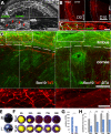
Figure 3.
Both denervation-induced and genetic ablation of corneal SCs caused the NK phenotype. (A) Corneal denervation completely ablated SCs in 48 hours, as shown by in vivo fluorescent imaging of tdT-positive SCs in Sox10-tdT mouse denervated cornea. The magnified area shows 3 and 48 hours after corneal denervation. Scale bar: 200 µm. (B) Fluorescent image of the limbal area of tamoxifen-induced tdT-positive SCs in Sox10-tdT (left panel) and Sox10-tdT;DTA mouse fixed corneas 10 days after tamoxifen administration (right panel). Scale bar: 500 µm. (C) Immunofluorescent image of the limbal area of whole-mount fixed cornea harvested from Sox10-DTA mouse shows an abundance of Sox10-positive SC nuclei (green, indicated by arrowheads) attached to βIII-tubulin–positive axons (red) at 4 hours (left panel), but SCs disappeared 10 days after tamoxifen administration (right panel). Scale bar: 200 µm. (D) Immunofluorescent 10-mImextended-depth focus image of Sox10-tdT (left panel) and Sox10-tdT;DTA mouse fixed cornea shows unmodified limbal and epithelial innervation by βIII-tubulin–positive (green) neurons in the SC-ablated, comparable to the SC-non-ablated corneas despite a significant reduction in volume of tdT-positive SCs (red) 10 days after tamoxifen administration, as shown in the magnified lower panel images. Scale bars: 100 µm and 250 µm in the main and magnified images, respectively. (E) In vivo photos of mouse corneas demonstrate opacification of the SC-ablated left (L) eye (arrows) compared to a clear cornea in the untreated right (R) eye. (F) In vivo photographs of de-epithelialized mouse corneas demonstrate the progress of corneal epithelial healing for 4 days after de-epithelialization in SC-ablated (SC abl, Sox10-DTA mice) and SC-present (SC pr) tamoxifen treated corneas. “0h” indicates the cornea condition immediately after epithelial removal, 10 days after tamoxifen administration. Fluorescein (yellow) stains the corneal area lacking epithelium. (G, H) Quantitative representation of SCs ablation in B to D (n = 3 per condition) and the corneal healing in F (n = 6 per condition), respectively. Error bars represent standard error. *P < 0.05; **P < 0.01; ***P < 0.005 (t-test in G and ANOVA in H).
Figure 4.
Figure 4.
The scRNA-seq identification of cell types populating the limbal niche in rat. scRNA-seq analysis was performed on dissociated limbi harvested from healthy rat corneas. The clusters of LSCs and MSCs were extracted and reanalyzed. (A) UMAP of resultant cell clusters, with colors and numbers denoting distinct clusters: LSCs (11); TACs (1 and 18); MSCs (14 and 15); epithelial cells (4, 6, 7, 8, 9, 12, and 13); conjunctival cells (0, 2, 3, 5, 10, and 18); Langerhans cells, macrophages, lymphocytes, mast cells, and NK cells (16 and 17); endothelial cells of lymphatic vessels (20); and SCs (19). The clusters of LSCs, MSCs, TACs, and SCs are highlighted. (B) Violin plot showing relative expression of the representative cell-type–specific gene markers as per A: Sox10 and Mbp for SCs; Abcg2 and Sox17 for LSCs; Pdgfra and Pdgfrb for MSCs; and Birc5 and Ki67 for TACs. (C) UMAP of colors and numbers represent transcriptionally distinct clusters of LSCs and MSCs which, as in A, were combined and reanalyzed. Subclusters 0, 3, 5, 8 and 10 outlined in black represent LSCs, and subclusters 1, 2, 6, 7, and 9 outlined in blue represent MSCs. Subcluster 4 shares mRNA representatives for both cell populations. (D) Violin plot showing relative expression of the selected genes in the indicated subclusters shown in C.
Figure 5.
Figure 5.
Expression changes following corneal de-epithelization or denervation allow prognosing of paracrine interactions regulating LSCs activity. (A) In vivo photographs of injured rat corneas demonstrate the progress of corneal epithelial healing for 3 days after de-epithelialization in denervated (den) and normally innervated (norm) corneas. “0h” indicates cornea condition immediately after epithelial removal. Fluorescein (yellow) stains the corneal area lacking epithelium. In the denervated cornea, de-epithelization was induced 5 days after a single electrocautery of the ophthalmomaxillary branch of the trigeminal nerve. (BF) A scRNA-seq–based quantitative representation of the indicated gene expression in clusters of SCs (B), MSCs (D), and LSCs (E), comparing dissociated limbi harvested from healthy (norm), de-epithelized (injured [inj]), and denervated (den) corneas. Note that, due to the absence of SCs in denervated corneas, no gene expression for this condition is presented in B or C. Expression of the Sox10/Sox2 ratio in limbal SCs comparing healthy (norm) versus de-epithelialized (inj). (F) Expression level of Ki67 in the three conditions in LSCs (upper chart) and MSCs (lower chart). (G) Transcriptome-based network model of computationally predicted paracrine communication between SCs and LSCs and between MSCs and LSCs, based on scRNA-seq analysis of the three experimental conditions in B to F. Arrows indicate the direction of communication. Green and yellow nodes indicate the expression of genes encoding for ligands of interest in both SCs and MCSs. Green nodes indicate genes with a notable expression change, and yellow nodes indicate genes with no or minor expression changes between normal, de-epithelialized (24 hours after de-epithelization), or denervation (5 days after denervation) conditions, as shown in A. The purple node indicates de novo expression of Osm by MSCs following corneal denervation of (inj) corneas.

References

    1. Hanna C, Bicknell DS, O'Brien JE. Cell turnover in the adult human eye. Arch Ophthalmol. 1961; 65: 695–698. - PubMed
    1. Cenedella RJ, Fleschner CR.. Kinetics of corneal epithelium turnover in vivo. Studies of lovastatin. Invest Ophthalmol Vis Sci. 1990; 31: 1957–1962. - PubMed
    1. Bentley AJ, Nakamura T, Hammiche A, et al. .. Characterization of human corneal stem cells by synchrotron infrared micro-spectroscopy. Mol Vis. 2007; 13: 237–242. - PMC - PubMed
    1. Chen Z, de Paiva CS, Luo L, Kretzer FL, Pflugfelder SC, Li DQ.. Characterization of putative stem cell phenotype in human limbal epithelia. Stem Cells. 2004; 22: 355–366. - PMC - PubMed
    1. Schlotzer-Schrehardt U, Kruse FE.. Identification and characterization of limbal stem cells. Exp Eye Res. 2005; 81: 247–264. - PubMed

Publication types

Grants and funding