. 2023 Apr 10;19(4):e1011215.
doi: 10.1371/journal.ppat.1011215. eCollection 2023 Apr.

MmuPV1 E7's interaction with PTPN14 delays Epithelial differentiation and contributes to virus-induced skin disease

Affiliations

MmuPV1 E7's interaction with PTPN14 delays Epithelial differentiation and contributes to virus-induced skin disease

James C Romero-Masters et al. PLoS Pathog. .

Abstract

Human papillomaviruses (HPVs) contribute to approximately 5% of all human cancers. Species-specific barriers limit the ability to study HPV pathogenesis in animal models. Murine papillomavirus (MmuPV1) provides a powerful tool to study the roles of papillomavirus genes in pathogenesis arising from a natural infection. We previously identified Protein Tyrosine Phosphatase Non-Receptor Type 14 (PTPN14), a tumor suppressor targeted by HPV E7 proteins, as a putative cellular target of MmuPV1 E7. Here, we confirmed the MmuPV1 E7-PTPN14 interaction. Based on the published structure of the HPV18 E7/PTPN14 complex, we generated a MmuPV1 E7 mutant, E7K81S, that was defective for binding PTPN14. Wild-type (WT) and E7K81S mutant viral genomes replicated as extrachromosomal circular DNAs to comparable levels in mouse keratinocytes. E7K81S mutant virus (E7K81S MmuPV1) was generated and used to infect FoxN/Nude mice. E7K81S MmuPV1 caused neoplastic lesions at a frequency similar to that of WT MmuPV1, but the lesions arose later and were smaller than WT-induced lesions. The E7K81S MmuPV1-induced lesions also had a trend towards a less severe grade of neoplastic disease. In the lesions, E7K81S MmuPV1 supported the late (productive) stage of the viral life cycle and promoted E2F activity and cellular DNA synthesis in suprabasal epithelial cells to similar degrees as WT MmuPV1. There was a similar frequency of lateral spread of infections among mice infected with E7K81S or WT MmuPV1. Compared to WT MmuPV1-induced lesions, E7K81S MmuPV1-induced lesions had a significant expansion of cells expressing differentiation markers, Keratin 10 and Involucrin. We conclude that an intact PTPN14 binding site is necessary for MmuPV1 E7's ability to contribute to papillomavirus-induced pathogenesis and this correlates with MmuPV1 E7 causing a delay in epithelial differentiation, which is a hallmark of papillomavirus-induced neoplasia.


Conflict of interest statement

The authors have declared that no competing interests exist.

Figures

Fig 1
Fig 1. In Silico analysis of amino acid sequence and protein structure identifies key amino acids for MmuPV1 E7’s interaction with PTPN14.
(A) Schematic of a representative HPV E7 protein. Papillomavirus E7 proteins contain conserved regions 1 and 2 (CR1, CR2) and a highly structured C-terminus organized around two zinc binding motifs (CxxC). Enlargement indicates an amino acid sequence alignment of the C-termini of several human and rodent papillomavirus E7 proteins. R84 and L91 are two amino acids in HPV18 E7 that make contact with PTPN14 as determined by the HPV18 E7-PTPN14 crystal structure [63]. Identity with these amino acids is indicated in red. An arginine corresponding to HPV18 R84 is conserved among many papillomavirus E7 but is replaced by lysine in at least two rodent papillomavirus E7 (yellow highlight). (B) Schematic of Homo sapiens (Hs)PTPN14 including its 4.1, ezrin, radixin, moesin (FERM) domain, protein tyrosine phosphatase (PTP) domain, and PxxY (PY) motifs identified in [87,88]. Enlargement indicates an amino acid sequence alignment of a segment of the PTP domains of human and selected rodent PTPN14 proteins. F1044, G1055, and E1095 are three amino acids in PTPN14 that make contact with HPV18 E7 as determined by the HPV18 E7-PTPN14 crystal structure [63], and these are highlighted in red. (C & D) Threading analysis using the structure information from the interaction between PTPN14 (white) and HPV18 E7 (blue) (C) was used to predict the structure of the PTPN14 (white) and MmuPV1 E7 (yellow) (D). Critical amino acids for the interaction between E7 and PTPN14 are highlighted in red with E1095 of PTPN14 indicated in both panels (C & D), and arginine (R) 84 in HPV18 E7 (C) and lysine (K) 81 in MmuPV1 (D). Red arrows point to the amino acids highlighted in the images. Only the regions within PTPN14 and E7 that interact are shown.
Fig 2
Fig 2. A lysine at amino acid position 81 in MmuPV1 E7 is critical for the interaction with PTPN14.
NIH 3T3 cells were transfected with either empty vector (N) or plasmid constructs that express N-terminally and C-terminally HA/FLAG epitope-tagged HPV16 E7, WT MmuPV1 E7, or MmuPV1 E7K81S mutant. MmuPV1 E7 constructs were either N- or C-terminally tagged as indicated. Cells were harvested and subjected to immunoprecipitation with HA antibodies. For HA IP, 1 mg of HPV16 E7 and 3 mg of other lysates was used and 100 ug WCL on 4–12% mini gels was used for input Western blot analysis. Whole cell lysates (Input) and HA IP eluates were subjected to western blot analysis using antibodies against PTPN14, pRb, FLAG (E7), Actin, or GFP (transfection control). Left panel on blot is the same images labeled Experiment 1 in S2 Fig.
Fig 3
Fig 3. MmuPV1 E7K81S mutant does not impair viral genome maintenance in basal keratinocytes.
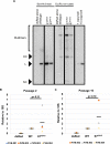
(A) Early passage mouse keratinocytes (MKs) isolated from neonates were transfected with dsRed alone or in combination with WT, E7D90A, or E7K81S re-circularized viral genomes. DNA was isolated and restriction digested with a linearizing enzyme (BamHI) or non-cutter (EcoRI). Southern blot analysis was performed using an MmuPV1 genome probe included a standard of 1 copy/cell and 10 copy/cell to estimate copy number level per cell. The positions of open circular (OC), linear (L), and supercoiled (SC) genomes are indicated. The different panels are derived from the same gel and have been sampled at the same exposure. An uncropped version of Fig 3A is provided in S3A Fig. (B and C) DNA isolated from transfected MKs were subjected to qPCR analysis using primers for the viral E2 gene and mouse 18S gene. Viral genome copy number was normalized to total DNA content as determined by levels of the 18S gene and relative copy number is indicated after normalization to the level of genomes in WT MmuPV1 transfected cells. Three different transfected MK cell populations are shown. Wilcoxon rank sum test was performed on the different conditions to assess statistical significance. Black bars indicate means for each condition.
Fig 4
Fig 4. E7K81S MmuPV1 induces lesions at a slower rate and the lesions are smaller in size than those induced by WT MmuPV1.
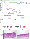
(A) Mice were monitored for papilloma development over a 6-month period every two weeks following infection. Each ear was considered a site of infection and the week post-infection that a lesion developed was recorded. A Kaplan-Meier curve was generated using the week post-infection versus fraction of lesion-free sites across all animals. Data was subjected to Log-Rank test to assess statistical significance (p<0.05 was considered significant). (B&C) Lesions were measured at the time of collection for area of lesion size (depth was excluded from measurements). Data was plotted as dot plots with each site measured represented by a single dot. Wilcoxon-rank sum analysis was performed on the data to assess statistical significance (p<0.05 was considered significant). Black bars indicate the mean for each condition. (D) Images of H&E-stained tissue sections at 2.5X magnification to show size differences of lesions at 6-months post-infection.
Fig 5
Fig 5. E7K81S MmuPV1 causes severe dysplasia comparable to WT MmuPV1.
(A&B) H&E tissue sections were scored for disease severity by two pathologists in a blinded fashion. Mock (uninfected) treated mice showed skin within normal limits (WNL). WT and E7K81S-infected mice all showed a range of mild dysplasia, severe dysplasia, and carcinoma in situ (CIS+) as indicated. Data was subjected to Wilcoxon-rank sum analysis to assess statistical significance (p<0.05 was considered significant). (C) Representative images of different disease severity are shown at low (4X) and high (20X) magnification. WNL: Normal ear skin with the arrow indicating a prior scarification site. At high magnification, no epithelial multilayering or cytologic atypia is observed. Mild Dysplasia: Mild thickening of surface epithelium (arrows). At high power, proliferation at the base of the epithelium with mild nuclear crowding, hyperchromasia and koliocytes is observed. There is preservation of epithelial maturation in the top half of the epithelium. Severe Dysplasia: Marked thickening and papillomatosis of surface epithelium. At high power, nuclear crowding and marked nuclear atypia and numerous mitotic figures (arrows) are present; surface maturation is minimal or absent. CIS+: In the background of severe dysplasia, an irregular area with bright eosinophilic keratinization is present. At high magnification, single epithelial cells in the stroma are present. These features are highly suspicious for nascent microinvasive squamous cell carcinoma.
Fig 6
Fig 6. MmuPV1 E7K81S mutant does not impair capsid protein production.
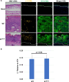
(A) FFPE tissue sections were subjected to immunofluorescence (IF) analysis using a tyramide signal amplification (TSA) system using antibodies against K14 (green) and the viral capsid protein L1 (yellow). Representative images are shown for K14, L1, and merged (which includes Hoechst stain) at 10X magnification. A 10X image of the H&E is also shown. Dashed white lines indicate basement membrane of epithelial tissues. (B) IF images were quantified using ImageJ to determine area of lesion that stained positive for L1 and K14 using 9 images per condition. L1 signal was normalized to K14 signal. Standard error is shown. Wilcoxon Rank Sum test was performed to assess statistical significance and p-value is shown.
Fig 7
Fig 7. MmuPV1 E7K81S mutant quasivirus produces viral E4 transcripts and genome amplification in lesions to the same degree as WT quasivirus.
(A) FFPE tissue were subjected to in situ hybridization with a probe that detects both E4 RNA and DNA using the RNAscope technology. 10X images are shown for H&E. Representative high (63X) and low (10X) magnification images are shown for RNA/DNAscope and DNAscope (RNAse treated section) to detect viral E4 transcripts and viral genome amplification. Dashed black lines indicate basement membrane of epithelial tissues. (B) The fraction of cells going through viral genome amplification within a lesion is shown by determining the number of nuclei and number of positive staining nuclei using an ImageJ plugin developed by the Lab of Dr. David Ornelles (Wake Forest) for 18 fields of view. Ratio of positive staining cells to the total number of nuclei was determined and average shown. Standard error is shown. Wilcoxon Rank Sum test was performed to assess statistical significance and p-value is shown. (C) Upon euthanasia, animals were examined for development of warts/lesions at secondary sites of infection (sites other than ears). The percentage of animals that developed warts/lesions was calculated and shown. Fischer’s Exact Test was used to assess statistical significance.
Fig 8
Fig 8. Similar expression of MCM7 and BrdU incorporation in the E7K81S- and WT induced lesions.
FFPE tissues were subjected to immunohistochemistry (IHC) using antibodies against MCM7 and BrdU (as indicated). H&E and stained tissue sections were imaged at both a low (10X) and high (20X) magnification as indicated.
Fig 9
Fig 9. Keratinocyte differentiation is less impaired in the E7K81S-induced lesions than in WT MmuPV1-induced lesions.
(A) FFPE tissue sections were subjected to Immunofluorescence (IF) using antibodies against K14 (green), K10 (cyan), and Involucrin (IVL, red). All sections were stained with the same secondary antibody (anti-mouse conjugated to AF488) and all images are pseudo colored for detection of AF-488. All images are 10X magnification. (B) IF images were quantified using ImageJ determining area with lesion that stained positive for K10 and IVL using 6 images per condition. K10 and IVL signal was normalized to K14 signal. Standard error is shown. Wilcoxon rank sum test was performed to assess statistical significance, and p-values are shown.

Similar articles

Cited by

References

    1. de Martel C, Ferlay J, Franceschi S, Vignat J, Bray F, Forman D, et al.. Global burden of cancers attributable to infections in 2008: a review and synthetic analysis. Lancet Oncol. 2012;13: 607–615. doi: 10.1016/S1470-2045(12)70137-7 - DOI - PubMed
    1. Forman D, de Martel C, Lacey CJ, Soerjomataram I, Lortet-Tieulent J, Bruni L, et al.. Global burden of human papillomavirus and related diseases. Vaccine. 2012;30 Suppl 5: F12–23. doi: 10.1016/j.vaccine.2012.07.055 - DOI - PubMed
    1. zur Hausen H. Papillomaviruses in the causation of human cancers—a brief historical account. Virology. 2009;384: 260–265. doi: 10.1016/j.virol.2008.11.046 - DOI - PubMed
    1. Liu L. Fields Virology, 6th Edition. Clinical Infectious Diseases. 2014;59: 613. doi: 10.1093/cid/ciu346 - DOI
    1. McBride AA. Human papillomaviruses: diversity, infection and host interactions. Nat Rev Microbiol. 2022;20: 95–108. doi: 10.1038/s41579-021-00617-5 - DOI - PubMed

Publication types

Substances