. 2023 Apr 18;120(16):e2219290120.
doi: 10.1073/pnas.2219290120. Epub 2023 Apr 10.

An SI3-σ arch stabilizes cyanobacteria transcription initiation complex

Affiliations

An SI3-σ arch stabilizes cyanobacteria transcription initiation complex

Liqiang Shen et al. Proc Natl Acad Sci U S A. .

Abstract

Multisubunit RNA polymerases (RNAPs) associate with initiation factors (σ in bacteria) to start transcription. The σ factors are responsible for recognizing and unwinding promoter DNA in all bacterial RNAPs. Here, we report two cryo-EM structures of cyanobacterial transcription initiation complexes at near-atomic resolutions. The structures show that cyanobacterial RNAP forms an "SI3-σ" arch interaction between domain 2 of σA2) and sequence insertion 3 (SI3) in the mobile catalytic domain Trigger Loop (TL). The "SI3-σ" arch facilitates transcription initiation from promoters of different classes through sealing the main cleft and thereby stabilizing the RNAP-promoter DNA open complex. Disruption of the "SI3-σ" arch disturbs cyanobacteria growth and stress response. Our study reports the structure of cyanobacterial RNAP and a unique mechanism for its transcription initiation. Our data suggest functional plasticity of SI3 and provide the foundation for further research into cyanobacterial and chloroplast transcription.

Keywords: RNA polymerase; cyanobacteria; gene transcription; transcription initiation; transcription initiation factor.


Conflict of interest statement

The authors declare no competing interest.

Figures

Fig. 1.
Fig. 1.
The cryo-EM structure of Syn6803 RPitc. (A) RNAP subunits of cyanobacteria Synechocystis sp. PCC 6803 (Left) and E. coli (Right). (B) The nucleic-acid scaffold used in the cryo-EM structure determination. (C) Front and back view orientations of Syn6803 RPitc cryo-EM map. (D) The front and back view orientations of Syn6803 RPitc structure. (E) The cryo-EM map and model of the nucleic-acid scaffold of Syn6803 RPitc. (F) The cryo-EM map and model of SI3, σ2, and the nontemplate -10 element DNA of Syn6803 RPitc. (G) Superimposition of the nucleic-acid scaffolds of Syn6803 RPitc and E. coli RPo. (H) Superimposition of the clamp domains of Syn6803 RPitc and E. coli RPo. RNAP subunits and nucleic-acid chains are colored as in the color scheme.
Fig. 2.
Fig. 2.
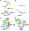
The structure of Syn6803 RNAP-SI3. (A) The overall structure of Syn6803 RNAP-SI3. The Trigger Helices connect to the SI3-tail and SI3-fin domains. (B) The overall structure of E. coli RNAP-SI3 (PDB: 4YLN). (C) SI3 encloses half of the RNAP surface. It extends from the secondary channel to the top of main cleft and makes interaction with σ2. (D) The SI3-fin and SI3-tail domains shield RNAP Rim Helices.
Fig. 3.
Fig. 3.
The structural and functional analysis of SI3-σ arch. (A) Surface presentation of the SI3-σ arch. (B) Electrostatic surface presentation of the SI3-σ arch. (C) Detailed interaction between SI3 and σ2. (D) The cryo-EM map of the SI3-σ2 interface. (E) The half-lives of RNAP-promoter open complex comprising galP1cons promoter and indicated holoenzymes challenged with heparin. Error bars represent SD from triplicate experiments. “Low stability” indicates that decay rates were too high to measure accurately. (F) Activity of EWT and EΔhead on different promoters. Top, promoter sequences with transcription start site shown in bold and consensus elements underlined; Bottom, representative gel (Left) and bar plot (Right) show amounts of runoff transcripts of EΔhead in % from that of EWT. Error bars represent SD from three independent experiments. (G) The half-lives of RNAP-promoter open complexes comprising WT/Δhead RNAP and different σ factors. Error bars represent SD from triplicate experiments. (H) Serial dilutions of cultures of S. elongatus 7942 WT strain and strain with genomic deletion of SI3 (Δhead) plated on BG-11 media and grown at conditions indicated below images with constant light. (I) Growth curves of WT and Δhead S. elongatus 7942 strains in 12 h light/12 h dark conditions. Error bars represent SD from three independently grown cultures. The Inset is an image of flasks after 33-d growth.
Fig. 4.
Fig. 4.
SI3 interacts with the rim helices. (A) Detailed interaction between the rim helices and SI3-tail. (B) The cryo-EM map of the SI3-rim interface. (C) The RTRHG loop deletion leads to increased pausing during elongation. Elongation complex is assembled with 14-nt long RNA labeled at 5′-end with 32P and template and nontemplate DNA oligonucleotides fully complementary to each other. NTPs were added to a final concentration of 10 μM, reaction stopped at indicated timepoints with addition of formamide-containing loading buffer. (D) The protein sequence alignment of the SI3-tail domain of various cyanobacterial RNAP and plant chloroplast PEPs.
Fig. 5.
Fig. 5.
TL refolding induces structural change in Syn6803 NTP-bound RPitc. (A) The cryo-EM map and model for the active site. CTP adopts an insertion state at the “i+1” site. (B) CTP induces refolding of TL into TH. (C) The Top panel shows the detailed interaction between CTP and RNAP residues; the Bottom panel shows the effect of alanine substitutions of CTP-contact residues on the efficiency of the first phosphoric diester bond formation during de novo initiation on galP1cons promoter. (D) The conformational changes of SI3 induced by TL refolding (cluster of arrows showing domain movement). (E) The comparison of SI3 movement upon TL refolding between Syn6803 RNAP (Left) and E. coli RNAP (Right).

References

    1. Decker K. B., Hinton D. M., Transcription regulation at the core: Similarities among bacterial, archaeal, and eukaryotic RNA polymerases. Annu. Rev. Microbiol. 67, 113–139 (2013). - PubMed
    1. Weiss A., Shaw L. N., Small things considered: The small accessory subunits of RNA polymerase in gram-positive bacteria. FEMS Microbiol. Rev. 39, 541–554 (2015). - PMC - PubMed
    1. Feklistov A., Sharon B. D., Darst S. A., Gross C. A., Bacterial sigma factors: A historical, structural, and genomic perspective. Annu. Rev. Microbiol. 68, 357–376 (2014). - PubMed
    1. Murakami K. S., Masuda S., Darst S. A., Structural basis of transcription initiation: RNA polymerase holoenzyme at 4 A resolution. Science 296, 1280–1284 (2002). - PubMed
    1. Li L., Fang C., Zhuang N., Wang T., Zhang Y., Structural basis for transcription initiation by bacterial ECF sigma factors. Nat. Commun. 10, 1153 (2019). - PMC - PubMed

Publication types

LinkOut - more resources