. 2023 Apr 18;120(16):e2220340120.
doi: 10.1073/pnas.2220340120. Epub 2023 Apr 10.

RPL26/uL24 UFMylation is essential for ribosome-associated quality control at the endoplasmic reticulum

Affiliations

RPL26/uL24 UFMylation is essential for ribosome-associated quality control at the endoplasmic reticulum

Francesco Scavone et al. Proc Natl Acad Sci U S A. .

Abstract

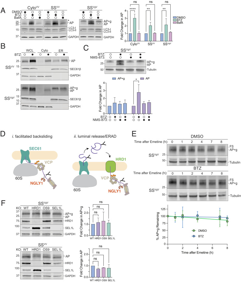
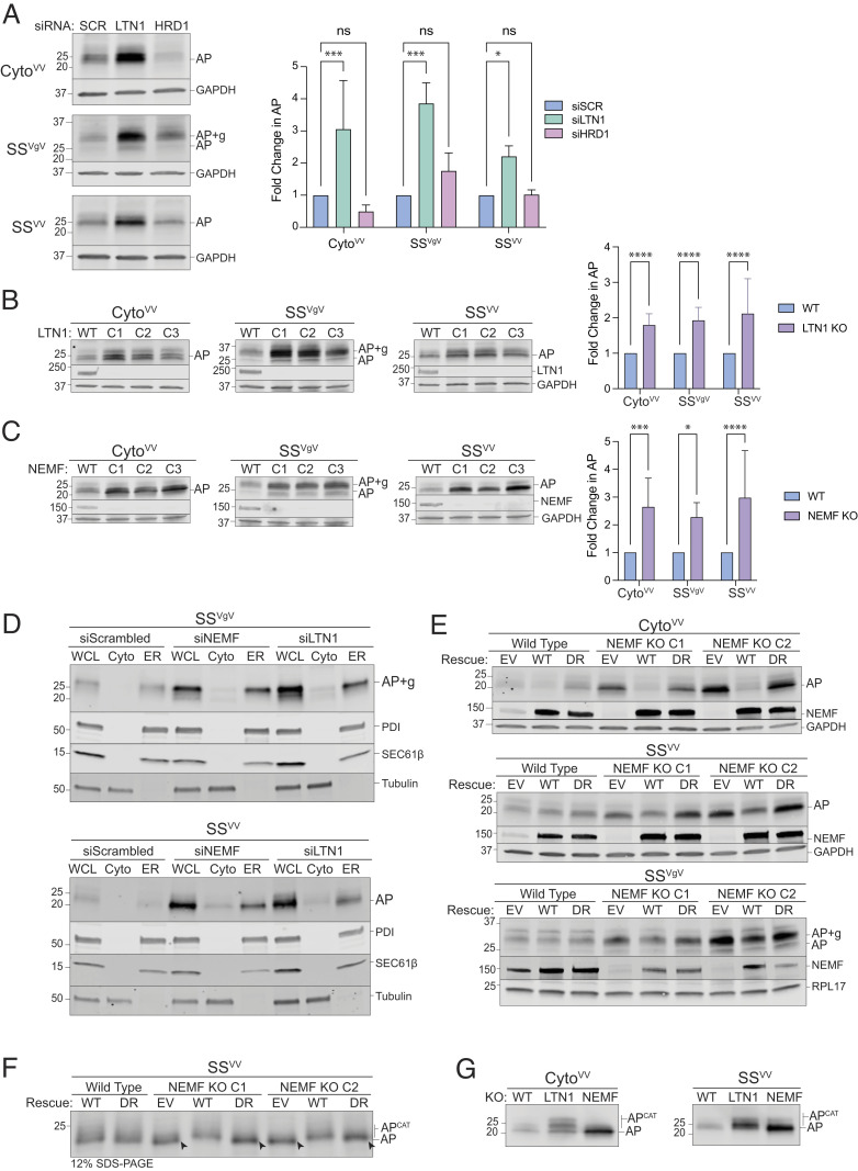
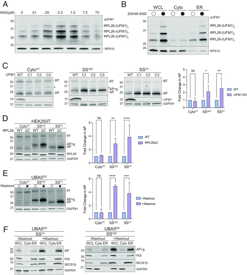
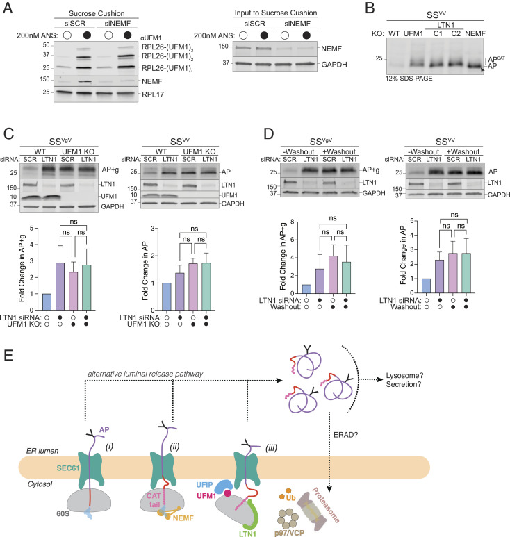
Ribosomes that stall while translating cytosolic proteins are incapacitated by incomplete nascent chains, termed "arrest peptides" (APs) that are destroyed by the ubiquitin proteasome system (UPS) via a process known as the ribosome-associated quality control (RQC) pathway. By contrast, APs on ribosomes that stall while translocating secretory proteins into the endoplasmic reticulum (ER-APs) are shielded from cytosol by the ER membrane and the tightly sealed ribosome-translocon junction (RTJ). How this junction is breached to enable access of cytosolic UPS machinery and 26S proteasomes to translocon- and ribosome-obstructing ER-APs is not known. Here, we show that UPS and RQC-dependent degradation of ER-APs strictly requires conjugation of the ubiquitin-like (Ubl) protein UFM1 to 60S ribosomal subunits at the RTJ. Therefore, UFMylation of translocon-bound 60S subunits modulates the RTJ to promote access of proteasomes and RQC machinery to ER-APs.

Keywords: UFM1; UFMylation; endoplasmic reticulum; ribosome-associated quality control; ubiquitin.


Conflict of interest statement

The authors declare no competing interest.

Figures

Fig. 1.
Fig. 1.
ER-targeted reporters used to investigate ribosome stalling at the ER. (A) Schematic of ribosome-associated quality control (RQC) in the mammalian cytoplasm. (B) Topological organization of 60S-AP-tRNA stalled at an ER translocon. APs on ER ribosomes are topologically segregated from the cytosol by the ribosome–translocon junction (RTJ). (C) Schematic of the stalling reporters used in this study. SSPPL, signal sequence from bovine preprolactin; FLAG, FLAG epitope tag; ± glyc, presence or absence of an N-glycosylation sequon; “Y”, N-glycan; VHP, villin headpiece domain; K20, polylysine stalling sequence of 20 lysine residues; GFP, superfolder green fluorescent protein; V5, epitope tag. Composition of each reporter shown below, with predicted MW (indicated in kDa) for arrest peptide (AP, black line) or readthrough (RT, black line + dashed black line) species produced by each stalling reporter. (D) Endoglycosidase H (endoH) treatment on SSVgV reporter demonstrates glycosylation of SSVgV by increase in AP mobility. HEK293 cells were transfected with the indicated reporters and products were analyzed by immunoblot of whole cell lysates (WCLs) with FLAG antibody. Labels indicate mobilities of glycosylated (+g) and nonglycosylated RT and APs; data shown are representative of three independent experiments. (E) ER-stalled ribosomes are recognized and split by ZNF598 and ASCC3, respectively. HEK293 cells were transfected with scrambled (SCR), ZNF598, or ASCC3 small interfering RNAs (siRNA) and stalling reporter constructs CytoVV, SSVV, SSVgV. Quantification of RT and AP species were calculated as a ratio of RT/AP. Data are the mean ± SD of at least three independent experiments. *P < 0.05, **P < 0.01, ****P < 0.0001 determined by two-way ANOVA. (F) Cell fractionation analysis of subcellular AP distribution shows that ER-APs cofractionate with ER markers. U2OS cells were transfected with the indicated reporters and subjected to cell fractionation. Reporter products were analyzed by immunoblot of WCL, cytosolic (Cyto), and ER fractions with FLAG antibody. GAPDH: cytosol marker; PDI: ER marker; data shown are representative of three independent experiments. (G) ER-APs predominantly localize in the ER lumen. HEK293 cells were transfected with SSgVgV. Cells were fractionated under conditions optimized to promote leakage of ER luminal contents while preserving membrane integrity as optimized in SI Appendix, Fig. S1E. Reporter products were analyzed by immunoblot of WCL, ER lumen (ER-Lum), and ER membrane (ER-Mem) fractions with FLAG antibody. PDI: ER lumen marker; SEC61β: ER membrane marker; data shown are representative of two independent experiments.
Fig. 2.
Fig. 2.
Proteasomal degradation of ER-APs requires dislocation to the cytosol via a p97/VCP-dependent and HRD1-independent pathway. (A) Cytosolic- and ER-APs are stabilized by proteasome but not lysosome inhibitors. Left panels, HEK293 cells expressing the indicated reporters were treated either with DMSO, 1 µM bortezomib (BTZ), or 100 nM Bafilomycin A1 (BafA) for 4 h. WCLs were analyzed by immunoblot with FLAG antibody to detect AP and AP+g species, and LC3 antibody to assess the effect of BafA treatment on LC3-II accumulation. Asterisks indicate a nonspecific immunoreactive band. GAPDH: loading control. Right panel, Quantification of immunoblot data as indicated. Fold change in AP relative to DMSO treated cells was calculated after normalization to GAPDH. Data are the mean ± SD of at least three independent experiments. **P < 0.01, ****P < 0.0001 determined by two-way ANOVA. (B) ER-APs are dislocated to the cytosol prior to degradation via the proteasome. U2OS cells were transfected with the indicated reporter and treated with DMSO or 1 µM BTZ for 4 h prior to cell fractionation. Reporter products were analyzed by immunoblot of WCL, Cyto, and ER fractions with FLAG antibody. GAPDH: cytosol marker; SEC61β: ER marker; data shown are representative of three independent experiments. (C) ER-AP dislocation to the cytosol depends on p97/VCP. Upper panel, HEK293 cells expressing SSVgV were treated with 1 µM BTZ, 5 µM NMS-873, or 1 µM BTZ and 5 µM NMS-873 for 4 h. Reporter products were analyzed by immunoblot with FLAG antibody. Tubulin: loading control. Lower panel, Quantification of immunoblot data as indicated. Data are the mean ± SD of two independent experiments. *P < 0.05 determined by two-way ANOVA. (D) Two models of ER-AP dislocation to the cytosol via p97/VCP and degradation by the proteasome. Details in text. (E) Turnover of glycosylated SSVgV is slow. Upper panels, HEK293 cells transfected with SSVgV (“Original” reporter, see SI Appendix, Fig. S2F) were treated with 20 µM emetine and either DMSO or 1 µM BTZ for the indicated times. Reporter products were analyzed by immunoblot with FLAG antibody. Tubulin: loading control. Lower panel, Quantification of immunoblot data as indicated. %AP+g remaining was determined by normalizing AP+g signal to tubulin signal, then calculating the fraction remaining relative to time = 0 h. Data are the mean ± SD of two independent experiments. (F) ER-AP degradation does not require the HRD1 retrotranslocon. Left panels, HEK293 WT, HRD1KO, OS9KO, and SEL1LKO cell lines were transfected with the indicated reporters. Reporter products were analyzed by immunoblot with FLAG antibody. Knockouts were confirmed by blotting with antibodies against endogenous HRD1 or SEL1L proteins. GAPDH: loading control. Right panels, Quantification of AP intensity for SSVV and AP+g intensity for SSVgV. Fold changes relative to WT cells were calculated after normalization to GAPDH. Data are the mean ± SD of at least two independent experiments. ns > 0.05, determined by one-way ANOVA.
Fig. 3.
Fig. 3.
ER-AP degradation requires RQC machinery. (A) Knockdown of LTN1 but not HRD1 stabilizes both cytosolic- and ER-APs. Left panels, HEK293 cells were transfected with the indicated siRNAs and the indicated stalling reporters. Reporter products were analyzed by immunoblot with FLAG antibody. GAPDH: loading control. Right panel, Quantification of AP intensity for CytoVV and SSVV and AP+g intensity for SSVgV. Fold change relative to WT cells was calculated after normalization to GAPDH. Data are the mean ± SD of at least three independent experiments. *P < 0.05, ***P < 0.001, determined by two-way ANOVA. (B) LTN1 is required for degradation of cytosolic- and ER-APs. Left panels, HEK293 WT and clonal LTN1KO cell lines (C1, C2, C3) were transfected with the indicated reporters. Reporter products were analyzed by immunoblot with FLAG antibody. Knockouts were confirmed by blotting with antibodies against endogenous LTN1 protein. GAPDH: loading control. Right panel, Quantification as in Fig. 3A. Fold change relative to WT cells was calculated after normalization to GAPDH. Data are the mean ± SD of at least three independent experiments. ****P < 0.0001, determined by two-way ANOVA. (C) NEMF is required for degradation of cytosolic- and ER-APs. Left panels, HEK293 WT and clonal NEMFKO cell lines (C1, C2, C3) were transfected with the indicated reporters. Reporter products were analyzed by immunoblot with FLAG antibody. Knockouts were confirmed by blotting with antibodies against endogenous NEMF protein. GAPDH: loading control. Right panel, Quantification as in Fig. 3A. Fold change relative to WT cells was calculated after normalization to GAPDH. Data are the mean ± SD of at least three independent experiments. *P < 0.05, ***P < 0.001, ****P < 0.0001, determined by two-way ANOVA. (D) Disruption of RQC favors luminal release of ER-APs. U2OS cells were transfected with indicated siRNAs and stalling reporters followed by cell fractionation. Reporter products were analyzed by immunoblot of WCL, Cyto, and ER fractions with FLAG antibody. Tubulin: cytosol marker; SEC61β and PDI: ER markers. (E) CATylation is required for ER-AP degradation. HEK293 WT or clonal NEMFKO cells (C1 and C2) were transfected with empty vector (EV), WT NEMF, or NEMF-DR and the indicated stalling reporters. Reporter products were analyzed by immunoblot with FLAG antibody. Endogenous and ectopic NEMF expression was validated by immunoblotting with anti-NEMF antibody. GAPDH or RPL17: loading controls. (F) ER-APs are CATylated. HEK293 WT or clonal NEMFKO cells (C1 and C2) were transfected with EV, WT, or DR NEMF and the indicated stalling reporters as in panel E. SSVV-transfected cell lysates were separated by 12% SDS-PAGE and analyzed by immunoblot with FLAG antibody. Unmodified APs are indicated by the label “AP” and by arrowheads; CATylated APs are indicated by “APCAT”; data shown are representative of two independent experiments. (G) CATylated ER-APs accumulate in LTN1-deficient cells. HEK293 cells were transfected with the indicated siRNAs and stalling reporters. Reporter products were analyzed by immunoblot with FLAG antibody. AP and APCAT labels indicate unmodified and CATylated APs, respectively; data shown are representative of two independent experiments.
Fig. 4.
Fig. 4.
ER-AP degradation requires UFMylation. (A) Global ribosome stalling promotes RPL26 UFMylation. U2OS cells were treated with the indicated concentrations of anisomycin (ANS) for 20 min treatment prior to harvesting. Cell lysates were sedimented through a sucrose cushion and the pellets were analyzed by immunoblot using an anti-UFM1 antibody. RPS10: loading control. (B) Stalling induced RPL26 UFMylation occurs primarily on ER-bound ribosomes. U2OS cells were treated with either DMSO or 200 nM ANS for 20 min prior to cell fractionation and immunoblotting with UFM1. RPS10: loading control; data shown are representative of two independent experiments. (C) Knockout of UFM1 stabilizes ER- but not cytosolic-APs. Left panels, HEK293 WT and clonal UFM1KO cell lines (C1, C2, C3) were transfected with the indicated reporters. Reporter products were analyzed by immunoblot with FLAG antibody. Asterisks indicate a nonspecific immunoreactive band. Knockouts were confirmed by blotting with antibodies against endogenous UFM1 protein. GAPDH: loading control. Right panel, Quantification of AP intensity for CytoVV and SSVV and AP+g intensity for SSVgV. Fold change relative to WT cells was calculated after normalization to GAPDH. Data are the mean ± SD of at least three independent experiments. ns > 0.05, *P < 0.05, **P < 0.01, determined by two-way ANOVA. (D) Specific UFMylation of RPL26 is required for ER-AP degradation. Left panel, HEK293T WT and clonal RPL26ΔC cell lines were transfected with the indicated reporters. Reporter products were analyzed by immunoblot with FLAG antibody. C-terminal deletion of RPL26 was confirmed by blotting with an antibody against endogenous RPL26 protein. GAPDH: loading control. Right panel, Quantification as in Fig. 4C. Fold change relative to WT cells was calculated after normalization to GAPDH. Data are the mean ± SD of at least three independent experiments. ns > 0.05, **P < 0.01, ****P < 0.0001 determined by two-way ANOVA. (E) Acute disruption of UFMylation stabilizes ER- but not cytosolic-APs. Left panel, U2OS UBA5DD cells were transfected with the indicated reporters and cultured in complete DMEM with TMP (“-Washout”) or washed to remove TMP from the media (“+Washout”). Reporter products were analyzed by immunoblot with FLAG antibody. GAPDH: loading control. Right panel, Quantification as in Fig. 4C. Fold change relative to WT cells was calculated after normalization to GAPDH. Data are the mean ± SD of at least three independent experiments. ns > 0.05, ***P < 0.001, ****P < 0.0001, determined by two-way ANOVA. (F) Acute disruption of UFMylation favors release of ER-APs into ER lumen. As in Fig. 4E, followed by cell fractionation. Reporter products were analyzed by immunoblot of WCL, Cyto, and ER fractions with FLAG antibody. GAPDH: cytosol marker; SEC61β and PDI: ER markers; data shown are representative of two independent experiments.
Fig. 5.
Fig. 5.
RQC machinery cooperates with UFMylation to degrade ER-APs. (A) NEMF is not required for RPL26 UFMylation. Left panel, Sucrose cushion sedimentation of WCLs derived from HEK293 cells transfected with either scrambled (SCR) or NEMF siRNA and treated with either DMSO or 200 nM ANS for 30 min to induce RPL26 UFMylation. Pellets were immunoblotted with anti-UFM1 and anti-NEMF antibodies. RPL17: loading control. Right panel, Effect of scrambled (SCR) or NEMF siRNA on endogenous protein levels, assayed by immunoblot for endogenous NEMF with anti-NEMF antibody in WCLs (input to sucrose cushion). GAPDH: loading control. (B) ER-AP CATylation is independent of UFMylation. HEK293 WT, UFM1KOLTN1KO (two independent clonal lines, C1 and C2), and NEMFKO cells were transfected with SSVV. WCLs were separated on 12% SDS-PAGE and analyzed by immunoblot with anti-FLAG antibody. Unmodified APs are indicated by the label “AP” and by arrowheads; CATylated APs are indicated by “APCAT”; data shown are representative of two independent experiments. (C) UFM1 and LTN1 act in the same pathway to degrade ER-APs. Upper panels, WT or UFM1KO HEK293 cells were transfected with either scrambled (SCR) or LTN1 siRNA and the indicated reporters. Reporter products were analyzed by immunoblot with FLAG antibody. Knockout or knockdown was confirmed by immunoblot for endogenous LTN1 or UFM1 proteins with anti-LTN1 or anti-UFM1 antibodies, respectively. GAPDH: loading control. Lower panels, Quantification of AP intensity for SSVV and AP+g intensity for SSVgV. Fold change relative to WT cells was calculated after normalization to GAPDH. Data are the mean ± SD of at least two independent experiments. ns > 0.05, determined by one-way ANOVA. (D) UBA5 and LTN1 act in the same pathway to degrade ER-APs. Upper panels, U2OS UBA5DD cells were transfected with either scrambled (SCR) or LTN1 siRNA and the indicated reporters. Cells were cultured in complete DMEM with TMP (“-Washout”) or washed to remove TMP from the media (“+Washout”). Reporter products were analyzed by immunoblot with FLAG antibody. Knockdown was confirmed by immunoblot for the endogenous LTN1 protein. GAPDH: loading control. Lower panels, Quantification as in Fig. 5C. Data are the mean ± SD of at least two independent experiments. ns > 0.05, determined by one-way ANOVA. (E) Model of ER-AP degradation. ER-APs partition between facilitated backsliding through SEC61 to the cytosol (steps i to iii) or release into the ER lumen (dashed lines). Once released into the ER lumen, ER-APs are stable but could be subject to trafficking or other degradation pathways. UFIP: UFM1 interacting protein. See text for details.

Update of

Similar articles

Cited by

References

    1. Brandman O., Hegde R. S., Ribosome-associated protein quality control. Nat. Struct. Mol. Biol. 23, 7–15 (2016). - PMC - PubMed
    1. Joazeiro C. A. P., Mechanisms and functions of ribosome-associated protein quality control. Nat. Rev. Mol. Cell Biol. 20, 368–383 (2019). - PMC - PubMed
    1. Sitron C. S., Brandman O., Detection and degradation of stalled nascent chains via ribosome-associated quality control. Annu. Rev. Biochem. 89, 417–442 (2020). - PMC - PubMed
    1. Schuller A. P., Green R., Roadblocks and resolutions in eukaryotic translation. Nat. Rev. Mol. Cell Biol. 19, 526–541 (2018). - PMC - PubMed
    1. Chandrasekaran V., et al. , Mechanism of ribosome stalling during translation of a poly(A) tail. Nat. Struct. Mol. Biol. 26, 1132–1140 (2019). - PMC - PubMed

Publication types

MeSH terms

LinkOut - more resources