Review
. 2023 Apr 10;8(1):152.
doi: 10.1038/s41392-023-01400-z.

Diabetic vascular diseases: molecular mechanisms and therapeutic strategies

Affiliations
Review

Diabetic vascular diseases: molecular mechanisms and therapeutic strategies

Yiwen Li et al. Signal Transduct Target Ther. .

Abstract

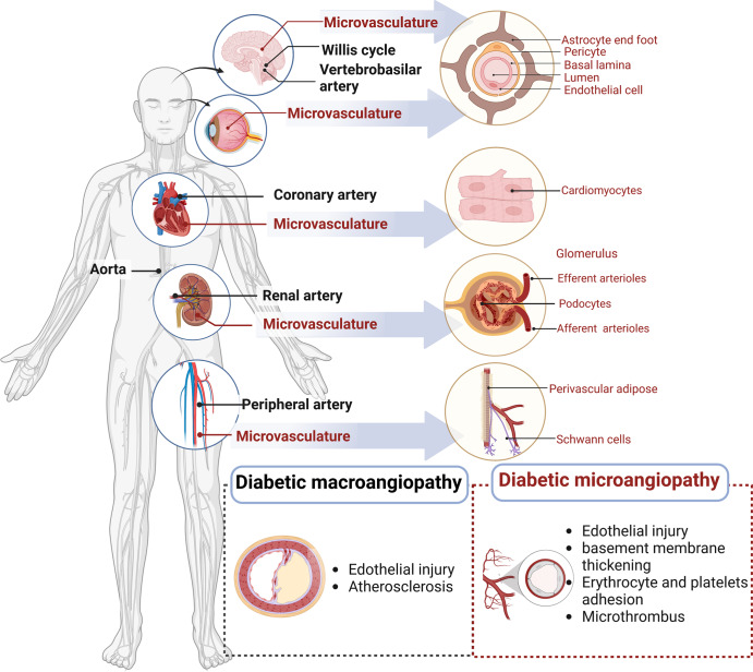
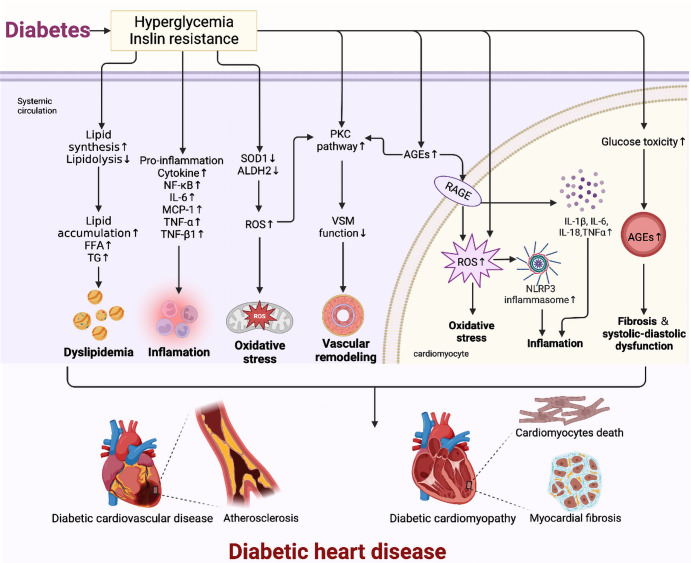
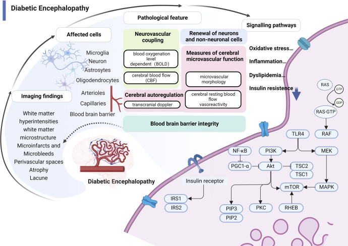
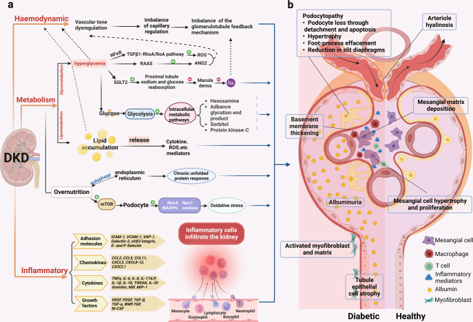
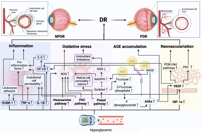
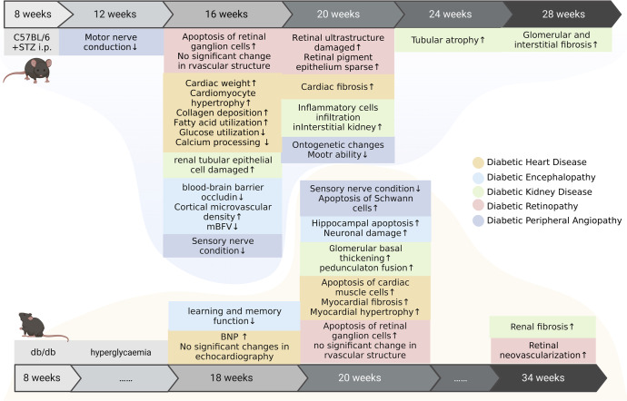
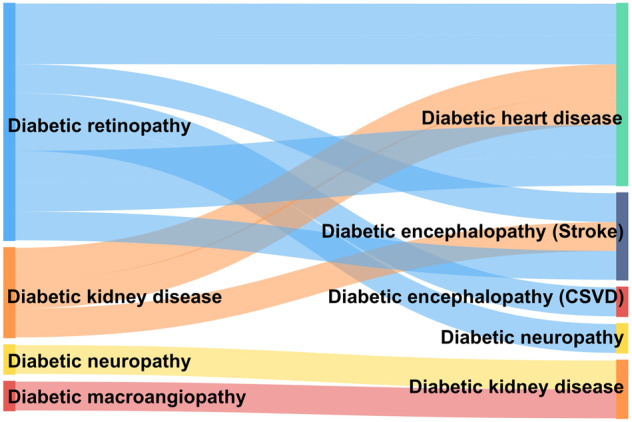
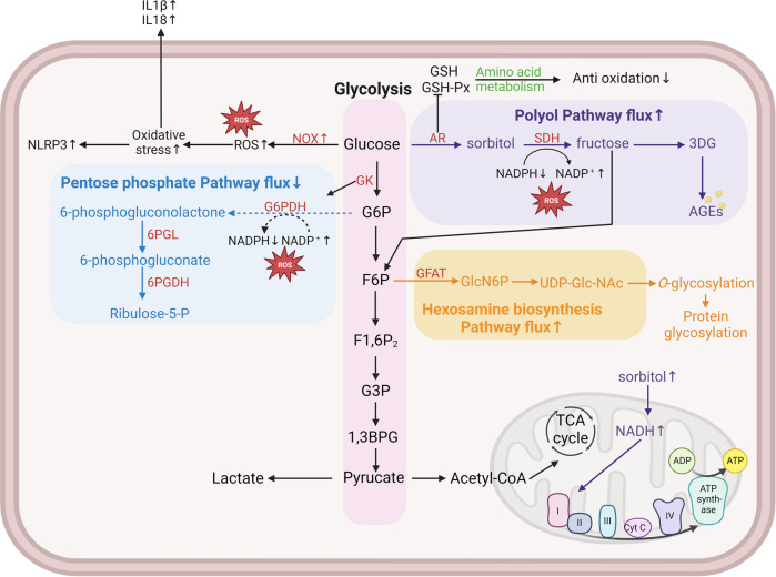
Vascular complications of diabetes pose a severe threat to human health. Prevention and treatment protocols based on a single vascular complication are no longer suitable for the long-term management of patients with diabetes. Diabetic panvascular disease (DPD) is a clinical syndrome in which vessels of various sizes, including macrovessels and microvessels in the cardiac, cerebral, renal, ophthalmic, and peripheral systems of patients with diabetes, develop atherosclerosis as a common pathology. Pathological manifestations of DPDs usually manifest macrovascular atherosclerosis, as well as microvascular endothelial function impairment, basement membrane thickening, and microthrombosis. Cardiac, cerebral, and peripheral microangiopathy coexist with microangiopathy, while renal and retinal are predominantly microangiopathic. The following associations exist between DPDs: numerous similar molecular mechanisms, and risk-predictive relationships between diseases. Aggressive glycemic control combined with early comprehensive vascular intervention is the key to prevention and treatment. In addition to the widely recommended metformin, glucagon-like peptide-1 agonist, and sodium-glucose cotransporter-2 inhibitors, for the latest molecular mechanisms, aldose reductase inhibitors, peroxisome proliferator-activated receptor-γ agonizts, glucokinases agonizts, mitochondrial energy modulators, etc. are under active development. DPDs are proposed for patients to obtain more systematic clinical care requires a comprehensive diabetes care center focusing on panvascular diseases. This would leverage the advantages of a cross-disciplinary approach to achieve better integration of the pathogenesis and therapeutic evidence. Such a strategy would confer more clinical benefits to patients and promote the comprehensive development of DPD as a discipline.


Conflict of interest statement

The authors report no commercial or financial relationships that could be construed as a potential conflict of interest.

Figures

Fig. 1
Fig. 1
Schematic overview of panvasculopathy in diabetes mellitus. Diabetic panvasculopathy involves the cardiac, cerebral, renal, ophthalmic and peripheral systems. The macrovascular lesions are in black text. The microvascular lesions are in red. The microvascular system varies in different organs, which affects vascular function
Fig. 2
Fig. 2
Pathology and molecular mechanisms of DHD. The mechanisms of diabetic heart disease are complex, including oxidative stress, inflammation, and altered metabolic pathways (advanced glycosylation end product (AGE) formation, PKC pathway), which intersect and work together to ultimately lead to myocardial remodeling and dysfunction
Fig. 3
Fig. 3
Pathology and molecular mechanisms of DE. (1) Structural brain changes from MRI studies in diabetes are the primary diagnostic basis of DE, including microinfarcts and microbleeds, perivascular spaces, white matter hyperintensities, white matter microstructure, lacunes, and atrophy; (2) Pathologies related to imaging findings include blood-brain barrier permeability, perfusion defects, hypoxia, and increased angiogenesis that can involve brain microvessels, multiple nerve cells, and the blood-brain barrier; (3) Microvascular dysfunction manifested by impaired neurovascular coupling and impaired neuronal function. Neurovascular coupling links transient local neural activity to subsequently increased blood flow; (4) The molecular pathways of hyperglycemic damage to brain microvasculature are closely related to oxidative stress, inflammation, abnormal lipid metabolism, and insulin resistance. RAS rat sarcoma protein, GTP guanosine triphosphate, GDP guanosine diphosphate, TLR4 Toll-like receptor 4, MEK mitogen-activated protein kinase, PI3K phosphoinositide 3-kinase, Akt protein kinase B, TSC tuberous sclerosis complex, MAPK mitogen-activated protein kinases, mTOR mammalian target of rapamycin, RHEB Ras homolog protein enriched in brain, PKC protein kinase C, PGC1-α PPAR-gamma co-activator-1 alpha, PIP2,3 phosphatidylinositol bisphosphate2, 3, IRS-1,2 insulin receptor substrate1, 2, NFκB nuclear factor kappa-B
Fig. 4
Fig. 4
Pathology of the glomerulus and tubules in DKD. a The classical pathological mechanisms of DKD. It mainly includes hemodynamic, metabolic disturbances, and inflammation, which often interact with each other. (1). Hemodynamic disturbances lead to dysregulation of tubulobulbar feedback balance. (2). Metabolic disorders are crucial to the pathogenesis of DKD. Hyperglycemia affects pathways such as TGFβ1-RhoA/Roa pathway, RAAS, proximal tubular sodium and glucose reabsorption, and intracellular metabolism; abnormal lipid metabolism can affect the release of mediators such as cytokines and ROS; in the presence of nutrient overload in the organism, endoplasmic reticulum autophagy leads to a chronic unfolded protein response, and mTOR also disturbs the podocytes leading to oxidative stress. (3). Inflammation promotes the release of inflammatory mediators such as adhesion molecules, chemokines, cytokines, and growth factors, causing renal infiltration of inflammatory cells. b Schematic representation of the pathological damage of DKD. Differences in structural changes of glomeruli and tubules in the diabetic setting and in the healthy state. Diabetic glomerulopathy is characterized by arterial hyalinization, thylakoid stromal deposition, basement membrane thickening, glomerular thylakoid cell hypertrophy and proliferation, podocytosis, proteinuria, tubular epithelial atrophy, activated myofibroblasts, and stromal accumulation. NFκB nuclear factor kappa-B, TGFβ transforming growth factor-β, ROS reactive oxygen species, RAAS renin-angiotensin-aldosterone system, ANG2 angiotensin II, SGLT2 sodium-dependent glucose transporters 2, mTOR mammalian target of rapamycin, NADPH nicotinamide adenine dinucleotide phosphate, NOX NADPH oxidase, ICAM-1 intercellular cell adhesion molecule-1, VCAM-1 vascular cell adhesion molecule-1, VAP-1 vascular adhesion protein-1, CCL CC chemokine ligand, CXCL C-X-C motif chemokine ligand, TNF tumor necrosis factor, IL interleukin, TWEAK tumor necrosis factor-like weak inducer of apoptosis, MIF macrophage migration inhibitory factor, MIP-1 macrophage inflammatory protein-1, VEGF vascular endothelial growth factor, PDGF platelet-derived growth factor, BMP bone morphogenetic protein, FGF fibroblast growth factor, M-CSF macrophage colony-stimulating factor
Fig. 5
Fig. 5
Pathology and molecular mechanisms of DR. Multiple mechanisms are involved in the pathogenesis of DR. Hyperglycemia can promote oxidative stress through the polyol pathway, accumulation of advanced glycosylation end-products (AGEs), the protein kinase C (PKC) pathway, and the hexosamine pathway, and exacerbate inflammation and abnormal angiogenesis by stimulating the secretion of inflammatory factors and vascular endothelial growth factor, inducing retinal dysfunction until vision loss
Fig. 6
Fig. 6
The natural course of diabetic vasculopathy in mice. Streptozotocin induced in C57BL/6 mice simulate type 1 diabetes in human; db/db mice simulate type 2 diabetes in human. Vasculopathy in different organs appears over a period of time, In C57BL/6 mice Streptozotocin induced in C57BL/6 mice develop typical microangiopathy mostly at 8‒10 weeks after the onset of DM and develop advanced microangiopathy at 12–16 weeks. db/db mice develop microangiopathy at about 20 weeks of age and develop advanced microangiopathy at 34 weeks
Fig. 7
Fig. 7
Predictive relationships among DPDs. CSVD cerebral small vessel diseases
Fig. 8
Fig. 8
Schematic diagram of cellar sugar metabolic pathways. Cells obtain energy through multiple gluconeogenic pathways. These include glycolysis, polyol, hexosamine, and pentose phosphate pathways. In the diabetic environment, excessive intracellular glucose causes abnormal activation of the polyol and hexosamine pathways, and inhibition of the major glycolytic and pentose phosphate pathways, resulting in continued accumulation of reactive oxygen species, which ultimately leads to increased oxidative stress loss in cells and induces the development of DPDs. Different metabolic pathways are distinguished by different colors, with pink representing the glycolytic pathway, purple representing the polyol pathway, blue representing the pentose phosphate pathway, and yellow representing the hexosamine pathway. All enzymes are indicated in red. Solid arrows indicate that this process is promoted and dashed arrows indicate that this process is inhibited. IL interleukin, GSH glutathione, GSH-px glutathione peroxidase, AR aldose reductase, SDH sorbitol dehydrogenase, 3DG 3-deoxyglucosone, AGE advanced glycosylation end, NOX reduced nicotinamide adenine dinucleotide phosphate oxidase, ROS reactive oxygen species, NLRP3 NOD-like receptor thermal protein domain associated protein 3, GK glucokinase, G6P glucose-6-phosphate, F6P d-fructose-6-phosphate disodium salt hydrate, F1,6P2 fructose 1,6-bisphosphate, G3P glyceraldehyde 3-phosphate, 1,3BPG 1,3-bisphosphoglycerate, G6PDH glucose-6-phosphate dehydrogenase, 6PGL 6-phosphogluconolactonase, 6PGDH 6-phosphogluconate dehydrogenase, GFAT glutamine-fructose-6-phosphate aminotransferase, TCA tricarboxylic acid cycle

References

    1. Shaw JE, Sicree RA, Zimmet PZ. Global estimates of the prevalence of diabetes for 2010 and 2030. Diabetes Res. Clin. Pr. 2010;87:4–14. doi: 10.1016/j.diabres.2009.10.007. - DOI - PubMed
    1. Harding JL, Pavkov ME, Magliano DJ, Shaw JE, Gregg EW. Global trends in diabetes complications: a review of current evidence. Diabetologia. 2019;62:3–16. doi: 10.1007/s00125-018-4711-2. - DOI - PubMed
    1. Jia W, et al. Standards of medical care for type 2 diabetes in China 2019. Diabetes Metab. Res. Rev. 2019;35:e3158. doi: 10.1002/dmrr.3158. - DOI - PubMed
    1. Bruno G, et al. Incidence of type 1 and type 2 diabetes in adults aged 30-49 years: the population-based registry in the province of Turin, Italy. Diabetes Care. 2005;28:2613–2619. doi: 10.2337/diacare.28.11.2613. - DOI - PubMed
    1. Avogaro A, Fadini GP. Microvascular complications in diabetes: a growing concern for cardiologists. Int J. Cardiol. 2019;291:29–35. doi: 10.1016/j.ijcard.2019.02.030. - DOI - PubMed

Publication types

MeSH terms

Substances