. 2023 Apr 10;13(1):5840.
doi: 10.1038/s41598-023-32161-y.

The effect of barium and strontium on activity of glucoamylase QsGH97a from Qipengyuania seohaensis SW-135

Affiliations

The effect of barium and strontium on activity of glucoamylase QsGH97a from Qipengyuania seohaensis SW-135

Kaijuan Wu et al. Sci Rep. .

Abstract

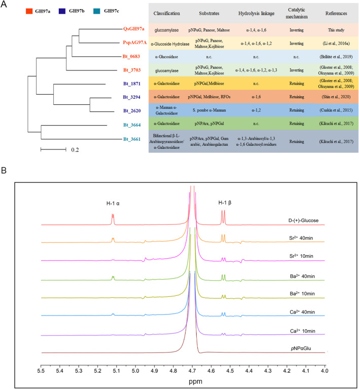
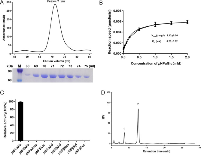
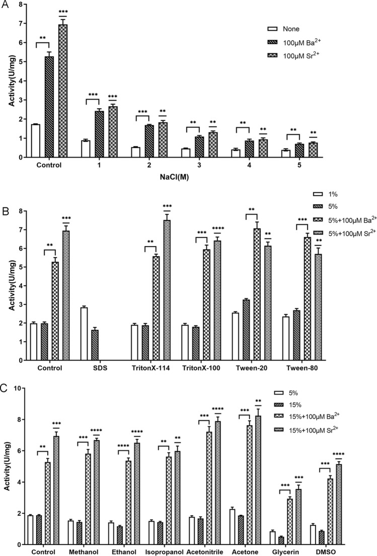
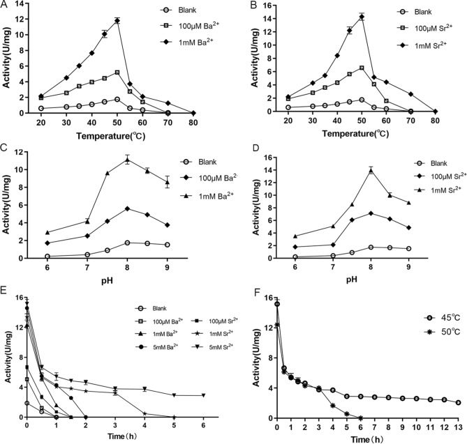
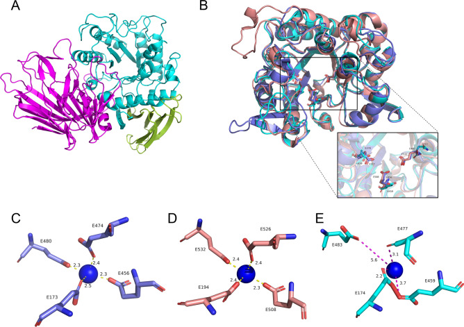
Glycoside hydrolases (GHs), the enzymes that break glycosidic bonds, are ubiquitous in the ecosystem, where they perform a range of biological functions. As an interesting glycosidase family, Glycoside hydrolase family 97 (GH97) contains α-glucosidase, α-galactosidase, and glucoamylase. Only ten members of GH97 have been characterized so far. It is critical to explore novel members to elucidate the catalytic mechanism and application potential of GH97 family. In this study, a novel glucoamylase QsGH97a from Qipengyuania seohaensis SW-135 was cloned and expressed in E. coli. Sequence analysis and NMR results show that QsGH97a is classified into GH97a, and adopts inverting mechanism. The biochemical characterization indicates that QsGH97a shows the optimal activity at 50 °C and pH 8.0. Ca2+ has little effect on the catalytic activity; however, the activity can be substantially increased by 8-13 folds in the presence of Ba2+ or Sr2+. Additionally, the metal content of QsGH97a assay showed a high proportion of Sr2+. The specific metal activity was initially revealed in glucoamylases, which is not found in other members. These results imply that QsGH97a not only is a new member of GH97, but also has potential for industrial applications. Our study reveals that Ba2+ or Sr2+ may be involved in the catalytic mechanism of glucoamylase, laying the groundwork for a more complete knowledge of GH97 and its possible industrial application.


Conflict of interest statement

The authors declare no competing interests.

Figures

Figure 1
Figure 1
The analysis of multiple sequence alignments in GH97. (A) The result of acid sequence alignments in inverting enzymes of GH97. Key Glu residues marked in blue (dark circle) are the conserved catalytic sites of inverting enzymes and Glu residues shown in black (dark star) are the sites coordinating with Ca2+. (B) The result of acid sequence alignments in retaining enzymes of GH97. Important residues marked in blue (dark circle) are the conserved catalytic sites of retaining enzymes. PspAG97A, α-glucoside hydrolase from Pseudoalteromonas sp. K8; Bt_0683, α-glucosidase from B. thetaiotaomicron VPI-5482; Bt_3703, glucoamylase SusB from B. thetaiotaomicron VPI-5482; Bt_3661, bifunctional β-L-arabinopyranosidase/α-galactosidase from B. thetaiotaomicron VPI-5482; Bt_3664, α-galactosidase from B. thetaiotaomicron VPI-5482; Bt_3294, α-galactosidase from B. thetaiotaomicron VPI-5482; Bt_2620, α-mannan α-galactosidase from B. thetaiotaomicron VPI-5482; Bt_1871, α-galactosidase from B. thetaiotaomicron VPI-5482.
Figure 2
Figure 2
Characteristic of GH97 family members and the catalytic mechanism of QsGH97a. (A) Evolutionary relationship and characteristic comparison between QsGH97a and the characterized GH97 family enzymes. QsGH97a is classified into GH97a subfamily according to the mini evolutionary tree in the left picture; the information related to other characterized members is shown in the right picture, including classification, substrates, hydrolysis linkage and catalytic mechanism. GH97-derived enzymes are categorized into inverting enzyme and retaining enzyme based on the catalytic mechanism. an.c.: not characterized. (B) The result of 1H NMR spectroscopy. 1H NMR analysis showed QsGH97a has inverting mechanism to produce β-glucose. The peaks of H-1 α and H-1 β were shown in the group of d-(+)-Glucose, “Ca2+ 10 min” represents that the reaction mixture is measured at 40 °C for 10 min, “Ca2+ 40 min” exhibits that the reaction mixture is detected at 40 °C for 40 min. Ba2+-treated groups and Sr2+-treated groups are identical to Ca2+-treated groups.
Figure 3
Figure 3
The SDS–PAGE result and the enzymatic activity analysis of QsGH97a. (A). Gel filtration chromatography of purified QsGH97a, the QsGH97a protein was eluted at the peak of 71.2 ml and SDS-PAGE result (bottom) of QsGH97a protein eluted from Superdex 200 16/600 column. Lanes from 1 to 9 were the molecular weight marker, and proteins with different elution volumes. Original blots/gels are presented in Supplementary Fig. S2B. (B) The kinetic curve on QsGH97a without addition metal. vmax = 2.13 ± 0.06 U mg−1; KM = 0.20 ± 0.02 mM, (C) The results of hydrolysis activity of QsGH97a on different substrates. (D) The result of hydrolysis activity of QsGH97a on maltose, peak 1, glucose; peak 2, maltose.
Figure 4
Figure 4
Enzymatic characterization of QsGH97a on pNPαGlu. (A) The activity of QsGH97a at various temperatures. The activity at 50 °C was regarded taken as 100%. (B) Effects of pH on enzyme activity. The activity at pH 8.0 was taken as 100%. (C) Thermal stability of QsGH97a incubated for 5–30 min at different temperatures. The activity incubated at 4 °C was taken as 100%. (D) pH stability of QsGH97a incubated for 2 h and 24 h at different pH values. The activity incubated at 4 °C was taken as 100%. (E) Effects of different metal ions on the QsGH97a activity. The activity without addition of metal ions was taken as 100%. ****Means a significant difference by comparison to blank group (p < 0.0001) (F) The results of the recovery experiment of the activity on EDTA-treated enzyme. The activity of untreated enzyme was taken as 100%.
Figure 5
Figure 5
Enzymatic characterization of QsGH97a on pNPαGlu without and with addition of Ba2+ and Sr2+. (A) Effects of 100 μM Ba2+or Sr2+ on QsGH97a activity in different NaCl concentrations (1 M, 2 M, 3 M, 4 M, 5 M). The activity of control group was detected without addition of NaCl (B) Effects of 100 μM Ba2+ and Sr2+ on QsGH97a activity in 5% various detergents. The activity of control group was detected without addition of detergents. (C) Effects of Ba2+ and Sr2+ on QsGH97a activity in 15% various organic solvents. The activity of control group was detected without addition of organic solvents. In figure, * means a significant difference by comparison to control group for each group (p < 0.05), ** means a significant difference of p < 0.01, *** means a significant difference of p < 0.001, **** means a significant difference of p < 0.0001.
Figure 6
Figure 6
Effects of temperature and pH on QsGH97a activity and stability with addition of Ba2+ and Sr2+. (A) The effect of different concentrations Ba2+ on the optimum temperature of QsGH97a. Ba2+ was not added in the blank group. (B) The effect of different concentrations Sr2+ on the optimum temperature of QsGH97a. Sr2+ was not added in the blank group. (C) The effect of different concentrations Ba2+ on the optimum pH of QsGH97a. Ba2+ was not added in the blank group. (D) The effect of different concentrations Sr2+ on the optimum pH of QsGH97a. Sr2+ was not added in the blank group. (E) The effect of different concentrations Ba2+ and Sr2+ on the thermal stability of QsGH97a at 45 °C. The blank group was incubated at 4 °C without addition of Ba2+ and Sr2+. (F) The effect of 5 mM Sr2+ and 10 mM Sr2+on the thermal stability of QsGH97a at 45 °C and 50 °C, respectively. The blank group was incubated at 4 °C without addition of Sr2+. The above substrate was pNPαGlu.
Figure 7
Figure 7
The predicted three-dimensional structure of QsGH97a. (A) The forecast overall structure of QsGH97a, the N-terminal domain is shown in magenta, the core (β/α)8domain in cyan, and the C-terminal domain in split pea. (B) The alignment of predicted QsGH97a (cyan) catalytic domain with PspAG97A (PDB ID: 5HQ4) (slate) and SusB (PDB ID: 2JKA) (salmon). The Glu residues in catalytic sites were marked. (CE) The distance between Ca2+ and interacting Glu residues. The blue ball represents calcium ion; picture (C) is QsGH97a; picture (D) is 5HQ4; and picture (E) is 2JKA.

Similar articles

References

    1. Cai Y, Wang M, Xiao X, Liang B, Fan S, Zheng Z, Cosnier S, Liu A. A membraneless starch/O(2) biofuel cell based on bacterial surface regulable displayed sequential enzymes of glucoamylase and glucose dehydrogenase. Biosens. Bioelectron. 2022;207:114197. doi: 10.1016/j.bios.2022.114197. - DOI - PubMed
    1. Sauer J, Sigurskjold BW, Christensen U, Frandsen TP, Mirgorodskaya E, Harrison M, Roepstorff P, Svensson B. Glucoamylase: Structure/function relationships, and protein engineering. Biochim. Biophys. Acta. 2000;1543:275–293. doi: 10.1016/S0167-4838(00)00232-6. - DOI - PubMed
    1. Coines J, Raich L, Rovira C. Modeling catalytic reaction mechanisms in glycoside hydrolases. Curr. Opin. Chem. Biol. 2019;53:183–191. doi: 10.1016/j.cbpa.2019.09.007. - DOI - PubMed
    1. Naumoff DG. GH97 is a new family of glycoside hydrolases, which is related to the alpha-galactosidase superfamily. BMC Genom. 2005;6:112. doi: 10.1186/1471-2164-6-112. - DOI - PMC - PubMed
    1. Okuyama M. Function and structure studies of GH family 31 and 97 α-glycosidases. Biosci. Biotechnol. Biochem. 2011;75:2269–2277. doi: 10.1271/bbb.110610. - DOI - PubMed

Publication types