. 2023 May;42(21):1763-1776.
doi: 10.1038/s41388-023-02686-7. Epub 2023 Apr 10.

UBXN2A suppresses the Rictor-mTORC2 signaling pathway, an established tumorigenic pathway in human colorectal cancer

Affiliations

UBXN2A suppresses the Rictor-mTORC2 signaling pathway, an established tumorigenic pathway in human colorectal cancer

Sanam Sane et al. Oncogene. 2023 May.

Abstract

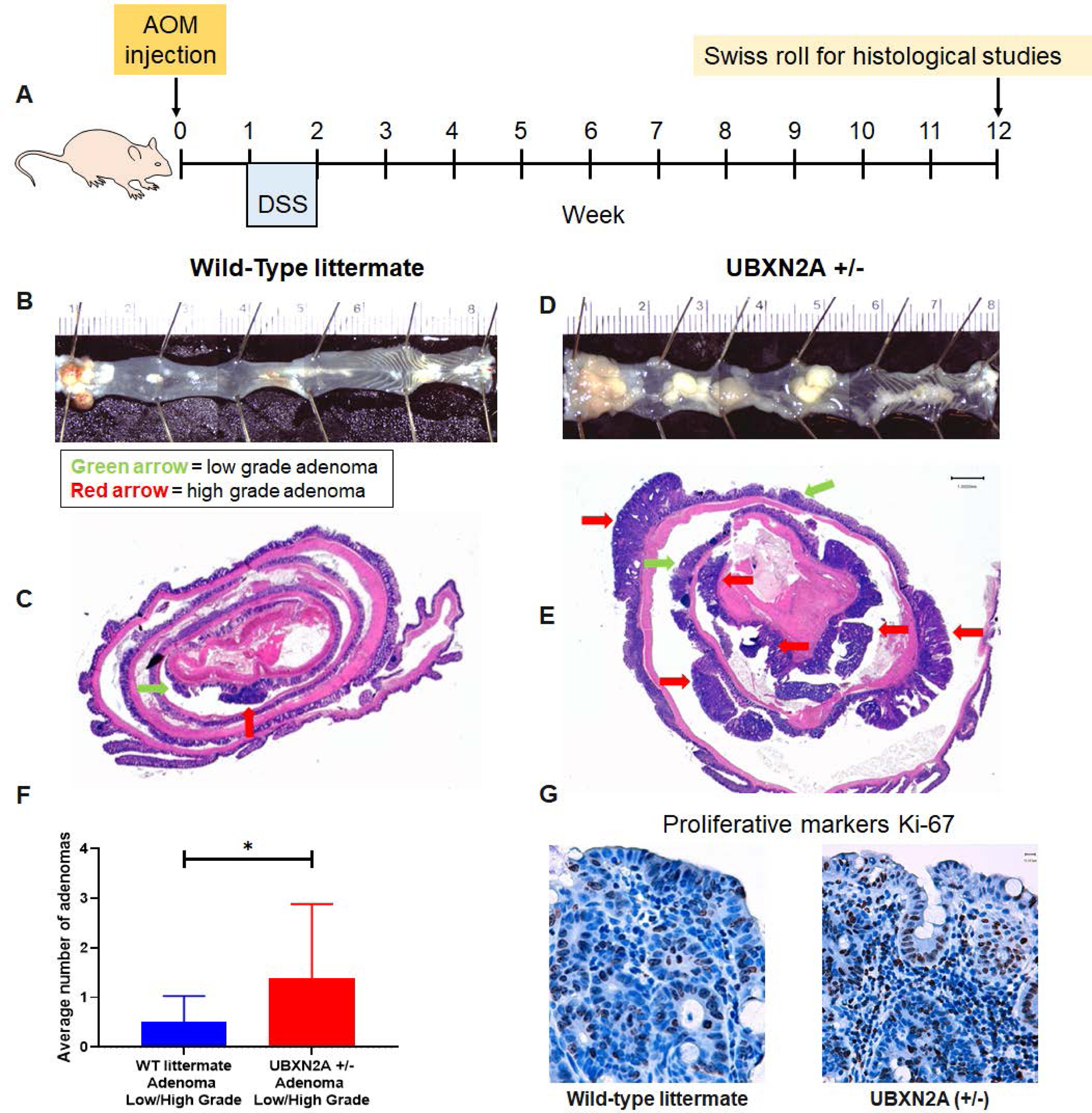
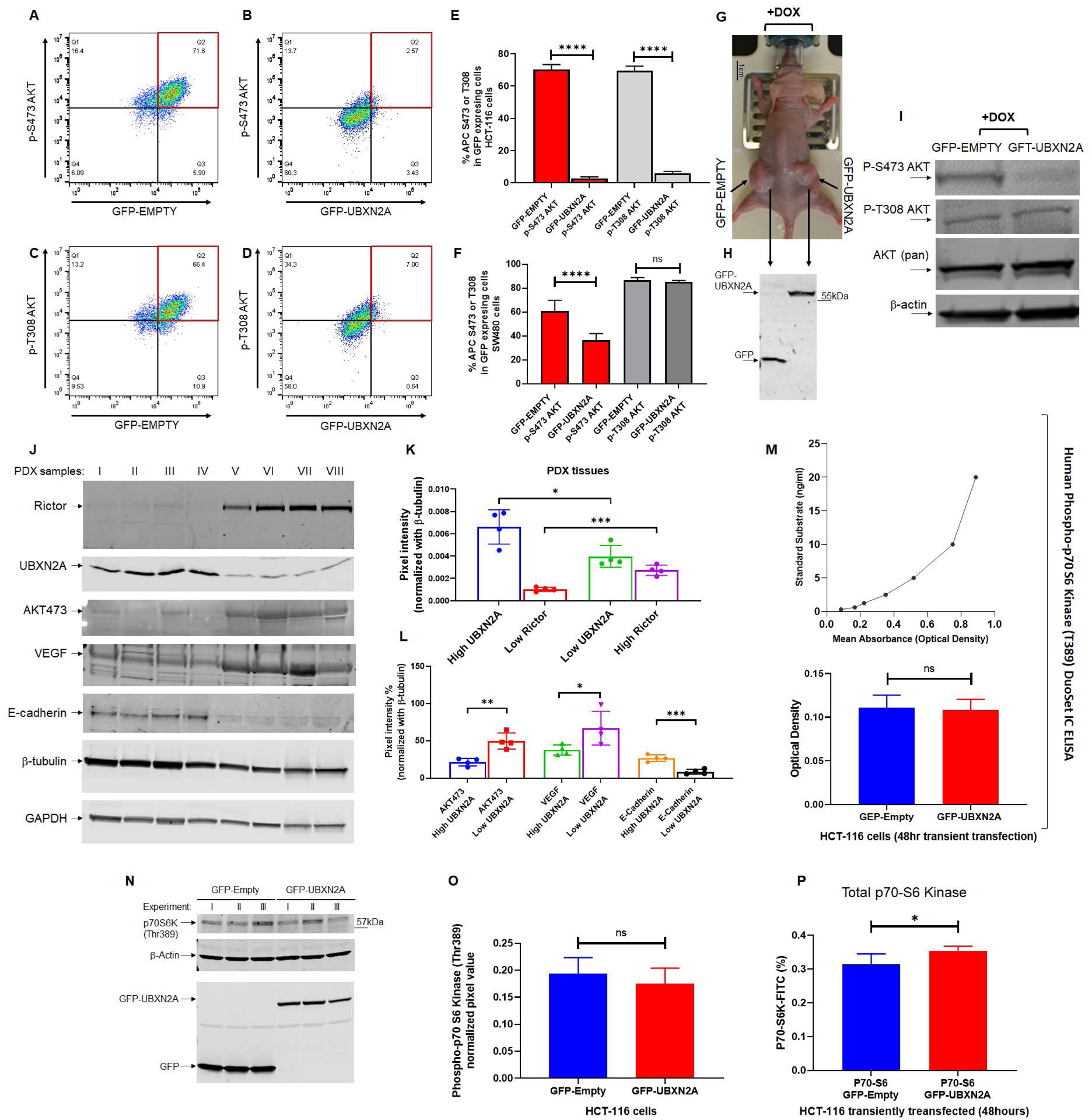
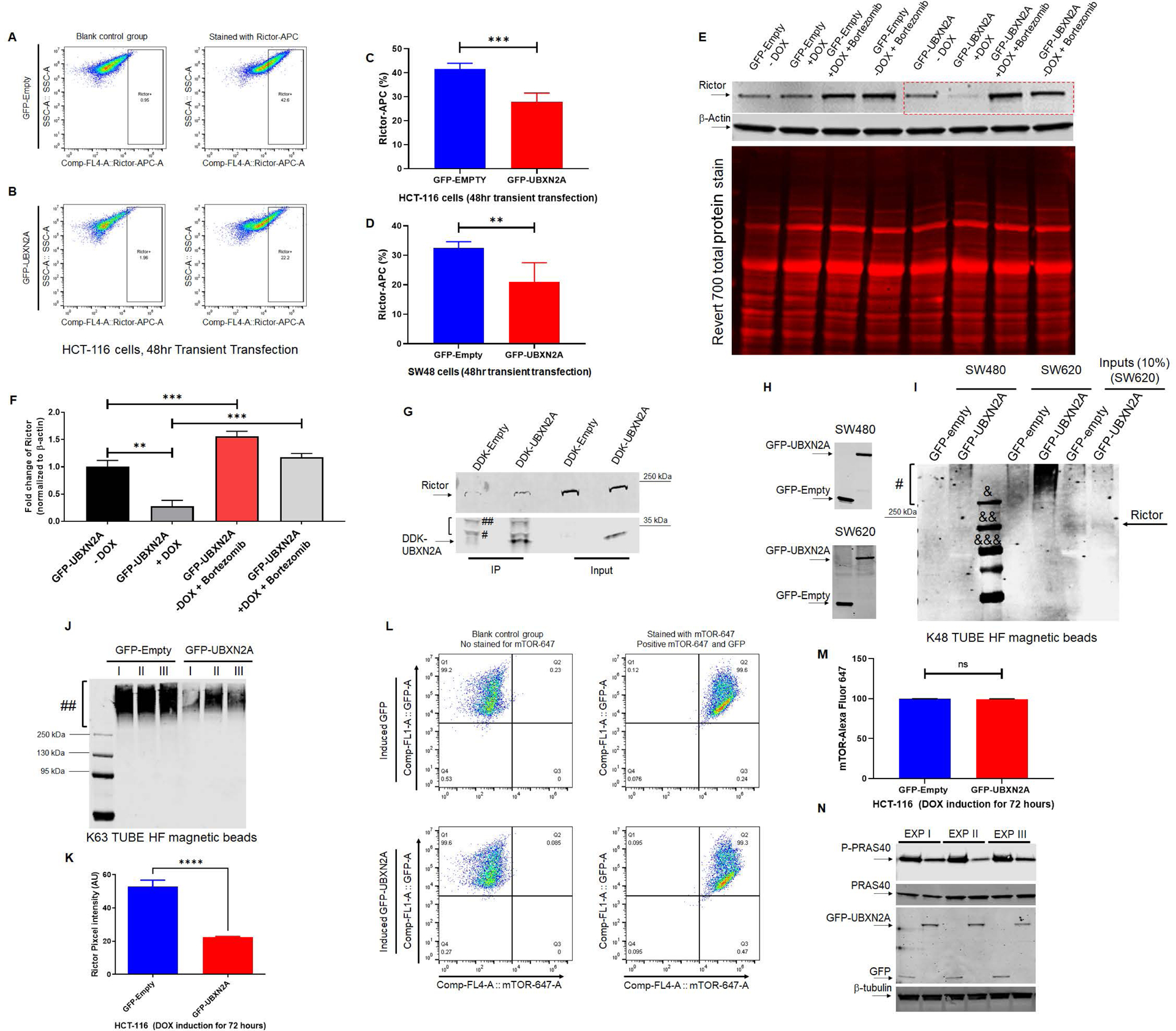
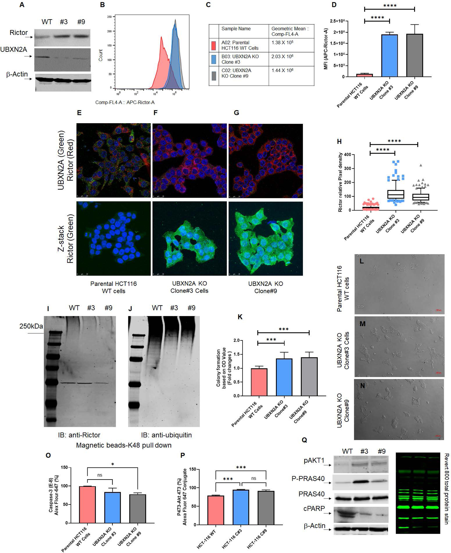
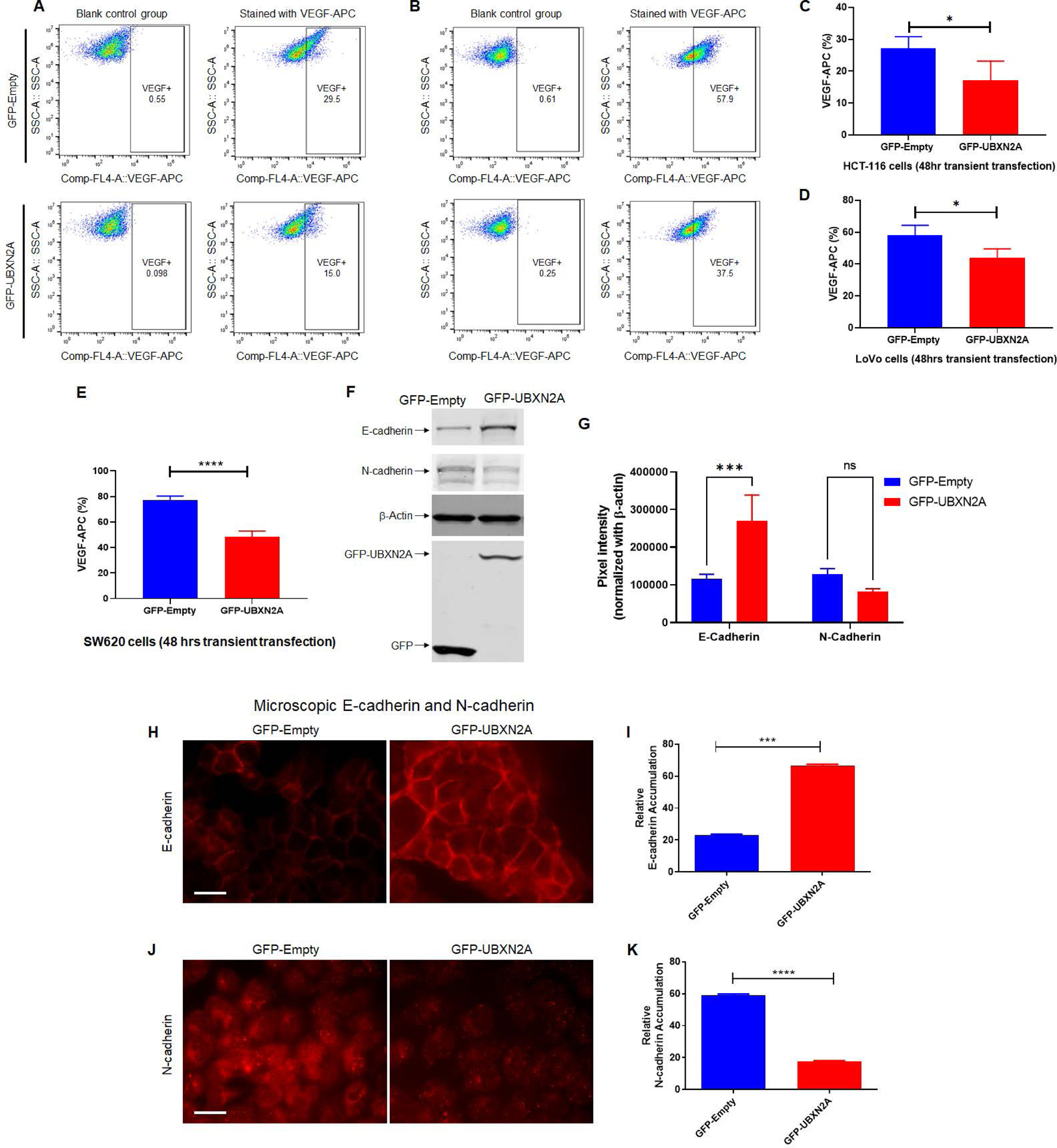
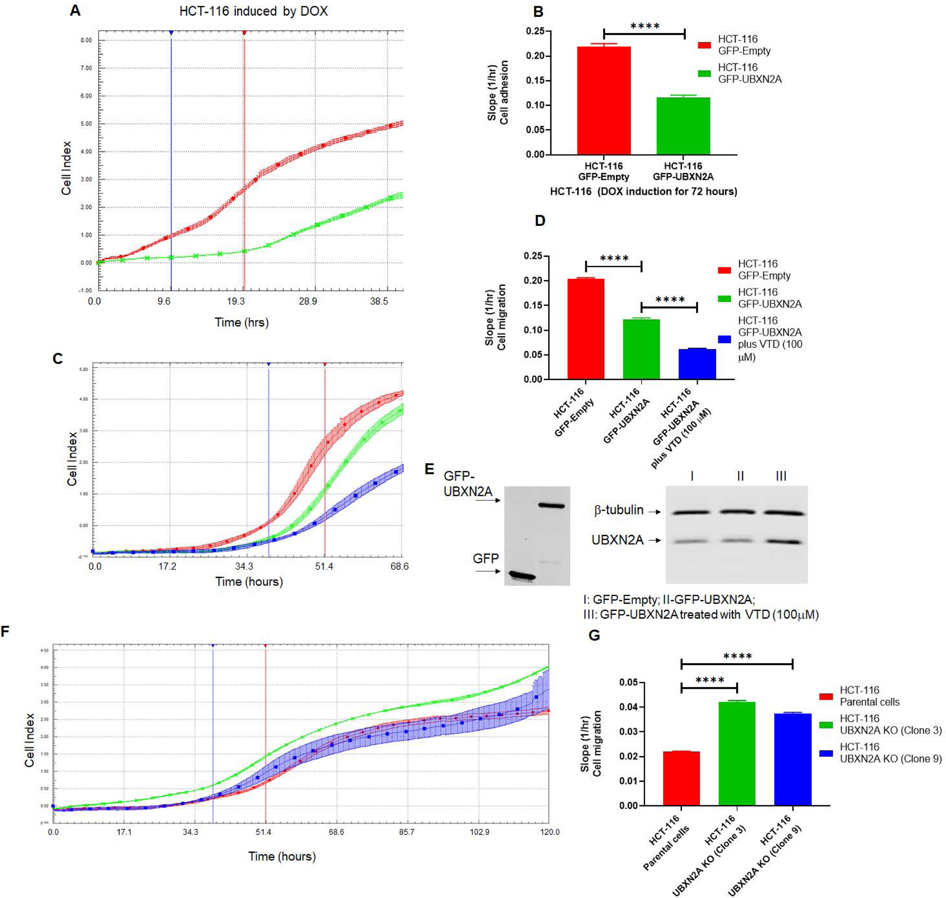
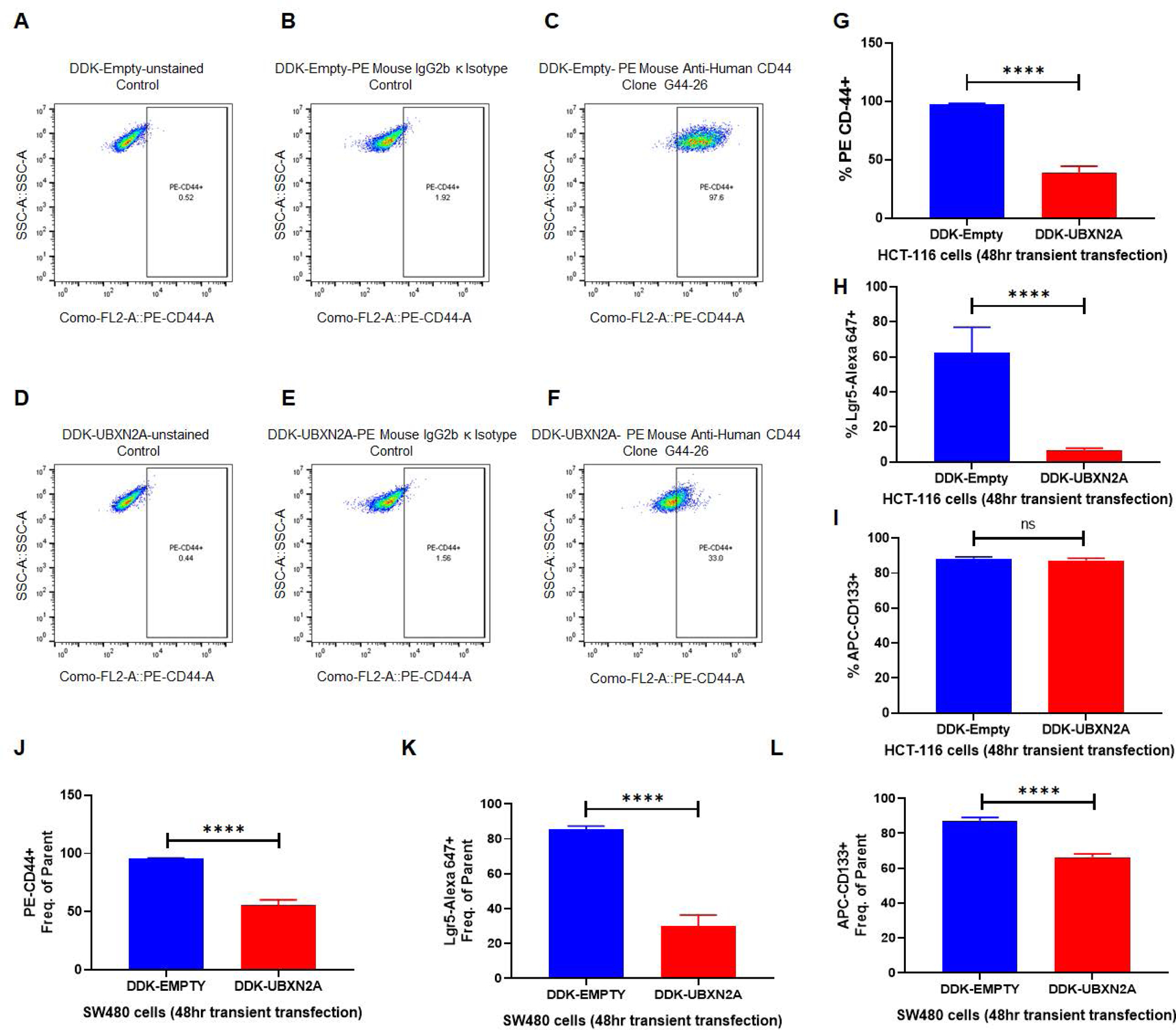
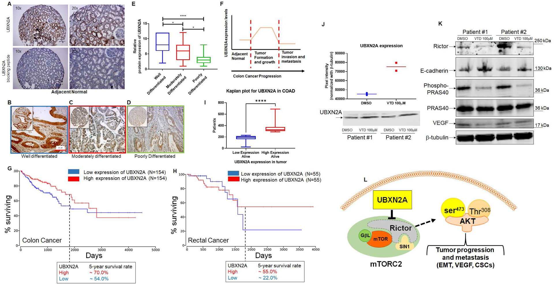
The mTORC2 pathway plays a critical role in promoting tumor progression in human colorectal cancer (CRC). The regulatory mechanisms for this signaling pathway are only partially understood. We previously identified UBXN2A as a novel tumor suppressor protein in CRCs and hypothesized that UBXN2A suppresses the mTORC2 pathway, thereby inhibiting CRC growth and metastasis. We first used murine models to show that haploinsufficiency of UBXN2A significantly increases colon tumorigenesis. Induction of UBXN2A reduces AKT phosphorylation downstream of the mTORC2 pathway, which is essential for a plethora of cellular processes, including cell migration. Meanwhile, mTORC1 activities remain unchanged in the presence of UBXN2A. Mechanistic studies revealed that UBXN2A targets Rictor protein, a key component of the mTORC2 complex, for 26S proteasomal degradation. A set of genetic, pharmacological, and rescue experiments showed that UBXN2A regulates cell proliferation, apoptosis, migration, and colon cancer stem cells (CSCs) in CRC. CRC patients with a high level of UBXN2A have significantly better survival, and high-grade CRC tissues exhibit decreased UBXN2A protein expression. A high level of UBXN2A in patient-derived xenografts and tumor organoids decreases Rictor protein and suppresses the mTORC2 pathway. These findings provide new insights into the functions of an ubiquitin-like protein by inhibiting a dominant oncogenic pathway in CRC.


Conflict of interest statement

COMPETING INTRESTS

The authors declare no competing financial interests.

Figures

Figure 1:
Figure 1:. Haploinsufficiency of the UBXN2A tumor suppressor gene promotes colon cancer progression.
A-D: Colorectal tumors developed in all AOM/DSS treated C57BL/6 mice. E-F: Tumors were stained with hematoxylin and eosin (H & E). The Swiss roll technique followed by IHC did not have access to all those rectal tumors (panels B and D) due to the nature of the technique. Unavoidably, sections are omitted at both ends of the Swiss roll during construction. Low expression of UBXN2A in heterozygote mice (+/−) significantly increased both low- and highgrade adenomas. Green arrow = low grade adenoma, red arrow = high grade adenoma (n = 11 per genotype, male and female, Welch’s t test, *p<0.05, mean ± SD). G: IHC showed a higher level of cell proliferation in UBXN2A +/− (scale bar, 1mm).
Figure. 2:
Figure. 2:. Induction of UBXN2A interferes with the mTORC2 pathway but not the mTORC1 signaling pathway.
Tet-On inducible HCT-116 cells were incubated with DOX to express GFP or GFP-UBXN2A and analyzed by flow cytometry using Alexa Fluor pAKT-Ser473 and pAKT-T308. Induction of UBXN2A significantly decreased pAKT-Ser473 and pAKT-Thr308 (A-E). Similarly, UBXN2A significantly decreased pAKT-Ser473 in SW480 cells. However, UBXN2A induction showed no effect on pAKT-Thr308 in SW480, suggesting UBXN2A affects mTORC2 in a cell-dependent manner (F). Xenograft mice carrying Tet-On inducible HCT-116 cells were fed doxycycline (G), followed by tumor extraction and WB experiments (H).Representative WB of three mice with similar results (one GFP-empty tumor and one GFP-UBXN32A tumor per mouse) confirmed UBXN2A decreases the level of pAKT-S473/T308 in xenograft tumors (I). PDX tissues were subjected to WB followed by quantitation of their corresponding bands. Results revealed that the high level of UBXN2A in PDXs significantly decreases Rictor protein (J-K), and it changes Rictor’s downstream target proteins, including AKT-473, VEGF, and E-Cadherin (L). ELISA assays (M) and WB of phospho-p70 S6 kinase (T389) in HCT-116 cells (N and O) combined with flow-cytometry analysis of the total level of p70 S6 kinase (P) revealed that elevated UBXN2A has no significant effect on phosphorylation and activation of phospho-p70 S6 kinase (T389), which is located downstream of mTORC1 (n=3, * p< 0.05, **** p< 0.0001, mean +/− SD).
Figure 3:
Figure 3:. UBXN2A binds to Rictor protein and promotes its proteasomal degradation.
Flowcytometry analysis of HCT-116 cells transiently transfected with GFP-empty or GFP-UBXN2A vectors for 48 hours revealed UBXN2A overexpression significantly decreases Rictor protein (A-C). Similar to the HCT-116 cell line, transient transfection of SW48 cells with GFP-empty or GFP-UBXN2A vectors for 48 hours followed by flow-cytometry analysis showed UBXN2A can significantly decrease the protein level of Rictor (D). Induction of UBXN2A in HCT-116 treated with DOX for 72 hours decreased the half-life of Rictor, while the presence of a proteasome inhibitor (bortezomib, 50nM) rescues rapid turnover of Rictor (red box in panel E; quantitated signals in panel F). A β-actin antibody and Revert 700 total protein stain were used as the loading control. HCT-116 cells were transfected with DDK-Empty or DDK-UBXN2A plasmid for 48 hours followed by immunoprecipitation using anti-DDK antibody immobilized on magnetic IgG beads. DDK-UBXN2A pulled down Rictor protein, indicating UBXN2A binds to Rictor protein. Two bands tagged with one # and two ## are light and heavy chains, respectively (G). Due to the high molecular weight of Rictor protein (220 kDa), we observed a low non-specific affinity of Rictor protein to magnetic beads in control. In another set of experiments, K48 TUBE HF magnetic beads were used to pull down ubiquitinated Rictor in SW480 and SW620 (a metastatic colon cancer cell line) transiently transfected with GFP-empty or GFP-UBXN2A vectors for 48 hours (H). GFP-UBXN2A increases the K48-linked ubiquitination ladder (#) of Rictor protein (I), particularly in SW620. The K48 pull-down experiment was repeated with the same results trend. &, &&, and &&& are protein molecular markers for 250 kDa, 130, kDa, and 95 kDa, respectively. In contrast, K63 magnetic beads revealed induced UBXN2A decreases pulled down K63 linked chain of Rictor protein (n=3, Panels J) due to the reduction of total Rictor protein in the presence of UBXN2A measured by immunofluorescent microscopy in HCT-116 cells (n=150 cells-Panel K). A set of flow-cytometry analyses in HCT-116 cells transiently overexpressing GFP-empty of GFP-UBXN2A proteins showed the presence of UBXN2A has no significant effect on mTOR protein, another protein member of the mTORC2 complex (L and M). Finally, the Tet-on inducible GFP-empty and GFP-UBXN2A were treated with DOX for 72 hours. Cell lysates were subjected to WB using anti-PRAS40 (total protein and P-PRAS40, a phosphorylated form of protein) as well as a GFP antibody. A triplicate WB revealed that UBXN2A decreases the protein level P-PRAS40, activated by the Rictor-mTORC2 signaling pathway (N). Together, these flow-cytometry, K48/K63 linked chains, and WB results indicate UBXN2A selectively targets and degrades Rictor proteins via ubiquitin-proteasome pathway in colon cancer cells (** p< 0.01, *** p< 0.001, mean +/− SD).
Fig. 4:
Fig. 4:. Absence of UBXN2A leads to elevation of Rictor, resulting in inhibition of mTORC2’s downstream protein targets.
Two stable CRISPR UBXN2A KO HCT-116 cells generated by CRISPR/Cas9 genome editing were validated by WB (A) and flow-cytometry analysis (B-D). Clone 3 and clone 9 UBXN2A KO were subjected to confocal microscopy study (E-H), TUBE K48-linked ubiquitin chain magnetic beads pull-down (I-J), and crystal violet cell viability assay (K-N). These data demonstrate that the absence of UBXN2A in HCT-116 (clones 3 and 9) significantly elevates the level of Rictor protein measured by flow- cytometry and immunocytochemistry (D and H). Furthermore, the absence of UBXN2A decreases the K48 ubiquitinated form of Rictor in HCT-116 cells (I-J). The absence of UBXN2A and simultaneous elevation of Rictor leads to higher cell proliferation and less apoptosis stained and quantitated by crystal violet assay (K-N). Flow-cytometry analysis of Caspase-3 (O) and the elevation of pAKT473 (P) further confirmed the inverse relationship between UBXN2A and Rictor protein levels and Rictor’s downstream pathways responses. A set of WB experiments revealed that the absence of UBXN2A (clones 3 and 9) leads to the elevation of pAKT1 and P-PRAS40, as well as the reduction of cleaved PARP (cPARP), which are regulated by the activated mTORC2 pathway (Q). A β-actin antibody and Revert 800 total protein stain were used as the loading control (* n≥3, p<0.05, ***p<0.001, ****p<0.0001, mean ± SD).
Figure. 5:
Figure. 5:. UBXN2A expression leads to the reduction of VEGF proteins in metastatic colon cancer cells.
To determine the effects of UBXN2A expression on the VEGF protein level, we transiently transfected HCT-116 (A and C) and LoVo (B and D) colon cancer cells with GFP-empty and GFP-UBXN2A. Flow-cytometry experiments revealed that GFP-UBXN2A and not GFP-Empty decreases the level of VEGF proteins (C and D). A similar significant reduction of VEGF was measured in SW620 metastatic colon cancer cells (n=4, E). Cell lysates of HCT-116 cells transiently transfected with GFP-empty or GFP-UBXN2A were subjected to WB and probed with anti-E-cadherin and anti-N-cadherin antibodies (F). The quantitation of E-cadherin and N-cadherin bands normalized by β-actin in WB results (n=3, G). To confirm that UBXN2A regulates functional E-cadherin and N-Cadherin at the plasma membrane, we conducted a set of immunocytochemistry experiments (H and J). Measured fluorescent signals (n=150 cells per group, I and K) revealed UBXN2A overexpression can switch the E-cadherin and N-cadherin which potentially suppress EMT. These results indicate that UBXN2A expression can directly interfere with the two major metastatic pathways (angiogenesis and EMT) activated by the Rictor-mRORC2 pathway (* p<0.05, ***p<0.001, ****p<0.0001, mean ± SD).
Figure. 6:
Figure. 6:. Genetic and pharmacological regulation of UBXN2A suppress colon cancer migration.
HCT-116 GFP-empty or GFP-UBXN2A treated with DOX for 72 hours, were plated in 16-wells E-plate or CIM-plate (xCELLigence Real-Time technology) and monitored in real-time for cell adhesion (A-B) and migration (C-D). A and C are representative graphs comparing the rate of adhesion and migration using the calculated cell index (see methods section). B and D show calculated slopes for these two events during critical time points marked with blue and red lines in the A and C diagrams. Interestingly, enhancement of UBXN2A endogenous protein by the pharmacological tool, veratridine, further decreases HCT-116 cancer cell migration (D, blue column versus green column). E shows equal overexpression of exogenous GFP and GFP-UBXN2A in HCT-116 with the Tet-on promoter system and elevated endogenous UBXN2A in veratridine-treated cells subjected to xCELLigence analysis. To examine the physiological effect of UBXN2A in cell migration, HCT-116 UBXN2A KO cells (clones 3 and 9) were subjected to a set of xCELLigence migration assays (F). Results shown in G indicate that the UBXN2A’s loss of function leads to significant cell migration indicating that the negative interaction of UBXN2A with Rictor affects downstream signaling pathways regulated by the mTORC2-Rictor pathway. Experiments were repeated two times with N of 4 per cell line per experiment (****P<0.0001, mean ± SD).
Fig. 7:
Fig. 7:. UBXN2A induction decreases cancer stem cell populations in human colon cancer cells.
CSCs positive for Lgr5, CD44, and CD133 are regulated by the mTORC2-hedgehog axis in cancer cells and are potentially responsible for the high recurrence rates of CRC. HCT-116 (A-I) and SW480 (J-L) colon cancer cells were transfected with DDK-tag empty vector or DDK-tag UBXN2A. After 48 hours, cells were subjected to flow cytometry analysis using three CSC markers (Lgr5, CD44, and CD133). As previously described for HCT-116 cells, overexpression of UBXN2A has no significant effect on CD133 (I). UBXN2A overexpression led to a significant reduction of CD44 (J) and Lgr5 (K) as well as CD133 (L), which exhibits increased tumor sphere-forming efficiency and increased tumorigenic potential in the SW480 cell line (N=4, **** p< 0.0001, mean +/− SD).
Fig. 8:
Fig. 8:. UBXN2A protein levels predominantly upgrade during the early stage of tumor development and improve survival rates in colorectal cancer patients
A shows UBXN2A’s medium protein expression level in normal colon tissues. A peptide-blocking assay confirmed the specificity of the antibody against the UBXN2A protein in the IHC study. B-D: IHC staining was used to stain cytoplasmic and nuclear UBXN2A in well-differentiated (n=26), moderately (n=74), and poorly differentiated (n=24 tumor tissues) human colon tumor tissues. E: UBXN2A expression levels of colon cancer tissues were manually scored. The quantitative scoring system revealed that UBXN2A, as a tumor suppressor protein, significantly upregulates in the early stage of colon cancer and shows a significant reduction in a higher stage of colon cancer. The reduction of UBXN2A is associated with a poorer prognosis in higher stages of CRC. (F). Kaplan–Meier’s analysis of extracted survival data from TCGA shows COAD, a subtype of colorectal cancer, indicates a correlation of higher survival rate with higher UBXN2A expression. The 5-year survival rate is ~20% higher than patients with low expression of UBXN2A in tumors (G). READ, a subtype of colorectal cancer Kaplan–Meier’s analysis, shows a favorable survival outcome in high UBXN2A expression. Patients with rectal cancer with a high level of UBXN2A show ~30% elevation of 5-year survival (H). Panel G and H were produced using the Human Protein Atlas and ONCL tools. For ONCL, we used 33 low and 33 high percentiles parameters. Further analysis shows a significantly larger portion of alive patients with COAD had higher UBXN2A expression compared to lower expression of UBXN2A, indicating a longer progression-free survival than those with low UBXN2A in the TCGA (I). PDOs generated from surgically removed CRC tumors (n=2) were treated with the UBXN2A enhancer Veratridine (VTD, 100μM) for 72 hours. Two individual PDOs showed VTD elevates the level of UBXN2A protein expression by approximately two-fold (J) and simultaneously decreases Rictor resulting in alteration of mTORC2 protein targets, including reduction of P-PRAS40 and VEGF proteins as well as elevation of E-cadherin (K). Schematic diagram showing the mechanistic inhibitory action of UBXN2A on mTORC2 tumorigenic pathway through selective proteasomal degradation of Rictor protein. The absence of a fully functional mTORC2 complex leads to the inhibition of several downstream metastatic pathways (L).

Similar articles

Cited by

References

    1. Roulin D, Cerantola Y, Dormond-Meuwly A, Demartines N, Dormond O. Targeting mTORC2 inhibits colon cancer cell proliferation in vitro and tumor formation in vivo. Molecular Cancer. 2010;9:57-. - PMC - PubMed
    1. Wong CK, Lambert AW, Ozturk S, Papageorgis P, Lopez D, Shen N, et al. Targeting RICTOR Sensitizes SMAD4-Negative Colon Cancer to Irinotecan. Mol Cancer Res. 2020. - PMC - PubMed
    1. Ponnurangam S, Standing D, Rangarajan P, Subramaniam D. Tandutinib inhibits the Akt/mTOR signaling pathway to inhibit colon cancer growth. Mol Cancer Ther. 2013;12(5):598–609. - PMC - PubMed
    1. Shiratori H, Kawai K, Okada M, Nozawa H, Hata K, Tanaka T, et al. Metastatic role of mTOR signaling activation by chemoradiotherapy in advanced rectal cancer. Cancer Sci. 2020. - PMC - PubMed
    1. Jhanwar-Uniyal M, Wainwright JV, Mohan AL, Tobias ME, Murali R, Gandhi CD, et al. Diverse signaling mechanisms of mTOR complexes: mTORC1 and mTORC2 in forming a formidable relationship. Advances in Biological Regulation. 2019;72:51–62. - PubMed

Publication types

MeSH terms

Substances