. 2023 May;7(5):732-744.
doi: 10.1038/s41559-023-02012-0. Epub 2023 Apr 10.

An ancient metalloenzyme evolves through metal preference modulation

Affiliations

An ancient metalloenzyme evolves through metal preference modulation

K M Sendra et al. Nat Ecol Evol. 2023 May.

Abstract

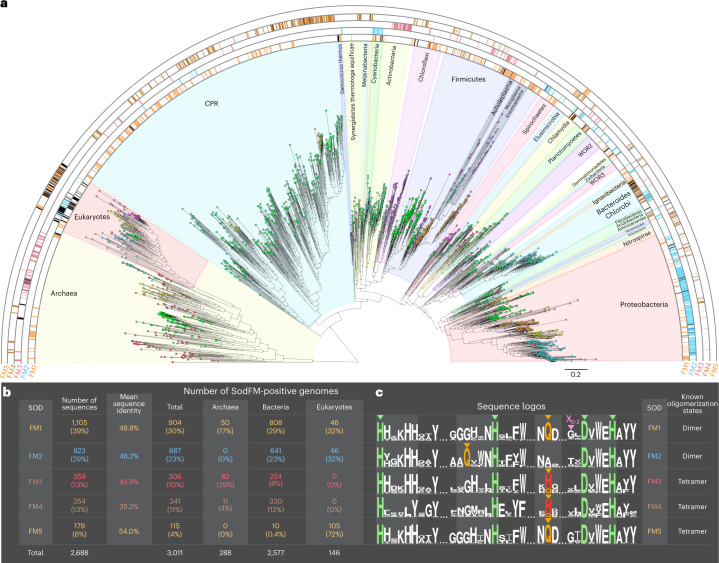
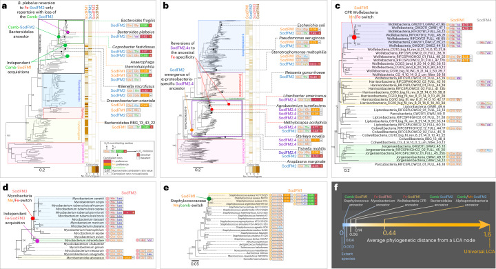
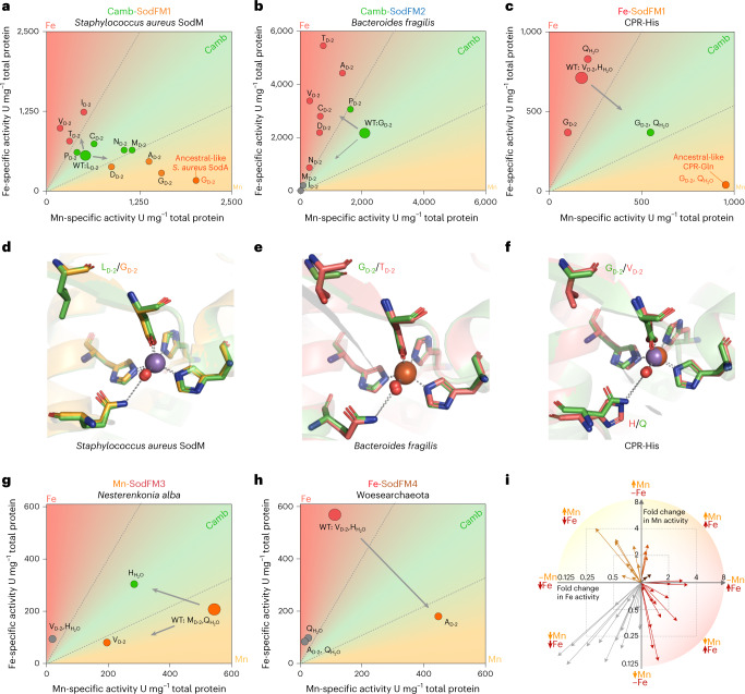
Evolution creates functional diversity of proteins, the essential building blocks of all biological systems. However, studies of natural proteins sampled across the tree of life and evaluated in a single experimental system are lacking. Almost half of enzymes require metals, and metalloproteins tend to optimally utilize the physicochemical properties of a specific metal co-factor. Life must adapt to changes in metal bioavailability, including those during the transition from anoxic to oxic Earth or pathogens' exposure to nutritional immunity. These changes can challenge the ability of metalloenzymes to maintain activity, presumptively driving their evolution. Here we studied metal-preference evolution within the natural diversity of the iron/manganese superoxide dismutase (SodFM) family of reactive oxygen species scavengers. We identified and experimentally verified residues with conserved roles in determining metal preference that, when combined with an understanding of the protein's evolutionary history, improved prediction of metal utilization across the five SodFM subfamilies defined herein. By combining phylogenetics, biochemistry and structural biology, we demonstrate that SodFM metal utilization can be evolutionarily fine tuned by sliding along a scale between perfect manganese and iron specificities. Over the history of life, SodFM metal preference has been modulated multiple independent times within different evolutionary and ecological contexts, and can be changed within short evolutionary timeframes.


Conflict of interest statement

The authors declare no competing interests.

Figures

Fig. 1
Fig. 1. Evolutionarily distinct SodFM subfamilies have wide and variable distribution across the tree of life.
a, Pattern of variable distribution of the five SodFM subfamilies (Extended Data Fig. 2) observed across the tree of life. The coloured semi-circles represent the distribution of SodFM1s (1,105 sequences, orange), SodFM2s (823 sequences, blue), SodFM3s (359 sequences, red), SodFM4s (354 sequences, brown) and SodFM5s (179 sequences, orange), mapped onto a phylogeny of 146 eukaryotes, 2,577 bacteria and 288 archaea. The presence of species with more than a single representative of a particular SodFM subfamily (black, Supplementary Data 18) reveals multiple independent lineage-specific SodFM1–5 subfamily expansions. b, Tables containing numbers and mean percentage of protein sequence identity of the SodFM1–5 subfamily members found within the analysis of 3,011 genomes sampled across the tree of life (a). c, Sequence logos illustrate frequencies of key residues found across the analysed SodFMs, including four universally conserved metal-coordinating residues, three histidines and a single aspartate (green triangles). The key distinction among SodFM1–5 subfamilies is the identity and position of a water-coordinating residue (orange triangles) spatially located within the enzymes’ catalytic centres: SodFM1/5s have a C-terminal Gln, SodFM2s have an N-terminal Gln, and most SodFM3–4s (75% sequences) have a C-terminal His and the rest of the SodFM3s and SodFM4s (25% each group) contained C-terminal Gln instead of His, often reflecting secondary His/Gln switches (Extended Data Fig. 2a). The key distinction between SodFM3s and SodFM4s is the loss of the conserved C-terminal His in the HXXXHH motif in SodFM4s. The key distinction between SodFM1s and SodFM5s is that the studied representatives of the former are homodimeric, whereas those of the latter are tetrameric (Extended Data Figs. 2a and 3a). Residue XD-2 represents an important determinant of Fe/Mn metal preference (pink triangle).
Fig. 2
Fig. 2. Enzymes with diverse metal preferences can be found across the SodFM subfamilies.
a, The aCR values for the 64 characterized SodFMs were mapped onto the protein tree inferred from 2,688 SodFM sequences identified across the tree of life (Fig. 1a). A range of aCRs, from higher Fe preference (aCR >2, red), through cambialistic (0.5 < aCR < 2, green), to higher Mn preference (aCR <0.5, orange), were found across the SodFM subfamilies. Sensitivity to peroxide inhibition (+) was detected for Fe-preferring and Fe-loaded cambialistic enzymes, but not in the Mn-preferring enzymes, which were resistant to peroxide treatment (−). Identity of the metal’s two key secondary coordination sphere residues, namely water-coordinating Gln/His and the XD-2 residue located close to the Asp ligand (Fig. 1c), are displayed next to aCRs. b, Distribution of the aCRs for the SodFM subfamilies is presented as box and whiskers plots (left), and the frequencies of different amino acid residues found at the XD-2 position are presented as pie charts (right). SodFM1s and SodFM5s were grouped together here as they both contained C-terminal Gln and displayed similar metal preference and XD-2 distribution patterns (SodFM5s XD-2, G: 89%; A: 7%; T: 1.7%). The high proportion of Mn-preferring SodFM1s and Fe-preferring SodFM2s, and the predicted Gln/His and the XD-2 residues in the reconstructed sequences of the ancestral nodes (a, dashed outlines), are consistent with the hypothesis that the LCAs of these two subfamilies were Mn and Fe preferring, respectively (Extended Data Fig. 2f,g). c, Distribution of the aCR values for SodFMs (top) with different water-coordinating residues (C-terminal Gln, N-terminal Gln or C-terminal His) and different XD-2 residues (bottom) are displayed as box and whiskers plots on a logarithmic scale, colour coded to reflect apparent metal specificities (Mn: orange; cambialistic (camb): green; Fe: red). Amino acid residues were colour coded to enable comparisons among a, b and c.
Fig. 3
Fig. 3. SodFM metal preference and peroxide sensitivity have been changed independently many times within different evolutionary contexts.
ae Distribution of SodFM1–4 subfamilies (orange–brown heat maps in a and b, or coloured circles in ce) was mapped onto the species trees of Bacteroidota (a), Proteobacteria (b), CPR Wolfebacteria (c), Mycobacteriaceae (d) and Staphylococcaceae (e). In Gram-negative bacteria, SodFM repertoires consisted mainly of SodFM2s in Proteobacteria (b) and SodFM1s in Bacteroidota (a). Within the Bacteroidota, a subset of the Bacteroidales have switched to SodFM2 (a). SodFM1s and SodFM4s were found in uncultured CPR Wolfebacteria (c). In Gram-positive Mycobacteriaceae only SodFM3s were identified (d), whereas Staphylococcaceae contained only SodFM1s (e). The identities of the water-coordinating Gln/His, the residue XD-2, and verified aCR and peroxide inhibition for the SodFMs characterized in this study were mapped onto the trees and annotated as previously described (Fig. 2). Inferred multiple independent evolutionary shifts towards higher Fe preference and peroxide sensitivity (red circles), higher Mn preference and peroxide resistance (orange circles), emergences of cambialism (green circles) and likely SodFM repertoire changes via LGT (magenta circles) were annotated onto the trees (ae). f, The average phylogenetic distances between the inferred LCA nodes (coloured circles in ae) and their descendent tree tips were calculated and presented on a colour-coded scale, where the minimum value represents the extant homologues (tree tips) and the maximum value represents the average distances of all studied archaea, bacteria and eukaryotes from their LCA (universal LCA). Emergence of alphaproteobacterial SodFM2.4 was the most ancient and that of the cambialistic SodFM1 within staphylococci the most recent of the identified metal-preference evolutionary modulation events. Scale bars represent the number of substitutions per site.
Fig. 4
Fig. 4. Functional studies reveal multiple molecular pathways to metal-preference modulation in SodFM metalloenzymes.
a,b, Residues at the XD-2 position in cambialistic S.aureus SodFM1 (LD-2, a) and B.fragilis SodFM2 (GD-2, b) were mutated to those found commonly at this position in natural SodFMs (Fig. 2b). The average specific activities of the WTs and mutants loaded with either Mn (x axis) or Fe (y axis) were plotted as colour-coded circles. In S.aureus (a), residues most commonly found in Mn-preferring SODs, AD-2 and GD-2 (Fig. 2a,b), shifted metal preference towards Mn (orange circles), whereas those found mainly in Fe-preferring SODs, VD-2 and TD-2 (Fig. 2a,b), shifted towards higher Fe preference (red circles). In the cambialistic SodFM2 from B.fragilis (b), all mutants displayed lower activity with Mn and the majority had higher activity with Fe, representing change towards its most likely ancestral Fe-preferring phenotype. c, For Fe-preferring CPR Wolfebacteria SodFM1 (CPR-His), mutations in both the XD-2 and water-coordinating H/Q residues were necessary to shift towards the inferred ancestral higher Mn activity. The likely ancestral phenotype was proxied by the closely related SodFM1 (Fig. 2a) from more basal Wolfebacteria (Fig. 3c). g,h, Single mutants, but not double mutants, shifted metal preference of N.alba SodFM3 (g, Q/H mutant) and Woesearchaeota SodFM4 (h, VD-2A). df, Crystal structures of S. aureus SodFM1 LD-2G (d), B. fragilis SodFM2 GD-2T (e) and CPR-His SodFM1 VD-2G H/Q double mutant (f) were analyzed to investigate structural effects of these mutations. The structures in e and f were solved in this study. Cartoon representation of the active sites were superimposed onto corresponding WT. Apart from the mutated residues, there were no significant changes to the position of metal (manganese, blue, and iron, orange spheres), catalytic water molecules (red spheres) and primary metal coordination bonds (dashed lines) within the resolution limit of the solved structures (backbone root mean squared deviation (RMSD) of 0.4 Å, and metal primary coordination sphere RMSD below 0.1 Å). i, Plot represents fold changes in activity with Mn and Fe of the tested SodFM mutants relative to the WTs (Supplementary Data 8), where arrows indicate significant increase in activity with only one metal (Fe, red; Mn, orange), or decline in activity with both metals (grey). Neither of the increases in activity with both metals in a single mutant (brown) were significant. The fold changes in activity are represented on a log2 scale.
Extended Data Fig. 1
Extended Data Fig. 1. SodFMs constitute the largest and most widely distributed family of superoxide-detoxifying enzymes.
a The coloured semi-circles represent the distribution of Fe/Mn SODs (SodFMs; magenta), Cu/Zn SODs (green), and Ni SODs (red), mapped onto a phylogeny of 146 Eukaryotes, 2577 Bacteria, and 288 Archaea. SORs (superoxide reductases, orange) were often found in the genomes that lack any detectable SOD sequences (SOD-negative, grey; and Supplementary Data 17): in Archaea, including representatives from Euryarchaeota, Proteoarchaeota, TACK, and DPANN Archaea; Bacteria, with notable examples from classes Erysipelotrichia and Negativicutes within phylum Firmicutes; and Eukaryotic unicellular microbes Spironucleus salmonicida and Giardia intestinalis, well-studied examples of SOR-only eukaryotes. Neither of the canonical superoxide-scavenging enzymes were detected in 389 out of 3011 analysed genomes (SOD/SOR-negative, black). This group included members of the phylum Tenericutes (highlighted in grey within Firmicutes), such as the human pathogen Mycoplasma pneumoniae, which provides a known example of an aerobic organism without any identifiable SOD homologues encoded in its genome and without any measurable SOD activity in protein extracts,. b Table displaying the number of the identified protein sequences, their mean sequence identity, and the number of genomes encoding at least one representative of each of the protein families. Fe/Mn SODs constitute the most highly conserved (35.7% mean sequence identity), and the most common (found in 67.4% of all analysed genomes) family of superoxide-detoxifying enzymes found in all three domains of life (Eukaryotes, Bacteria, Archaea). c Each of the analysed protein families (Fe/Mn SOD, CuSOD, NiSOD, and SOR) utilises different metal cofactors and is structurally distinct, consistent with independent evolution of the capacity for superoxide detoxification within evolutionarily distinct protein folds.
Extended Data Fig. 2
Extended Data Fig. 2. SodFM protein family can be subdivided into five distinct subfamilies based on phylogenetics, protein sequence, and 3D structure comparative analyses.
a. The rings, coloured to represent the presence of either of the three alternative water-coordinating residues, HCterm/QCterm/QNterm, within SOD catalytic centres (three innermost rings) and the oligomerisation state identified in available crystal structures (two outermost rings), were mapped onto the maximum likelihood protein tree of all SodFMs identified in our sampling of genomes from across the tree of life (Extended Data Fig. 1). N-terminal Gln (QNterm: blue) is exclusively found in dimeric (green) SodFM2s, C-terminal water-coordinating Gln (QCterm: orange) is found mainly in dimeric SodFM1s and tetrameric (brown) SodFM5s, and C-terminal His (HCterm: red) is most commonly (Fig. 2b) found in tetrameric (purple) SodFM3s and SodFM4s. b. An unrooted version of the SodFM protein tree presented in (a) c. Each cartoon represents a structural alignment of monomers of all SodFM1-5s with an available crystal structure in the protein data bank (PDB). The architecture of the two N-terminal α-helices (blue/turquoise/green) within monomers distinguishes the dimeric SodFM1s and SodFM2s from the tetrameric SodFM3s, SodFM4s, and SodFM5s. d. The tree represents a structural comparison of SodFMs based on crystal structures available in the PDB, generated using a Distance matrix alignment (DALI). The DALI analysis reveals that SodFM subfamilies identified through phylogenetics (b) and amino acid sequence analysis (a) also form distinct groups when only their 3D structures are compared. e. Maximum likelihood phylogenetic protein tree generated using amino acid sequences of the SodFMs with available crystal structures used in the DALI analysis (d). f. The fastAnc estimates of the ancestral aCR states (black numbers, and tree colour coding) were mapped onto a consensus phylogeny of a representative sample of extant SodFMs with experimentally verified aCRs. Ultrafast bootstrap support values were presented for the key nodes (grey numbers) g. The frequency distributions of the ancestral SodFM1 (left panel) and SodFM2 (right panel) aCR state estimates from Bayesian analyses were presented as density plots on a log scale. Bayesian MCMC estimates from anc.Bayes function (PhyTools, bottom panel) were estimated from the consensus phylogeny (f). To account for the uncertainty of the phylogeny (f) BayesTraits analysis (top panel) was performed using 20,000 locally optimal trees generated with IQtree.
Extended Data Fig. 3
Extended Data Fig. 3. Position and identity of the conserved water-coordinating residue is not the sole determinant of SodFM1-5 phylogenetic grouping.
a. A key fragment of the multiple sequence alignment of representative amino acid sequences from all five SodFM subfamilies. The rectangles outline sequence regions that were removed in order to test the robustness of the SodFM1-5 groupings. This enabled confirmation that the distinct features of SodFM1-5s that are responsible for their grouping in protein phylogenies are encoded in sequence regions outside of the water coordinating Gln or His (Q/HH20) and their immediate surroundings (black), and of the sequence region corresponding to the variable structural feature within the first two N-terminal α-helices (green) that distinguishes dimeric and tetrameric SodFMs (Extended Data Fig. 2a). Arrowheads indicate metal-coordinating residues (turquoise), water-coordinating N-terminal Gln, QNterm (blue); C-terminal Gln, QCterm (orange); C-terminal His, HCterm (red). b. The protein tree of SodFMs generated using a protein sequence alignment containing all regions of interest is the same as that used in previous figures (Extended Data Fig. 2a and b). c-e. Protein phylogenies generated after the removal of the regions containing the water-coordinating residue (c), the region encompassing the tetramerisation interface within two N-terminal helices (d), or all these regions at the same time (e) from the amino acid alignments (a) used to generate the trees. The overall SodFM protein tree topology has been retained in all trees (b-e), indicating that SodFM1-5 subfamily sequence-determinants are encoded within regions outside of the most apparent group-specific features.
Extended Data Fig. 4
Extended Data Fig. 4. Investigating metal-preference and peroxide-sensitivity of natural SodFMs.
a-h, In-gel (a, c, e, g, i, k) and liquid (b, d, f, h, j, l) activity assay on soluble protein extracts from E. coli BL21(DE3) ΔsodAΔsodB over-expressing SodFM1 and SodFM5s (a, b), SodFM2s (c,d), SodFM3s (e,f), SodFM4s (g,h). i-l. Experiments performed on expanded sampling of Bacteroidia SodFM1s and SodFM2s (i,j), and alphaproteobacterial SodFM2s (k,l), of interest identified in the bioinformatics analyses of metal-preference evolution. Proteins were expressed following 4 h IPTG induction in M9 medium supplemented with either 200 μM Fe (red) or 1 mM Mn (orange). Band patterns can be interpreted following the examples presented in the Extended Data Fig. 10. Negative controls consisted of the expression strain transformed with an empty vector (pET22). Some SodFMs (grey) were expressed at higher levels in E. coli BL21(DE3) ΔsodAΔsodB transformed with pRARELysS plasmid over-night (O/N) at 16 °C. All in-gel activity assays (in-gel, and in-gel + H2O2) are native polyacrylamide gels, and all Coomassie blue-stained protein gels (SDS) are denaturing gels. The identity of the water-coordinating residue (H2Ocoord) and that of the residue XD-2 for each of the tested enzymes were annotated under the gels. Liquid SOD activity assay (b, d, f, h, j, l) was performed on the samples presented in (a, c, e, g, i, k). Bars represent average enzyme activities from assay performed in triplicate and are presented in a stacked format, therefore the total bar hight represents the sum of Fe- and Mn-activities, and the line separating the top bars (Mn activity, orange) from the bottom bars (Fe activity, red) can be interpreted as representing a relative proportion of the activity with each metal (cambialism ratio). Data are presented as mean values + /- standard deviations of three replicates. The presented values are expressed in U of SOD activity per mg of total protein measured using BCA assay. Full gels including molecular weight markers were included in Supplementary Data 10.
Extended Data Fig. 5
Extended Data Fig. 5. Cambialism has evolved multiple independent times from two distinct SodFM subfamilies in Bacteroidales.
a. An enlarged version of the species tree of Bacteroidales (Fig. 3a) extracted from the tree of life (Fig. 1a). Colour-coded circles represent the presence of SodFM1 (pink), SodFM2 (blue), CuSOD (yellow), NiSOD (green), or SOR (orange). Coloured rectangles display experimentally verified aCRs (Fig. 2a, and Extended Data Fig. 4) for SodFM1 and SodFM2s. Red dashed line (a) outlines the lineages with SodFM2s containing a hallmark of higher Mn-preference, residue GD-2. Coloured triangles indicate inferred ancestral Fe/Camb- (red), or Mn/Camb-switching (blue), and likely LGT (green) events. The likely source (dashed black line) and direction (arrowhead) of the LGTs were inferred based on the SodFM2 protein tree topology (b). Selected species names were colour coded to enable comparisons between the species tree (a) and the protein trees (b and d). b-d. Enlarged fragments of the tree of all SodFMs (Fig. 2a) containing Bacteroidia SodFM2s (b) and SodFM1s (c and d). Annotations correspond to those used in the species tree (A). Branches corresponding to SodFM1 sequences from classes of Bacteroidota and those from closely related Ignavibacteria were colour coded (c and d), whereas the black branches correspond to SodFM1s from other taxa. The regions of the SodFM1 tree containing Bacteroidia sequences of interest were outlined with green dashed lines (c), and their magnified versions were presented in (d).
Extended Data Fig. 6
Extended Data Fig. 6. Alphaproteobacteria-specific SodFM2.4 was acquired in their last common ancestor.
a. An enlarged fragment of the species tree of Alphaproteobacteria (Fig. 3b) extracted from the tree of life (Fig. 1a). Colour-coded circles represent the presence of SodFM1 (pink), SodFM2.4 (light blue), a SodFM2 other than SodFM2.4 (dark blue), SodFM4 (red), CuSOD (yellow), NiSOD (green), or SOR (orange). Coloured rectangles display experimentally verified aCRs for SodFM1 and SodFM2s (Fig. 2a, and Extended Data Fig. 8). Coloured triangles indicate inferred acquisitions of higher Mn-preference (red), or reversions back to the most likely ancestral Fe-preference (blue). The likely acquisition of the ancestral SodFM2.4 in the last common ancestor of the Alphaproteobacteria following the split from Ricketsiales was indicated (large pink triangle). Selected species names were colour-coded to enable comparisons between the species tree (a) and the protein tree (b). b. Zoomed in fragments of the tree of all SodFMs (Fig. 2a) containing all SodFM2.4s. Annotations correspond to those used in the species tree (a). The letters mapped onto the protein tree (positions 147, 160, and 167; numbering as described in Extended Data Fig. 8) correspond to the SodFM2.4-specific residues identified in the amino acid correlation analysis (Extended Data Fig. 8a). The overall topology of the protein tree (b) resembles that of the species tree (a), consistent with ancient acquisition of a SodFM2.4 (large pink triangle, and Fig. 3F) with an increased Mn-preference followed by vertical inheritance. Grouping of the Fe-preferring SodFM2.4s (blue triangles) alongside the closely related canonical SodFM2.4s provides further support for the hypothesis that the Fe-preferring SodFM2.4s represent evolutionary reversion to their ancestral Fe-preference.
Extended Data Fig. 7
Extended Data Fig. 7. Structural investigation of SodFM mutants with changed metal-preference.
a-h. Overall architecture (a, c, e, g) and enlarged view of the catalytic centre (b, d, f, h) of B. fragilis wild type (WT) camb-SodFM2 (a, b), its Fe-GD-2T,FD-1C mutant (c, d), and CPR Wolfebacteria Fe-SodFM1 (e, f) and its camb-H/Q,VD-2G mutant (g, h). The grey mesh surrounding stick representations of residues of interest correspond to 2Fo-Fc electron density map in the solved structures. Spheres represent iron (orange), manganese (blue), and water (red) molecules. B. fragilis structure was solved for double GD-2T,FD-1C mutant for consistency with previously solved structures of S. aureus SodFM mutants (PDB 6qv8, and 6qv9). The mutation FD-1C had no apparent effect on metal-preference compared to that of the B. fragilis GD-2T single mutant (Extended Data Fig. 9c,d). i. General secondary structure topology diagram of SodFMs displaying the position of the key residues involved in: metal coordination (H/DFM, black), metal-preference determination XD-2 (yellow), and two alternative locations of catalytic water coordinating H/Q within either C-terminal (HCterm/QCterm, magenta) or N-terminal (QNterm, blue) domains. Cylinders represent α-helices and arrows represent β-strands. j. Analytical size-exclusion chromatography of WT CPR Wolfebacteria Fe-SodFM1. The protein elution pattern (j) is consistent with the homodimeric architecture observed in its crystal structure (e) despite containing water-coordinating His residue common in homotetrameric SodFM3s/SodFM4s.
Extended Data Fig. 8
Extended Data Fig. 8. Signatures of metal-preference switching can be detected in amino acid correlation analysis of SodFM2 subfamily members.
a. Residues identified in amino acid correlation analysis of SodFM2s were colour coded and mapped onto the SodFM2 protein phylogeny as separate semi-circles. Multiple amino acid correlation groups with distinct distribution patterns across the SodFM2 tree were identified. The alphaproteobacterial SodFM2.4s constituted the most distinct subgroup of SodFM2s, which grouped closely with eukaryotic chloroplast SodFM2.3s and Cyanobacterial SodFM2.2s. Alongside Bacteroidales SodFM2.6, SodFM2.4s also represented one of the two groups with strong patterns of evolutionary metal-preference switching (black elliptical outlines) identified in our dataset. Characteristically in these two groups, residues from the first correlation group (innermost magenta semi-circles) were often replaced with those from the second group (the innermost green semi-circle), one of which was the key metal-preference determinant XD-2 (Figs. 1c and 2c). The top ring (black) indicates SODs with available crystal structures. Species names and the outermost semi-circle (orange) indicated the SODs with aCR values experimentally verified in this study (Fig. 2a). b. Sequence logos of each SodFM2 sub-group, with amino acid correlation groups mapped onto the logos as coloured semi-circles. Within the analysed SodFM2 alignment residue XD-2 is at position 160. Black arrows indicated two highly distinct His residues identified in SodFM2.5, which contained many sequences from diverse Eukaryotic microbial pathogens including Plasmodium falciparum, Toxoplasma gondii, Trypanosoma brucei, and Cryptosporidium parvum. Arrowheads under each logo signify metal coordinating residues (magenta), catalytic water coordinating QNterm (orange), and the residue XD-2 (green).
Extended Data Fig. 9
Extended Data Fig. 9. Testing the effects of mutagenesis of key secondary coordination sphere residues on metal-preference of SodFMs.
a-l, In-gel (a, c, e, g, i, k) and liquid (b, d, f, h, j, l) activity assay on soluble protein extracts from E. coli BL21(DE3) ΔsodAΔsodB over-expressing SodFM mutants and wild type (WT) controls. a,b S. aureus cambialistic SodFM1 (SodM), and its XD-2 mutants, and canonical S. aureus Mn-SodFM1 (SodA) representing likely ancestral state of the SodA/SodM last common ancestor. c,d B. fragilis cambialistic SodFM2, and its XD-2 mutants. The XD-2 residues substituted in B. fragilis and S. aureus SodFM mutants (a,d) were selected based on residues found in other natural enzymes at this position (Fig. 2b). e-l, Mutants of: SodFM1s and Homo sapiens SodFM5 (e,f), SodFM2s (g,h), SodFM4s (i,j), SodFM3s (k,l). m, Mutagenesis of SodFM2.4-specific residues (Extended Data Fig. 8) in A. tumefaciens Mn-SodFM2.4. The results were analysed and presented as described in Extended Data Fig. 4. Identity of the mutated residues were indicated above the gels. Full gels including molecular weight markers were included in Supplementary File 10.
Extended Data Fig. 10
Extended Data Fig. 10. Combination of a standardised SOD activity assay with SodFM-free protein expression strain provides a reliable method for metal-preference determination.
a. Examples of in-gel activity assay results (in-gel) for Fe-, Mn-, and cambialistic-SodFMs with common indicators of each metal-preference and resistance to peroxide (H2O2). b. In-gel SOD activity assay on total protein extracts from wild-type (WT) E. coli BL21(DE3) overexpressing SodFMs(EcSodA and EcSodB, and SaSodA and SaSodM) in the presence of Fe (red) or Mn (orange). Samples contain endogenous EcSodA (turquoise arrowheads) and EcSodB (purple) expressed from the WT genome, as well as numerous heterodimeric forms (magenta) assembled from monomers of EcSodA, EcSodB, and the overexpressed enzymes. c,d. To address the issue of the contaminating endogenous E. coli SodFMs, we generated E. coli BL21(DE3) ΔsodAΔsodB clean double knock out strain (ΔΔsodAB). No endogenous SodFM activity bands were observed in total protein extracts from ΔΔsodAB (c, right lane) and in those from ΔΔsodAB overexpressing SodFM isozymes (d, E. coli SodA and S. aureus SodM) in the presence of Fe (red) or Mn (orange), and a negative control transformed with empty plasmid (pET22a). e. In-gel activity assay on soluble protein extracts from SodFM (SaSodA, and SaSodM) expressing ΔΔsodAB cultured in the presence of varying metal concentrations added to the M9 medium indicates that metal supplementation is required to metal-load overexpressed SodFMs. Experiments 7a-7d constituted routine controls and were reproduced more than three times, experiment 7e was independently reproduced two times. f. Comparison of aCR values estimated from the liquid SOD assay on soluble protein extract (liquid aCR), aCR from in-gel band intensities using the same samples (in-gel aCR), and ‘true’ CR samples calculated using metal-verified protein preparations generated here (this study) or from the previously published data (literature data, where ‘?’ represents unreported values).

References

    1. Jayaraman V, Toledo-Patino S, Noda-Garcia L, Laurino P. Mechanisms of protein evolution. Protein Sci. 2022;31:e4362. doi: 10.1002/pro.4362. - DOI - PMC - PubMed
    1. Huang R, et al. Enzyme functional evolution through improved catalysis of ancestrally nonpreferred substrates. Proc. Natl Acad. Sci. USA. 2012;109:2966–2971. doi: 10.1073/pnas.1019605109. - DOI - PMC - PubMed
    1. Dean P, et al. Transporter gene acquisition and innovation in the evolution of Microsporidia intracellular parasites. Nat. Commun. 2018;9:1709. doi: 10.1038/s41467-018-03923-4. - DOI - PMC - PubMed
    1. Waldron KJ, Rutherford JC, Ford D, Robinson NJ. Metalloproteins and metal sensing. Nature. 2009;460:823–830. doi: 10.1038/nature08300. - DOI - PubMed
    1. Andreini C, Bertini I, Cavallaro G, Holliday GL, Thornton JM. Metal ions in biological catalysis: from enzyme databases to general principles. J. Biol. Inorg. Chem. 2008;13:1205–1218. doi: 10.1007/s00775-008-0404-5. - DOI - PubMed

Publication types