. 2023 Apr 10;6(1):392.
doi: 10.1038/s42003-023-04741-1.

Topography of associations between cardiovascular risk factors and myelin loss in the ageing human brain

Affiliations

Topography of associations between cardiovascular risk factors and myelin loss in the ageing human brain

Olga Trofimova et al. Commun Biol. .

Abstract

Our knowledge of the mechanisms underlying the vulnerability of the brain's white matter microstructure to cardiovascular risk factors (CVRFs) is still limited. We used a quantitative magnetic resonance imaging (MRI) protocol in a single centre setting to investigate the cross-sectional association between CVRFs and brain tissue properties of white matter tracts in a large community-dwelling cohort (n = 1104, age range 46-87 years). Arterial hypertension was associated with lower myelin and axonal density MRI indices, paralleled by higher extracellular water content. Obesity showed similar associations, though with myelin difference only in male participants. Associations between CVRFs and white matter microstructure were observed predominantly in limbic and prefrontal tracts. Additional genetic, lifestyle and psychiatric factors did not modulate these results, but moderate-to-vigorous physical activity was linked to higher myelin content independently of CVRFs. Our findings complement previously described CVRF-related changes in brain water diffusion properties pointing towards myelin loss and neuroinflammation rather than neurodegeneration.


Conflict of interest statement

The authors declare no competing interests.

Figures
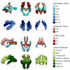
Fig. 1
Fig. 1. Group-average (n = 1104) white matter tracts projected in standard space.
From left to right: sagittal view from the left, axial view from the bottom, coronal view from the front. AF arcuate fasciculus, CG cingulum bundle, CST cortico-spinal tract, FX fornix, IFOF inferior fronto-occipital fasciculus, ILF inferior longitudinal fasciculus, MLF middle longitudinal fasciculus, OR optic radiation, SLF superior longitudinal fasciculus, UF uncinate fasciculus.
Fig. 2
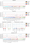
Fig. 2. White matter tract microstructure associations with cardiovascular risk factors (n = 1104).
Models were adjusted for age, age2, sex and total intracranial volume. MRI indices of tract-specific axonal density (ICVF), free water (ISOVF), myelin content (MTsat) and tract volume were analysed. Standardised βs are shown as filled circles for significant associations (FDR-corrected p < 0.05) and as crosses for non-significant associations (FDR-corrected p ≥ 0.05). The x-axis contains the 31 tracts-of-interest coloured and grouped, as in Fig. 1, by association tracts (shades of blue), projection tracts (brown), limbic tracts (pink/red) and corpus callosum (yellow/green). AF arcuate fasciculus, BMI body mass index, CC corpus callosum (1 = rostrum, 2 = genu, 3 = rostral body, 4 = anterior midbody, 5 = posterior midbody, 6 = isthmus, 7 = splenium), CG cingulum bundle, CST cortico-spinal tract, FX fornix, ICVF intra-cellular volume fraction, IFO inferior fronto-occipital fasciculus, ILF inferior longitudinal fasciculus, ISOVF isotropic volume fraction, MLF middle longitudinal fasciculus, MTsat magnetization transfer saturation, OR optic radiation, SLF superior longitudinal fasciculus, UF uncinate fasciculus, WHR waist-to-hip ratio.
Fig. 3
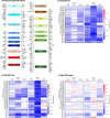
Fig. 3. Clustering of tracts based on their associations with cardiovascular risk factors.
a Hierarchical clustering of tracts based on the Jaccard distance between patterns of association with CVRFs (significant vs. non-significant). Blue (cluster 1), orange (cluster 2), green (cluster 3) and red (cluster 4) indicate clusters of tracts with inter-tract distance <65% of the maximum distance. Grey indicates all other (i.e., not clustered) tracts. b Mean percentage of CVRFs significantly associated with each of the four clusters shown in a, for volume, ICVF, ISOVF and MTsat separately. Each dot represents a tract. AF arcuate fasciculus, CC corpus callosum (1 = rostrum, 2 = genu, 3 = rostral body, 4 = anterior midbody, 5 = posterior midbody, 6 = isthmus, 7 = splenium), CG cingulum bundle, CST cortico-spinal tract, FX fornix, ICVF intra-cellular volume fraction, IFO inferior fronto-occipital fasciculus, ILF inferior longitudinal fasciculus, ISOVF isotropic volume fraction, MLF middle longitudinal fasciculus, MTsat magnetization transfer saturation, OR optic radiation, SLF superior longitudinal fasciculus, UF uncinate fasciculus.
Fig. 4
Fig. 4. White matter tract microstructure associations with the aggregate cardiovascular risk score (n = 1104).
Models were adjusted for age, age2, sex and total intracranial volume. MRI indices of tract-specific axonal density (ICVF), free water (ISOVF), myelin content (MTsat) and tract volume were analysed. Standardised βs are shown as filled circles for significant associations (FDR-corrected p < 0.05) and as crosses for non-significant associations (FDR-corrected p ≥ 0.05). The x-axis contains the 31 tracts-of-interest coloured and grouped, as in Fig. 1, by association tracts (shades of blue), projection tracts (brown), limbic tracts (pink/red) and corpus callosum (yellow/green). AF arcuate fasciculus, CC corpus callosum (1 = rostrum, 2 = genu, 3 = rostral body, 4 = anterior midbody, 5 = posterior midbody, 6 = isthmus, 7 = splenium), CG cingulum bundle, CST cortico-spinal tract, CVRF cardiovascular risk factor, FX fornix, ICVF intra-cellular volume fraction, IFO inferior fronto-occipital fasciculus, ILF inferior longitudinal fasciculus, ISOVF isotropic volume fraction, MLF middle longitudinal fasciculus, MTsat magnetization transfer saturation, OR optic radiation, SLF superior longitudinal fasciculus, UF uncinate fasciculus.
Fig. 5
Fig. 5. For models with significant CVRF × sex interaction, white matter microstructure associations with CVRFs shown separately for males (squares, n = 543) and females (triangles, n = 561).
MRI indices of tract-specific axonal density (ICVF), free water (ISOVF), myelin content (MTsat) and tract volume were analysed. Sex-specific models included age, age2 and total intracranial volume as covariates. The x-axis contains the 31 tracts-of-interest coloured and grouped, as in Fig. 1, by association tracts (shades of blue), projection tracts (brown), limbic tracts (pink/red) and corpus callosum (yellow/green). AF arcuate fasciculus, BMI body mass index, CC corpus callosum (1 = rostrum, 2 = genu, 3 = rostral body, 4 = anterior midbody, 5 = posterior midbody, 6 = isthmus, 7 = splenium), CG cingulum bundle, CST cortico-spinal tract, CVRF cardiovascular risk factor, FX fornix, ICVF intra-cellular volume fraction, IFO inferior fronto-occipital fasciculus, ILF inferior longitudinal fasciculus, ISOVF isotropic volume fraction, MLF middle longitudinal fasciculus, MTsat magnetization transfer saturation, OR optic radiation, SLF superior longitudinal fasciculus, UF uncinate fasciculus, WHR waist-to-hip ratio.
Fig. 6
Fig. 6. CVRF associations with volume and MTsat in white matter tracts and their grey matter seed and target regions (n = 1104).
Standardised βs are marked with an asterisk for significant associations (FDR-corrected p < 0.05). a Constituent sub-regions of the seed and target of each tract. b GM and WM associations with hypertension. c, d GM and WM associations with high BMI in c male and d female participants. A/PCG anterior/posterior cingulate gyrus, AF arcuate fasciculus, Amyg amygdala, AnG angular gyrus, A/L/M/POrG anterior/lateral/middle/posterior orbital gyrus, BMI body mass index, Calc calcarine cortex, CC corpus callosum (1 = rostrum, 2 = genu, 3 = rostral body, 4 = anterior midbody, 5 = posterior midbody, 6 = isthmus, 7 = splenium), CG cingulum bundle, CST cortico-spinal tract, Ent. area entorhinal area, FRP frontal pole, FX fornix, GM grey matter, GRe gyrus rectus, Hippoc hippocampus, IFO inferior fronto-occipital fasciculus, ILF inferior longitudinal fasciculus, I/M/SOG inferior/middle/superior occipital gyrus, I/M/S/TTG inferior/middle/superior/transverse temporal gyrus, LiG lingual gyrus, MFC medial frontal cortex, M/SFG middle/superior frontal gyrus, MLF middle longitudinal fasciculus, (M)Po/PrG post/precentral gyrus (medial segment), MTsat magnetization transfer saturation, Op/Or/TrIFG opercular/orbital/triangular part of inferior frontal gyrus, OCP occipital pole, OFuG occipital fusiform gyrus, OR optic radiation, PHG parahippocampal gyrus, PO parietal operculum, Precun precuneus, SCA subcallosal area, SLF superior longitudinal fasciculus, SMC supplementary motor cortex, SMG supramarginal gyrus, SPL superior parietal lobule, TMP temporal pole, UF uncinate fasciculus, Ventral DC ventral diencephalon, WM white matter.

Similar articles

Cited by

References

    1. Jorgensen DR, et al. A population neuroscience approach to the study of cerebral small vessel disease in midlife and late life: an invited review. Am. J. Physiol.-Heart Circulatory Physiol. 2018;314:H1117–H1136. doi: 10.1152/ajpheart.00535.2017. - DOI - PMC - PubMed
    1. Prins ND, Scheltens P. White matter hyperintensities, cognitive impairment and dementia: an update. Nat. Rev. Neurol. 2015;11:157–165. doi: 10.1038/nrneurol.2015.10. - DOI - PubMed
    1. Alber J, et al. White matter hyperintensities in vascular contributions to cognitive impairment and dementia (VCID): Knowledge gaps and opportunities. Alzheimer’s Dement.: Transl. Res. Clin. Interventions. 2019;5:107–117. doi: 10.1016/j.trci.2019.02.001. - DOI - PMC - PubMed
    1. Brugulat-Serrat A, et al. Patterns of white matter hyperintensities associated with cognition in middle-aged cognitively healthy individuals. Brain Imaging Behav. 2020;14:2012–2023. doi: 10.1007/s11682-019-00151-2. - DOI - PMC - PubMed
    1. ter Telgte A, et al. Cerebral small vessel disease: from a focal to a global perspective. Nat. Rev. Neurol. 2018;14:387–398. doi: 10.1038/s41582-018-0014-y. - DOI - PubMed

Publication types