. 2023 May;8(5):819-832.
doi: 10.1038/s41564-023-01354-6. Epub 2023 Apr 10.

Autophagy restricts Mycobacterium tuberculosis during acute infection in mice

Affiliations

Autophagy restricts Mycobacterium tuberculosis during acute infection in mice

Guillaume R Golovkine et al. Nat Microbiol. 2023 May.

Erratum in

Abstract

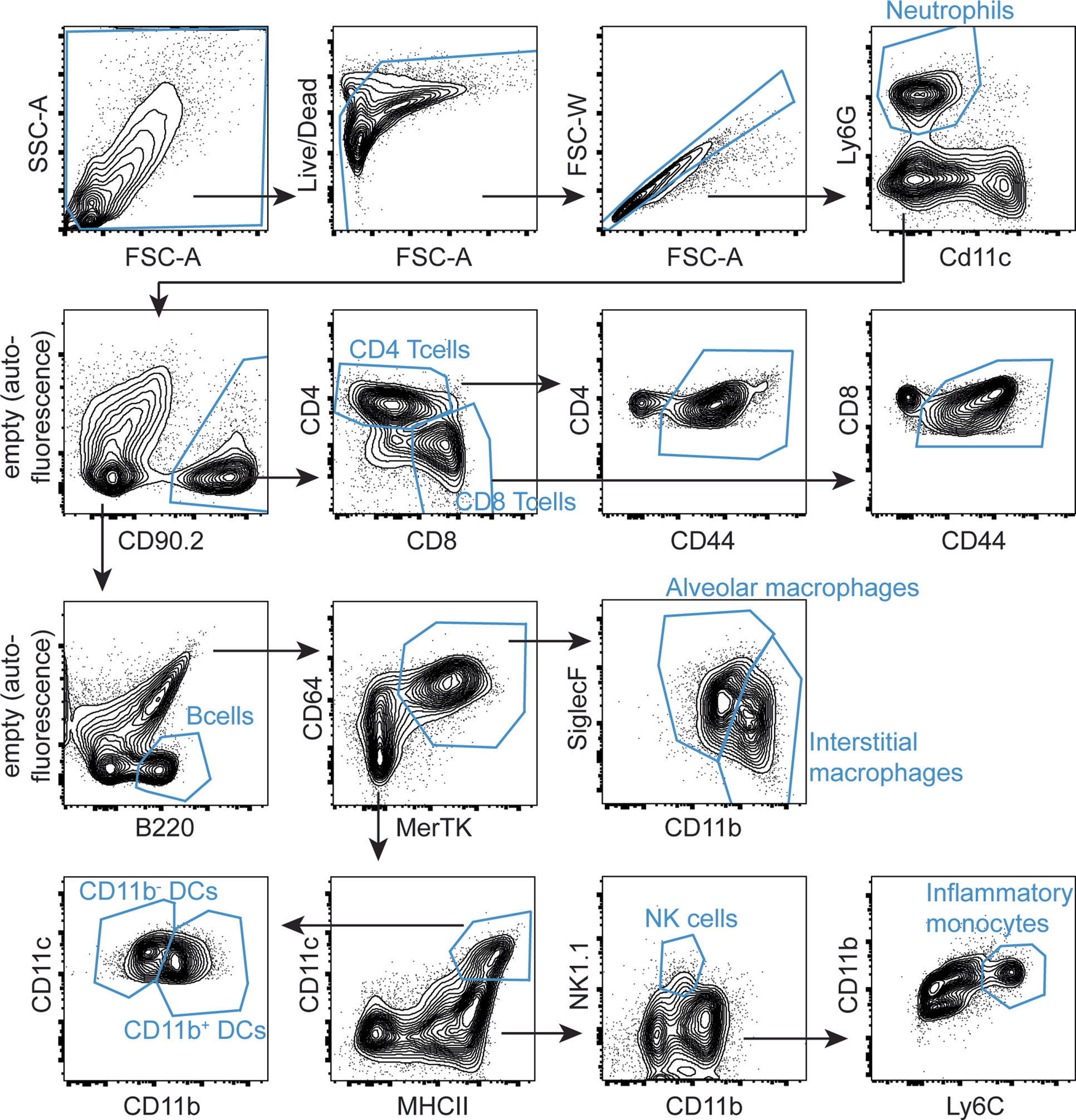
Whether or not autophagy has a role in defence against Mycobacterium tuberculosis infection remains unresolved. Previously, conditional knockdown of the core autophagy component ATG5 in myeloid cells was reported to confer extreme susceptibility to M. tuberculosis in mice, whereas depletion of other autophagy factors had no effect on infection. We show that doubling cre gene dosage to more robustly deplete ATG16L1 or ATG7 resulted in increased M. tuberculosis growth and host susceptibility in mice, although ATG5-depleted mice are more sensitive than ATG16L1- or ATG7-depleted mice. We imaged individual macrophages infected with M. tuberculosis and identified a shift from apoptosis to rapid necrosis in autophagy-depleted cells. This effect was dependent on phagosome permeabilization by M. tuberculosis. We monitored infected cells by electron microscopy, showing that autophagy protects the host macrophage by partially reducing mycobacterial access to the cytosol. We conclude that autophagy has an important role in defence against M. tuberculosis in mammals.


Conflict of interest statement

Competing interests

The authors declare no competing interests.

Figures

Extended data Fig. 1.
Extended data Fig. 1.. Sequences of LoxP sites, map of LoxP site insertions and measurement of Cre expression levels.
a, Sequences of LoxP sites in Atg5fl/fl, Atg16L1fl/fl and Atg7fl/fl mice. b, Map of LoxP sites and Neomycin resistance cassettes insertions in Atg5fl/fl, Atg16L1fl/fl and Atg7fl/fl mice. Grey boxes denote exons. c, Measurement of mRNA levels of Cre in bone marrow-derived macrophages differentiated for 10 days. Results are the means ± SD from 3 biological replicates. n.d. = not detected. Statistical analysis performed with two-sided t-test.
Extended data Fig. 2.
Extended data Fig. 2.. Gating strategy used to isolate lung populations in M. tuberculosis-infected mice.
Lungs from infected mice were harvested after 21 or 70 days of infection. Single cells from lung homogenates were sorted into different immune cell populations according to the strategy outlined in this figure.
Extended data Fig. 3.
Extended data Fig. 3.. Additional in vivo data on autophagy mutant mice infected with M. tuberculosis.
a-c, Mice (n=12–17 per group) were infected with approximately 100 colony-forming units (c.f.u.) of M. tuberculosis and tracked for weight change (a-c). Data is presented as means ± SD. Mice that lost 15% body weight were sacrificed are counted at -15% for subsequent time points. d-e, CFU counts from the lungs of mice (n=5 per group) infected with approximately 100 c.f.u of M. tuberculosis. Spleens were harvested at 21 days post infection (d) or 70 days post infection (e). Data is presented as means ± SD. f-g, Proportion of immune cell populations in the lungs of Atg7 (f) and Atg16L1 (g) mutant mice infected for 70 days with M. tuberculosis. Results are the means ± SD from 2–7 mice per condition. Nt = Neutrophils, Tc = T cells, Bc = B cells, Mac = Macrophages, DC = Dendritic cells, Mono = Ly6Chi inflammatory monocytes. For statistical analysis for CFU and histology experiments, LysMcre+/− and LysMcre+/+ conditions were compared to the respective LysMwt/wt control. * P ≤ 0.05, ** P ≤ 0.01 by two-sided Mann Whitney test.
Extended data Fig. 4.
Extended data Fig. 4.. Statistics of cell death and M. tuberculosis growth during time-course macrophage infection with M. tuberculosis.
a-b, Same data as in Fig. 4b–c but each day is presented as an individual graph. Data represents number of macrophages (a) and Mean Fluorescent Intensity of fluorescent bacteria (b). Results are the means ± SD from 4 technical replicates. For statistical analysis, LysMcre+/− and LysMcre+/+ conditions were compared to the respective LysMwt/wt control. * P ≤ 0.05 by two-sided t-test/.
Extended data Fig. 5.
Extended data Fig. 5.. Growth of M. tuberculosis in autophagy-mutant BMMs and susceptibility of Atg5-flox hemizygous macrophages.
a, BMMs from autophagy mutant mice were infected with M. tuberculosis at a MOI of 2. CFU were enumerated at day 0, day 2 and day 4 post infection. Results are the means ± SD from 3 technical replicates per condition. For statistical analysis, LysMcre+/− and LysMcre+/+ conditions were compared to the respective LysMwt/wt control for the day 4 condition. * P ≤ 0.05 by two-sided t-test. Grey star denotes conditions were the macrophage monolayer was disrupted. b, Macrophages from Atg5fl/wt, Atg5fl/wt LysMcre+/−, Atg5fl/wt LysMcre+/+ and Atg5fl/fl LysMcre+/+ were infected with M. tuberculosis at a MOI of 1 and macrophages were enumerated by imaging every day for 7 days. Macrophages carrying one wild-type allele of Atg5 and one recombined allele (Atg5fl/wt LysMcre+/−, Atg5fl/wt LysMcre+/+) were indistinguishable from wild-type cells. Each condition was tested in technical triplicates.
Extended data Fig. 6.
Extended data Fig. 6.. Absence of autophagy promotes phagosome escape of Mycobacteria in macrophages.
a, Illustrative images of wild-type or ΔeccC M. tuberculosis bacteria in Atg5fl/fl or Atg5fl/fl LysMcre+/+ macrophages after 24 hours of infection. Orange asterisks denote bacteria fully enclosed in phagosomes, blue asterisks show bacteria with poor membrane preservation, red asterisks denote bacteria in the cytosol. Scale bar = 0.4 μm. b, Quantification of bacteria fully enclosed in phagosomes, present directly in the cytosol, or with poor membrane preservation. Data is from a single experiment. A minimum of 40 bacteria per condition were quantified. c, Actin tail formation by M. marinum expressing mCherry in Atg5fl/fl or Atg5fl/fl LysMcre+/+ macrophages was measured at 6.5h, 8h, 9.5h and 11 hours post infection. Results are the means ± SD for 15 images from two technical replicates. * P ≤ 0.05 by two-sided t-test. d, Autophagy-mutant macrophages were infected with M. tuberculosis expressing GFP or ΔeccC M. tuberculosis expressing GFP. Colocalization with Gal-3 was measured at 10h post-infection. Results are the means ± SD from three technical replicates. For statistical analysis, LysMcre+/− and LysMcre+/+ conditions were compared to the respective LysMwt/wt control. * P ≤ 0.05, ** P ≤ 0.01, * P ≤ 0.001 by two-sided Mann Whitney test.
Extended data Fig. 7.
Extended data Fig. 7.. Survival of uninfected autophagy-deficient macrophages and statistics of cell death during infection with ΔeccC M. tuberculosis.
a-b, Quantification of number of uninfected macrophages over time. Data is averaged from two independent experiments with each condition tested in technical duplicates. Data presented as a time course (a) or as selected timepoints for statistical analysis (b). Data is presented as means ± SD. c, Same data as in Fig. 5c but with selected timepoints presented as an individual graphs. Results are the means ± SD from two independent experiments with each condition tested in technical duplicates. For statistical analysis, number of macrophages in cre-positive conditions were compared to the respective LysMwt/wt control. * P ≤ 0.05 by two-sided t-test.
Extended data Fig. 8.
Extended data Fig. 8.. RNA-seq on autophagy mutant macrophages infected with M. tuberculosis. RNA was collected from uninfected BMMs and BMMs infected with M. tuberculosis for 48h.
a-c, Volcano plot showing differentially expressed genes in Atg5fl/fl LysMcre+/+. Grey line demarcates adj.p.value of 0.05. (a), Atg16L1fl/fl LysMcre+/+ (b) and Atg7fl/fl LysMcre+/+ (c) macrophages compared to their respective flox controls. d-f, Venn diagrams showing genes with significant transcriptional changes (adj.p.value ≤ 0.05) for the cre-positive conditions compared to their respective LysMwt/wt control. Venn diagram for all DE genes in uninfected conditions (d), all DE genes in infected conditions (e) and genes exclusively differentially expressed during infection (i.e. with genes with significant DE in uninfected conditions filtered out) (f). g-i, Gene enrichment analysis showing pathways enriched in M. tuberculosis-infected cells. Genes with significant transcriptional changes (adj.p.value ≤ 0.05) were used for pathway enrichment using the hypergeometric test (HGT). Datasets for enrichment included: KEGG, REACTOME, MSigDB C2_CP and C3, GO, CORUM, and HALLMARK. Top 15 pathways commonly enriched in Atg5fl/fl LysMcre+/+, Atg16L1fl/fl LysMcre+/+ and Atg7fl/fl LysMcre+/+ (g), Top 15 pathways commonly enriched in all 3 genotypes specifically during infection (h) and top 15 pathways enriched exclusively in Atg5fl/fl LysMcre+/+ macrophages (i).
Extended data Fig. 9.
Extended data Fig. 9.. M. tuberculosis infection induces necrosis-like cell death and efferocytosis in autophagy-deficient macrophages.
a, Western blot for pRIP3, RIP3, BCL-xL and β-actin on cell lysates from uninfected BMMs and BMMs infected for 48 hours with M. tuberculosis. b, Measurement by ELISA of IL-1β secretion in supernatant of macrophages infected for 48 hours with M. tuberculosis. Results are the means ± SD from two independent experiments, in which each condition was tested in technical triplicates. * P ≤ 0.05 by two-sided t-test. c-d, Atg5fl/fl LysMcre+/+ macrophages were infected with GFP-expressing M. tuberculosis and imaged every 30 minutes over a 6-day period (Supplementary Video 3). Time and mode of cell death were recorded for 145 Atg5fl/fl LysMcre+/+ macrophages infected with GFP-expressing M. tuberculosis from 3 independent experiments. The average time of death by necrosis-like cell death (c) and the delay between efferocytosis and subsequent cell death (d) were measured. Center line: median; box limits: upper and lower quartiles; whiskers: 1.5x interquartile range. n = 80 and 65, respectively.
Extended data Fig. 10.
Extended data Fig. 10.. Illustrative images and statistics of PI and CellEvent staining of macrophages infected with M. tuberculosis.
a, Illustrative images of experiment quantified in Fig. 6a–b. Autophagy-deficient macrophages were infected with M. tuberculosis at a MOI of 1. Propidium iodide and CellEvent Caspase-3/7 reagent were added to the media at the beginning of the infection. Cells were imaged every 30 minutes over a period of 8 days. Scale bar denotes 100 μm. b, Same data as in Fig. 6a–b but with selected timepoints presented as an individual graphs. Results are the means ± SD with each condition tested in technical triplicates. For statistical analysis, number of CellEvent (b) and propodium iodide (c) positive macrophages in cre-positive conditions were compared to the respective LysMwt/wt control. * P ≤ 0.05, ** P ≤ 0.01, * P ≤ 0.001 by two-sided t-test.
Fig. 1 |
Fig. 1 |. Cre–Lox recombination efficiency in macrophages varies between different autophagy factors and is influenced by Cre dosage.
ac, Analysis of Cre–Lox recombination efficiency in BMMs differentiated for 7, 10 or 12 days. Percentage of intensity of deleted band over combined intensities of floxed (Flox) and deleted (Del) bands is indicated by ‘Percentage recombination’. ac, Samples extracted from Atg5fl/fl, Atg5fl/fl LysMcre+/− and Atg5fl/fl LysMcre+/+ mice (a), Atg16L1fl/fl, Atg16L1fl/fl LysMcre+/− and Atg16L1fl/fl LysMcre+/+ mice (b) and Atg7fl/fl, Atg7fl/fl LysMcre+/− and Atg7fl/fl LysMcre+/+ mice (c). df, Measurement of mRNA levels of autophagy factors in BMMs differentiated for 10 days from Atg5fl/fl, Atg5fl/fl LysMcre+/− and Atg5fl/fl LysMcre+/+ mice (d), Atg16L1fl/fl, Atg16L1fl/fl LysMcre+/− and Atg16L1fl/fl LysMcre+/+ mice (e) and Atg7fl/fl, Atg7fl/fl LysMcre+/− and Atg7fl/fl LysMcre+/+ mice (f). Results are the means ± s.d. from three biological replicates. *P ≤ 0.05 by two-sided t-test. g,h, Western blot analysis of LC3 and β-actin in BMMs differentiated for 12 days: samples are from untreated cells (g) or cells treated for 8 h with EBSS and 2 h with bafilomycin (h). Results are representative from two biological replicates.
Fig. 2 |
Fig. 2 |. ATG16L1 and ATG7 are required for control of M. tuberculosis at later stages of infection but the mouse phenotype is undermined by limitations of the Cre–Lox system.
af, Mice were infected with ~100 c.f.u. of M. tuberculosis and tracked for weight change (ac) and survival (df). Each group was composed of 10–12 mice, except LysMcre+/+ mice (n = 5). ac, Weight change over the first 12 weeks of infection of Atg5fl/fl, Atg5fl/fl LysMcre+/− and Atg5fl/fl LysMcre+/+ mice (a), Atg16L1fl/fl, Atg16L1fl/fl LysMcre+/− and Atg16L1fl/fl LysMcre+/+ mice (b) and Atg7fl/fl, Atg7fl/fl LysMcre+/−, Atg7fl/fl LysMcre+/+ and LysMcre+/+ mice (c). df, Survival of Atg5fl/fl, Atg5fl/fl LysMcre+/− and Atg5fl/fl LysMcre+/+ mice (d), Atg16L1fl/fl, Atg16L1fl/fl LysMcre+/− and Atg16L1fl/fl LysMcre+/+ mice (e) and Atg7fl/fl, Atg7fl/fl LysMcre+/−, Atg7fl/fl LysMcre+/+ and LysMcre+/+ mice (f). Statistical significance was tested against the flox control group with a two-sided log-rank (Mantel–Cox) test. ***P ≤ 0.001. gi, Proportion of immune cell populations in the lungs of Atg5 (g), Atg16L1 (h) and Atg7 (i) mutant mice infected for 21 days with M. tuberculosis. Results are the means ± s.d. from 2–7 mice per condition. Nt, neutrophils; Tc, T cells; Bc, B cells; AMO, alveolar macrophages; MDM, monocyte-derived macrophages; IM, interstitial macrophages; DC, dendritic cells; Mono, Ly6Chi inflammatory monocytes. For statistical analysis, LysMcre+/− and LysMcre+/+ conditions were compared to the respective LysMwt/wt control. *P ≤ 0.05, **P ≤ 0.01, ***P ≤ 0.001 by Mann–Whitney test.
Fig. 3 |
Fig. 3 |. Atg16L1 and Atg7 mutant mice display lung lesions at later stages of infection with M. tuberculosis.
a,b, The c.f.u. counts from the lungs of mice infected with ~100 c.f.u of M. tuberculosis. Lungs were collected at 21 days (a) or 70 days (b) postinfection. Results are the means ± s.d. from 2–6 mice per condition. cf, Segments of the lungs from the same mice as in a and b were processed for histology: average portion of the lungs presenting lesions was measured with ImageScope at day 21 (c) and day 70 (d) postinfection; results are the means ± s.d. from 2–6 mice per condition. e,f, Representative sections at day 21 (e) and day 70 (f) postinfection are presented. Scale bar, 2 mm. For statistical analysis for c.f.u. and histology experiments, LysMcre+/− and LysMcre+/+ conditions were compared to the respective LysMwt/wt control. *P ≤ 0.05, **P ≤ 0.01, ***P ≤ 0.001 by Mann–Whitney test.
Fig. 4 |
Fig. 4 |. Macrophages deficient in different autophagy factors are susceptible to M. tuberculosis infection.
a, Representative images from two independent experiments of a 6 days long time-lapse sequence of autophagy-deficient macrophages infected with GFP-expressing M. tuberculosis (Mtb) at an MOI of 2 (Supplementary Video 1). Scale bar, 100 μm. b,c, Macrophages infected with GFP-expressing M. tuberculosis at an MOI of 1 were imaged every day for 7 days by high-content microscopy. b,c, Number of cells (b) and integrated fluorescence intensity of GFP signal (c) were quantified over time. Data are the average of four technical replicates from an experiment representative of two independent experiments. Statistics in Extended Data Fig. 4a,b.
Fig. 5 |
Fig. 5 |. The autophagy-deficient macrophage susceptibility to M. tuberculosis is ESX-1-dependent and linked to phagosome escape.
a, Illustrative images of actin tail formation by M. marinum (Mm) expressing mCherry inside macrophages at 11 h postinfection. Actin tails were stained with AlexaFluor 488 Phalloidin (green) and nuclei with dapi (blue). Arrowheads indicate examples of bacteria with actin tails. Scale bar, 20 μm. b, Quantification of the percentage of mCherry-expressing M. marinum colocalizing with actin tails after 11 h of infection of autophagy-deficient macrophages. Results are the means ± s.d. for ten images from two technical replicates. *P ≤ 0.05 by two-sided t-test. c, Quantification of number of macrophages over time during time-course infection with GFP-expressing ΔeccC M. tuberculosis at an MOI of 1. Black curve shows survival of Atg5fl/fl LysMcre+/+ macrophages infected with wild-type M. tuberculosis. Data are averaged from two independent experiments with each condition tested in technical duplicates. Statistics in Extended Data Fig. 7c.
Fig. 6 |
Fig. 6 |. M. tuberculosis infection induces necrosis-like cell death and efferocytosis in autophagy-deficient macrophages.
a,b, Autophagy-deficient macrophages were infected with M. tuberculosis at an MOI of 1. PI and CellEvent Caspase-3/7 reagent were added to the media at the beginning of the infection (Supplementary Video 2): number of PI-positive cells (a) and CellEvent-positive cells (b) per field of view was quantified over time. Results are the average of three technical replicates. Statistics are given in Extended Data Fig. 10b,c. cf, Atg5fl/fl LysMcre+/+ macrophages were infected with GFP-expressing M. tuberculosis and imaged every 30 min over a 6 day period (Supplementary Video 3). c, Illustrative images of necrosis-like cell death. Yellow arrowhead shows dying cell, yellow asterisk denotes time of cell death. d, Illustrative images of efferocytosis followed by cell death. Macrophage (red arrow) dies (red asterisk) after phagocytosing an adjacent dead cell (orange arrowhead). e, Modes of cell death were recorded for 145 Atg5fl/fl LysMcre+/+ macrophages infected with GFP-expressing M. tuberculosis from three independent experiments. The number of cells dying of necrosis-like cell death or after efferocytosis was quantified and presented as means ± s.d. (c). f, Model depicting that autophagy forestalls phagocyte cell death during infection and leads to non-inflammatory apoptosis. In the absence of autophagy, phagosome disruption mediated by bacterial ESX-1 induces rapid necrosis, followed by a cascade of efferocytosis and cell death that promotes inflammation.

Comment in

References

    1. Levine B & Kroemer G Biological functions of autophagy genes: a disease perspective. Cell 176, 11–42 (2019). - PMC - PubMed
    1. Chang C, Jensen LE & Hurley JH Autophagosome biogenesis comes out of the black box. Nat. Cell Biol. 23, 450–456 (2021). - PMC - PubMed
    1. Deretic V & Lazarou M A guide to membrane atg8ylation and autophagy with reflections on immunity. J. Cell Biol. 221, e202203083 (2022). - PMC - PubMed
    1. Lu Q et al. Homeostatic control of innate lung inflammation by Vici syndrome gene Epg5 and additional autophagy genes promotes influenza pathogenesis. Cell Host Microbe 19, 102–113 (2016). - PMC - PubMed
    1. Park S et al. Autophagy genes enhance murine gammaherpesvirus 68 reactivation from latency by preventing virus-induced systemic inflammation. Cell Host Microbe 19, 91–101 (2016). - PMC - PubMed

Publication types

Substances