. 2023 Apr 10;14(1):3.
doi: 10.1186/s13100-023-00291-9.

Telomeric retrotransposons show propensity to form G-quadruplexes in various eukaryotic species

Affiliations

Telomeric retrotransposons show propensity to form G-quadruplexes in various eukaryotic species

Pavel Jedlička et al. Mob DNA. .

Abstract

Background: Canonical telomeres (telomerase-synthetised) are readily forming G-quadruplexes (G4) on the G-rich strand. However, there are examples of non-canonical telomeres among eukaryotes where telomeric tandem repeats are invaded by specific retrotransposons. Drosophila melanogaster represents an extreme example with telomeres composed solely by three retrotransposons-Het-A, TAHRE and TART (HTT). Even though non-canonical telomeres often show strand biased G-distribution, the evidence for the G4-forming potential is limited.

Results: Using circular dichroism spectroscopy and UV absorption melting assay we have verified in vitro G4-formation in the HTT elements of D. melanogaster. Namely 3 in Het-A, 8 in TART and 2 in TAHRE. All the G4s are asymmetrically distributed as in canonical telomeres. Bioinformatic analysis showed that asymmetric distribution of potential quadruplex sequences (PQS) is common in telomeric retrotransposons in other Drosophila species. Most of the PQS are located in the gag gene where PQS density correlates with higher DNA sequence conservation and codon selection favoring G4-forming potential. The importance of G4s in non-canonical telomeres is further supported by analysis of telomere-associated retrotransposons from various eukaryotic species including green algae, Diplomonadida, fungi, insects and vertebrates. Virtually all analyzed telomere-associated retrotransposons contained PQS, frequently with asymmetric strand distribution. Comparison with non-telomeric elements showed independent selection of PQS-rich elements from four distinct LINE clades.

Conclusion: Our findings of strand-biased G4-forming motifs in telomere-associated retrotransposons from various eukaryotic species support the G4-formation as one of the prerequisites for the recruitment of specific retrotransposons to chromosome ends and call for further experimental studies.

Keywords: Drosophila; G-quadruplex; Het-A; Retrotransposon; TAHRE; TART; Telomere.


Conflict of interest statement

The authors declare that they have no competing interests.

Figures

Fig. 1
Fig. 1
G4 distribution and experimental validation in the reference telomeric elements of D. melanogaster. A Schematic localization of G4 forming motifs inside Het-A, TART and TAHRE elements. Each triangle represents one in vitro verified G4. Orientation of the triangle indicates the PQS orientation (all G-rich sequences are on the template strand) and color indicates the topology (red for parallel, green for antiparallel and purple for 3 + 1 hybrid G4). Note that PQS8 in TART originates from TART-A1 (AY561850) at the respective position. The elements are not to scale. B CD spectra of all G4-forming sequences in 150 mM K+, melting temperature range as well as a visual representation of strand orientation is indicated for each topology
Fig. 2
Fig. 2
Identification of main PQS carriers among repetitive DNA in D. melanogaster genome. A Genomic proportion and PQS content of 5 main repeat types. B Analysis of the main interspersed repeats. Top: genomic proportion and PQS counts in individual types of interspersed elements. Bottom: mutual orientation of individual types of interspersed elements in the genome with respect to PQS orientation. The orange/yellow colors represent elements with sense PQS orientation—the G-rich sequence is on the coding strand, the blue colors represent elements with antisense PQS orientation (the repeat orientation is with respect to the chromosome, the orientation of PQS is with respect to the element in which PQS resides). C Analysis (as in B) of HTT harboring Jockey-clade elements. D GAG protein domain based phylogram shows the PQS distribution per reference full-length Jockey elements (red numbers, Additional file 2: Tab. S3). Note that G2 is not included due to the lack of GAG domain. Maximum likelihood phylogenetic tree was constructed using PhyML v3.0 with BioNJ used to build up the starting tree. Bootstrap support values higher than 50 are given
Fig. 3
Fig. 3
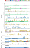
Visualization of LINE retrotransposons arrays with PQS in telomeric sequences of various Drosophila species. Mutual strand-specific orientation of LINEs and PQS is depicted. The order and taxonomic division of given species was adapted from the Drosophila genus consensus phylogenetic tree in [24]. The gray colored boxes in D. virilis are formed by tandem repeats TTR321 and TTR712. The PQS rich region in D. willistoni is formed by 392 bp long tandem repeat. Note that in each species there is at least one element with antisense PQS
Fig. 4
Fig. 4
Analysis of DNA-AA identity bias with respect to PQS distribution. A The scheme of non-LTR retrotransposon shows the average pairwise DNA and AA identity or similarity calculated as an average of each group with respect to functional regions of non-LTR retrotransposon. The charts above them show the difference of average DNA-AA identity and similarity as defined by BLOSUM62 for gag and pol genes for different telomeric retrotransposon groups. The distribution of PQS in functional regions is indicated by a pie chart. B Dissection of DNA-AA identity and similarity in gag gene that can be divided based on protein alignment to conserved area and the rest that shows little conserved motifs. The gag is divided by the conserved region to 5´fragment (N-terminal) and 3´fragment (see Additional file 1: Fig. S6; C-terminal; major homology region—MHR; zinc knuckle motifs—C2HC). As for A) the average DNA and AA pairwise similarities and identities derived from all elements are shown for corresponding regions as well as the DNA-AA difference for the transposon groups. The pie chart shows PQS distribution in the 3 gag regions, note that 5 PQS are on the border of the conserved region and the 5´fragment creating a difference of 10 between gag located PQS A) and sum of PQS in gag regions B). The difference is 10 since these 5 PQS were counted for both regions
Fig. 5
Fig. 5
Telomeres with intercalated transposable elements containing PQS. Schematic representation of telomeric sequence structure in various species in which specific retrotransposons insert into canonical short telomeric repeats. All the elements belong to 4 LINE clades (indicated on the right). The PQS orientation is in relation to the respective element (PQS + means that the G-rich sequence is on the coding strand). The sequences in parenthesis represent both the telomerase-synthesized short tandem repeats as well as the terminal 3´ ssDNA overhang. TAS stands for telomere associated sequences
Fig. 6
Fig. 6
PQS counts in selected families of LINEs with the telomeric elements. Mutual strand-specific orientation of PQS and TE in 4 groups of LINEs showed in dark- and light gray for antisense and sense, respectively. PQS in telomeric elements of all species (stated in Fig. 5) are highlighted and tend to be the most PQS-rich elements in each group. Number of analyzed elements for each TE group is given

Similar articles

Cited by

References

    1. Kubo Y, Okazaki S, Anzai T, Fujiwara H. Structural and phylogenetic analysis of TRAS, telomeric repeat-specific non-LTR retrotransposon families in Lepidopteran insects. Mol Biol Evol. 2001;18:848–857. doi: 10.1093/oxfordjournals.molbev.a003866. - DOI - PubMed
    1. Osanai-Futahashi M, Fujiwara H. Coevolution of telomeric repeats and telomeric repeat-specific non-LTR retrotransposons in insects. Mol Biol Evol. 2011;28:2983–2986. doi: 10.1093/molbev/msr135. - DOI - PubMed
    1. Takahashi H, Okazaki S, Fujiwara H. A new family of site-specific retrotransposons, SART1, is inserted into telomeric repeats of the silkworm. Bombyx mori Nucleic Acids Res. 1997;25:1578–1584. doi: 10.1093/nar/25.8.1578. - DOI - PMC - PubMed
    1. Arkhipova IR, Morrison HG. Three retrotransposon families in the genome of Giardia lamblia: two telomeric, one dead. Proc Natl Acad Sci USA. 2001;98:14497–14502. doi: 10.1073/pnas.231494798. - DOI - PMC - PubMed
    1. Higashiyama T, Noutoshi Y, Fujie M, Yamada T. Zepp, a LINE-like retrotransposon accumulated in the Chlorella telomeric region. EMBO J. 1997;16:3715–3723. doi: 10.1093/emboj/16.12.3715. - DOI - PMC - PubMed

LinkOut - more resources