. 2023 Apr 27;97(4):e0018823.
doi: 10.1128/jvi.00188-23. Epub 2023 Apr 11.

The Papain-Like Protease of Porcine Reproductive and Respiratory Syndrome Virus Impedes STING Translocation from the Endoplasmic Reticulum to the Golgi Apparatus by Deubiquitinating STIM1

Affiliations

The Papain-Like Protease of Porcine Reproductive and Respiratory Syndrome Virus Impedes STING Translocation from the Endoplasmic Reticulum to the Golgi Apparatus by Deubiquitinating STIM1

Feifei Diao et al. J Virol. .

Abstract

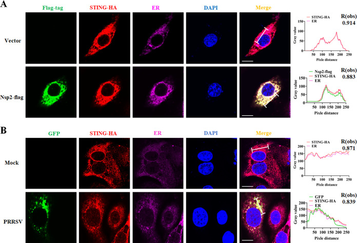
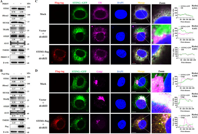
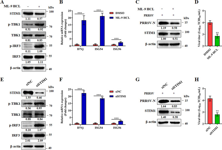
Stimulator of interferon (IFN) genes (STING) was recently pinpointed as an antiviral innate immune factor during the infection of RNA viruses. Porcine reproductive and respiratory syndrome virus (PRRSV), the swine arterivirus, is an enveloped RNA virus which has evolved many strategies to evade innate immunity. To date, the interactive network between PRRSV and STING remains to be fully established. Herein, we report that STING suppresses PRRSV replication through type I interferon signaling. However, PRRSV impedes STING trafficking from the endoplasmic reticulum (ER) to the Golgi apparatus, leading to the decreased phosphorylation of TANK-binding kinase 1 (TBK1) and interferon regulatory factor 3 (IRF3). Furthermore, PRRSV nonstructural protein 2 (Nsp2) colocalizes with STING, blocks STING translocation, and disrupts the STING-TBK1-IRF3 complex. Mechanistically, PRRSV Nsp2 retains STING at the ER by increasing the level of Ca2+ sensor stromal interaction molecule 1 (STIM1) protein. Functional analysis reveals that PRRSV Nsp2 deubiquitinates STIM1 by virtue of its papain-like protease 2 (PLP2) deubiquitinating (DUB) activity. Finally, we demonstrate that loss of STIM1 is associated with an elevated IFN response and restricts PRRSV replication. This work delineates the relationship between PRRSV infection and STING signaling and the importance of papain-like proteases (PLPs) in interfering in this axis. IMPORTANCE Porcine reproductive and respiratory syndrome virus (PRRSV), a member of the family Arteriviridae, is responsible for reproductive disorders in pregnant sows and respiratory problems in piglets, resulting in huge losses in the swine industry worldwide. Of note, PRRSV infection causes immunosuppression, of which the mechanism is not completely understood. Here, we demonstrate for the first time that STING, a protein typically associated with the antiviral response in DNA viruses, plays a critical role in controlling PRRSV infection. However, PRRSV utilizes its encoded protein Nsp2 to inhibit STING activity by blocking its translocation from the ER to the Golgi apparatus. In particular, Nsp2 retains STING at the ER by interacting with and further deubiquitinating STIM1. For this process, the activity of the viral PLP2 DUB enzyme is indispensable. The study describes a novel mechanism by which PLP2 plays a critical role in suppressing the innate immune response against arteriviruses and potentially other viruses that encode similar proteases.

Keywords: Nsp2; PRRSV; STIM1; STING; deubiquitinate; papain-like protease.


Conflict of interest statement

The authors declare no conflict of interest.

Figures

FIG 1
FIG 1
STING suppresses PRRSV replication through the type I interferon pathway. (A to C) The STING agonist diABZI inhibits PRRSV infection. Marc-145 cells were mock or PRRSV infected (MOI = 0.1) for 24 h with diABZI (15 μM) or DMSO (vehicle). (A) Western blot of PRRSV-N protein. (B) Viral titers measured by TCID50. (C) Cell viability of Marc-145 cells treated with diABZI at the indicated concentrations or DMSO for 24 h. (D to F) The STING inhibitor SN-011 increases PRRSV replication. Marc-145 cells were mock or PRRSV infected (MOI = 0.1) for 24 h with SN-011 (5 μM) or DMSO. (D) Western blot of PRRSV-N protein. (E) Viral titers measured by TCID50. (F) Cell viability of Marc-145 cells treated with SN-011 at the indicated concentrations or DMSO for 24 h. (G to I) STING deficiency promotes PRRSV infection. Marc-145 cells were mock or PRRSV infected (MOI = 0.1) for 24 h with control siRNA (siCtrl) or siRNA targeting STING (siSTING). (G) Western blot of PRRSV-N protein. (H) Viral titers measured by TCID50. (I) Cell viability of Marc-145 cells treated with siSTING (50 nM) or siCtrl (50 nM) for 24 h. (J to L) Marc-145 cells were transfected with control siRNA (siNC) or siRNA targeting IFN-αR (siIFN-αR1) for 24 h and then infected with PRRSV (MOI = 0.1) for another 24 h with diABZI (15 μM) or DMSO (vehicle). (J) PRRSV-N and IFN-αR1 protein levels determined by Western blotting. (K) TCID50 of PRRSV in cell supernatants. (L) Viability of Marc-145 cells transfected with siIFN-αR1. The protein levels were quantified by ImageJ and normalized to β-actin levels. Data are means and SD. *, P < 0.05; **, P < 0.01. The experimental data are representative of results from three independent experiments.
FIG 2
FIG 2
PRRSV infection inhibits STING-mediated antiviral immunity. (A) PRRSV blocks ISG induction stimulated by the STING agonist diABZI. Marc-145 cells were mock or PRRSV infected (MOI = 0.1) for 24 h with diABZI (15 μM) or DMSO (vehicle). Cells were then harvested for RNA extraction, and the mRNA expression of IFN-β, ISG54, and ISG56 was detected by RT-PCR. (B) PRRSV inhibits the STING-mediated downstream pathway. Marc-145 cells were mock or PRRSV infected (MOI = 0.1) for 24 h with diABZI (15 μM) or DMSO (vehicle). Cells were then harvested for Western blotting with antibodies against phosphorylated TBK1 (p-TBK1), TBK1, phosphorylated IRF3 (p-IRF3), IRF3, PRRSV-N, and β-actin. (C) PRRSV inhibits IRF3 translocated into the nucleus. Marc-145 cells were transfected with GFP-IRF3 for 12 h and then mock or PRRSV infected (MOI = 0.1) for 24 h with diABZI (15 μM) or DMSO (vehicle). Cells were stained with DAPI and examined under a confocal microscope. Bars, 5 μm. Images are representative of three biological replicates that included at least 20 technical replicates. (D) Cytosolic and nuclear distribution of IRF3 after PRRSV infection. Marc-145 cells were mock or PRRSV infected (MOI = 0.1) for 24 h with diABZI (15 μM) or DMSO (vehicle). Cells were then harvested for extraction of proteins in the cytoplasm or nucleus. The extracted proteins were determined by Western blotting with antibodies against IRF3, PRRSV-N, β-actin, and histone H3. The protein levels were quantified by ImageJ and normalized to β-actin levels. Data are means and SD. ***, P < 0.001. The experimental data are representative of results from three independent experiments.
FIG 3
FIG 3
PRRSV infection impedes STING trafficking from the ER to the Golgi apparatus. (A and B) Marc-145 cells expressing GFP-STING were treated in three different groups: mock (DMSO treatment only), diABZI (diABZI treatment only), and PRRSV + diABZI (PRRSV infection together with diABZI treatment). After 24 h, cells were stained with DAPI and incubated with antibodies specific for PRRSV-N protein, calreticulin (ER marker) (A), and GORASP2 (Golgi marker) (B). Samples were examined under a confocal microscope. The colocalization of STING and the ER or Golgi apparatus was analyzed by ImageJ software. Bars, 5 μm. Images are representative of 3 biological replicates, with at least 20 technical repeats. The degree of colocalization is shown as Robs for that image.
FIG 4
FIG 4
Nsp2 of PRRSV interacts with STING. (A) Colocalization of PRRSV-encoded proteins and STING. Marc-145 cells were cotransfected HA-STING together with the indicated plasmid-encoded Flag-tagged PRRSV viral proteins (Nsp1α, Nsp1β, Nsp2, Nsp4, Nsp5, Nsp7, Nsp9, Nsp10, Nsp11, Nsp12, GP2, GP3, GP4, GP5, M, and N) for 24 h, fixed, and subjected to immunofluorescence analysis by using rabbit anti-HA antibody (green) and mouse anti-Flag antibody (red). The nuclei were stained with DAPI. The fluorescence intensity profile of DAPI (blue), Flag (red), and HA (green) was measured along the line drawn by ImageJ. Bars, 5 μm. (B) PRRSV Nsp2 interacts with STING. Marc-145 cells were cotransfected HA-STING together with indicated plasmids (vector) for 24 h, then harvested for immunoprecipitation by using mouse anti-Flag antibody, and whole-cell lysates were subjected to Western blotting by using rabbit anti-STING antibody or mouse anti-Flag antibody, along with β-actin as a loading control. The data shown represent three independent experiments.
FIG 5
FIG 5
Nsp2 is colocalized with STING at the ER. (A) Marc-145 cells were cotransfected with plasmid-encoded Nsp2-Flag and HA-STING. After 24 h, cells were stained with DAPI and incubated with antibodies specific for the Flag tag, the HA tag, and calreticulin (ER marker). (B) Marc-145 cells transfected with STING-HA were mock infected or infected with PRRSV-GFP at an MOI of 0.1. After 24 h, cells were stained with DAPI and incubated with antibodies specific for the HA tag and calreticulin (ER marker). Samples were examined under a confocal microscope. The degree of colocalization (Robs) was determined using ImageJ software as Pearson’s correlation coefficient (r) via the Costes method. Bars, 5 μm.
FIG 6
FIG 6
PRRSV Nsp2 blocks STING trafficking and its downstream pathway. (A) Nsp2 blocks STING trafficking from the ER to the Golgi apparatus. (A and B) Expression of STING-GFP in Marc-145 cells. Cells were transfected with vector or Nsp2 expression plasmid and then treated with or without diABZI as for Fig. 5A and B. Cells were stained with DAPI and incubated with antibodies specific for Flag, GORASP2 (Golgi marker), and calnexin (ER marker). Samples were examined under a confocal microscope. The degree of colocalization (Robs) was determined using ImageJ software as Pearson’s correlation coefficient (r) via the Costes method. Bars, 5 μm. (C) Nsp2 disrupts the formation of the STING-TBK1-IRF3 complex. Marc-145 cells were cotransfected with Myc-STING together with TBK1-flag, IRF3-HA, Nsp2-flag, or vector for 24 h. Cell lysates were immunoprecipitated with anti-Myc antibody and then analyzed by Western blotting for Myc, Flag, HA, and β-actin. (D and E) Nsp2 blocks STING downstream signaling. (D) Nsp2 inhibits the activation of TBK1 and IRF3. Marc-145 cells were transfected with pCI-Nsp2-flag or empty vector (pCI-neo) for 24 h with diABZI (15 μM) or DMSO (vehicle); then, cell lysates were analyzed by Western blotting for Flag, p-TBK1, TBK1, p-IRF3, IRF3, PRRSV-N, and β-actin. (E) Nsp2 blocks ISG induction stimulated by the STING agonist diABZI. Marc-145 cells were transfected with pCI-Nsp2-flag or empty vector (pCI-neo) for 24 h with diABZI (15 μM) or DMSO (vehicle). mRNA expression of IFN-β, ISG54, and ISG56 was examined by RT-qPCR. The protein levels were quantified by ImageJ and normalized to β-actin levels. Data are means and SD. ***, P < 0.001. The experimental data are representative of results from three independent experiments.
FIG 7
FIG 7
Determination of proteins associated with STING translocation. (A and B) Marc-145 cells were mock or PRRSV infected (A) and transfected with vector plasmid or plasmid encoding Nsp2 (B). After 24 h, cells were then harvested for Western blotting with antibodies against STIM1, iRhom2, SEC61β, TRAPβ, STEEP, ALG2, TMED2, β-actin, and Flag or PRRSV-N. The protein levels were quantified by ImageJ and normalized to β-actin levels. The experimental data are representative of results from three independent experiments. (C and D) Influence of STIM1 overexpression on STING trafficking. Marc-145 cells expressing GFP-STING were treated in three different groups: mock (vector plasmid transfection only); vector + diABZI (vector plasmid transfection and diABZI treatment), and STIM1-Flag + diABZI (STIM1-Flag transfection and diABZI treatment). After 24 h, cells were stained with DAPI and incubated with antibodies specific for the Flag tag, calreticulin (ER marker) (C), and GORASP2 (Golgi marker) (D). Samples were examined under a confocal microscope. The colocalization of STING and ER or Golgi was analyzed by ImageJ software. Bars, 5 μm. Images are representative of 3 biological replicates, with at least 20 technical repeats. The degree of colocalization is shown as Robs for that image.
FIG 8
FIG 8
PRRSV Nsp2 impedes STING translocation by increasing STIM1 expression. (A to D) STIM1 is essential for Nsp2 to impede STING translocation. (A and B) STING-GFP expression in Marc-145 cells. Cells were transfected with Nsp2-flag or vector and treated with ML-9 HCl (15 μM) plus diABZI (15 μM) for 24 h. After 24 h, cells were stained with DAPI and incubated with antibodies specific for Flag protein, calreticulin (ER marker) (A), and GORASP2 (Golgi marker) (B). Samples were examined under a confocal microscope. (C and D) STIM1 deficiency promotes STING translocation. Expression of STING-GFP in Marc-145 cells. Cells were transfected with siRNA targeting STIM1 (siSTIM1) for 24 h and then transfected with Nsp2-flag for another 24 h with diABZI (15 μM). Colocalization of STING and the ER (C) or Golgi apparatus (D) was assessed. (E and F) Colocalization of Nsp2 and STIM1 at the ER. (E) Marc-145 cells were transfected with Flag-Nsp2 and then stained for Flag, STIM1, and calnexin (ER marker), and nuclei were stained with DAPI. (F) Marc-145 cells were infected with PRRSV-GFP (MOI = 0.1) and then stained for Flag, STIM1, and calnexin (ER marker), and nuclei were stained with DAPI; cells were observed by confocal microscopy. Bar, 5 μm. The colocalization of STING and the ER or Golgi apparatus was analyzed by ImageJ software. Images are representative of 3 biological replicates, with at least 20 technical repeats. The degree of colocalization is shown as Robs for that image. (G) Ubiquitination of STIM1 was blocked by Nsp2. Marc-145 cells were cotransfected with HA-Ub-K48, Nsp2-flag or vector. Twenty-four hours later, the cell extracts were subjected to immunoprecipitation and immunoblotting by using antibodies to STIM1, Flag, HA, and β-actin. The experimental data are representative of results from three independent experiments.
FIG 9
FIG 9
The PLP2 DUB enzyme in Nsp2 is critical for blocking STING translocation. (A) Schematic diagram of PRRSV Nsp2 domain mutations. A mutant was constructed in the pCI-Nsp2 expression plasmid. Protein fragments are drawn to scale, except where a thinner line was used. The putative catalytic residues are indicated with red stars, and the predicted PLP2 DUB domain is in blue. Numbering is based on amino acid positions of the respective replicase polyproteins. (B and C) The mutations (C55A/H224A) of Nsp2 were critical for STIM1 expression. (B) Marc-145 cells were transfected with pCI-Nsp2-flag, pCI-Nsp2(C55A/H224A)-flag, or empty vector (pCI-neo) for 24 h; then, cell lysates were analyzed by Western blotting for Flag, STIM1, and β-actin. (C) Marc-145 cells were cotransfected with HA-Ub-K48 and Nsp2-flag, Nsp2(C55A/H224A)-flag or vector for 24 h, and then the cell extracts were subjected to immunoprecipitation and immunoblotting as described in the materials and methods. (D and E) The mutation (C55A/H224A) of Nsp2 did not hinder STING translocation. Marc-145 cells expressing STING-GFP were transfected with Nsp2(C55A/H224A) expression plasmid and then treated with diABZI. Cells were stained with DAPI and incubated with antibodies specific for Flag, GORASP2 (D), and calnexin (E). Samples were examined under a confocal microscope. The degree of colocalization (Robs) was determined using ImageJ software as Pearson’s correlation coefficient (r) via the Costes method. Bar, 5 μm. (F and G) Rescue of PRRSV carrying mutations in the Nsp2 PLP2. (F) DNA sequencing of PRRSV cDNA clones of wild-type and mutated Nsp2 PLP2. (G) Plaques of rescued PRRSVs. Harvested transfected cells were infected with Marc-145 cells, and then plaque morphology of mock, rBB0907, and rBB0907(Nsp2-C55A/H224A) infection was visualized by crystal violet staining. The protein levels were quantified by ImageJ and normalized to β-actin levels. The experimental data are representative of results from three independent experiments.
FIG 10
FIG 10
STIM1 deficiency inhibits PRRSV replication by increasing type I interferon pathway. (A and B) Marc-145 cells were treated with DMSO or 15 μM ML-9 HCl for 24 h. (A) Immunoblotting was performed to assess the protein expression levels with the indicated antibodies. Representative blots of three independent experiments are shown. (B) qPCR analysis of IFN-β, ISG54, and ISG56 mRNA. (C and D) Marc-145 cells were treated with DMSO or 15 μM diABZI, followed by inoculation with PRRSV (MOI of 0.1) for 24 h. (C) Cell lysates were analyzed by Western blotting for PRRSV-N, STIM1, and β-actin. (D) TCID50 of PRRSV in cell supernatants. (E and F) Marc-145 cells were transfected with siRNA targeting STIM1 or siNC for 48 h. (E) Cell lysates were prepared and analyzed by immunoblotting using anti-STIM1, anti-p-TBK1, anti-TBK1, anti-p-IRF3, anti-IRF3, and anti-β-actin antibodies. (F) Cells were harvested for RNA extraction, and the mRNA expression of IFN-β, ISG54, and ISG56 was detected by RT-PCR. (G and H) STIM1 deficiency restrains PRRSV infection. Marc-145 cells were mock or PRRSV infected (MOI = 0.1) for 24 h with control siRNA (siCtrl) or siRNA targeting siSTIM1. (G) Western blot of PRRSV-N, STIM1, and β-actin. (H) Viral titers measured by TCID50. The protein levels were quantified by ImageJ and normalized to β-actin levels. Data are means and SD. **, P < 0.01; ***, P < 0.001. The experimental data are representative of results from three independent experiments.
FIG 11
FIG 11
Schematic diagram illustrating how the PRRSV Nsp2 protein’s DUB activity regulates innate immunity mediated by STING. Specifically, PRRSV Nsp2 DUB activity deubiquitinates STIM1, which prevents the translocation of STING. This leads to the disruption of the STING-TBK1-IRF3 complex and the subsequent inhibition of type I interferon expression, which ultimately facilitates the replication of PRRSV.

References

    1. Kuhn JH, Lauck M, Bailey AL, Shchetinin AM, Vishnevskaya TV, Bào Y, Ng TFF, LeBreton M, Schneider BS, Gillis A, Tamoufe U, Diffo JLD, Takuo JM, Kondov NO, Coffey LL, Wolfe ND, Delwart E, Clawson AN, Postnikova E, Bollinger L, Lackemeyer MG, Radoshitzky SR, Palacios G, Wada J, Shevtsova ZV, Jahrling PB, Lapin BA, Deriabin PG, Dunowska M, Alkhovsky SV, Rogers J, Friedrich TC, O’Connor DH, Goldberg TL. 2016. Reorganization and expansion of the nidoviral family Arteriviridae. Arch Virol 161:755–768. doi: 10.1007/s00705-015-2672-z. - DOI - PMC - PubMed
    1. Lunney JK, Fang Y, Ladinig A, Chen N, Li Y, Rowland B, Renukaradhya GJ. 2016. Porcine reproductive and respiratory syndrome virus (PRRSV): pathogenesis and interaction with the immune system. Annu Rev Anim Biosci 4:129–154. doi: 10.1146/annurev-animal-022114-111025. - DOI - PubMed
    1. Li Y, Tas A, Sun Z, Snijder EJ, Fang Y. 2015. Proteolytic processing of the porcine reproductive and respiratory syndrome virus replicase. Virus Res 202:48–59. doi: 10.1016/j.virusres.2014.12.027. - DOI - PubMed
    1. Kappes MA, Faaberg KS. 2015. PRRSV structure, replication and recombination: origin of phenotype and genotype diversity. Virology 479–480:475–486. doi: 10.1016/j.virol.2015.02.012. - DOI - PMC - PubMed
    1. Liu G, Gack MU. 2020. Distinct and orchestrated functions of RNA sensors in innate immunity. Immunity 53:26–42. doi: 10.1016/j.immuni.2020.03.017. - DOI - PMC - PubMed

Publication types

MeSH terms