. 2023 Apr 1;136(7):jcs260629.
doi: 10.1242/jcs.260629. Epub 2023 Apr 5.

Activity-dependent post-translational regulation of palmitoylating and depalmitoylating enzymes in the hippocampus

Affiliations

Activity-dependent post-translational regulation of palmitoylating and depalmitoylating enzymes in the hippocampus

Danya Abazari et al. J Cell Sci. .

Abstract

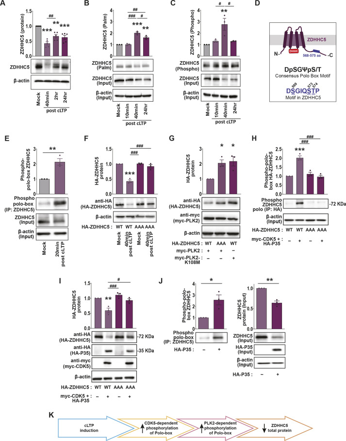
Activity-induced changes in protein palmitoylation can regulate the plasticity of synaptic connections, critically impacting learning and memory. Palmitoylation is a reversible post-translational modification regulated by both palmitoyl-acyl transferases that mediate palmitoylation and palmitoyl thioesterases that depalmitoylate proteins. However, it is not clear how fluctuations in synaptic activity can mediate the dynamic palmitoylation of neuronal proteins. Using primary hippocampal cultures, we demonstrate that synaptic activity does not impact the transcription of palmitoylating and depalmitoylating enzymes, changes in thioesterase activity, or post-translational modification of the depalmitoylating enzymes of the ABHD17 family and APT2 (also known as LYPLA2). In contrast, synaptic activity does mediate post-translational modification of the palmitoylating enzymes ZDHHC2, ZDHHC5 and ZDHHC9 (but not ZDHHC8) to influence protein-protein interactions, enzyme stability and enzyme function. Post-translational modifications of the ZDHHC enzymes were also observed in the hippocampus following fear conditioning. Taken together, our findings demonstrate that signaling events activated by synaptic activity largely impact activity of the ZDHHC family of palmitoyl-acyl transferases with less influence on the activity of palmitoyl thioesterases.

Keywords: Chemical long-term potentiation; Palmitoylation; Post-translational modification; Synapse activity; ZDHHC enzymes.


Conflict of interest statement

Competing interests The authors declare no competing or financial interests.

Figures

Fig. 1.
Fig. 1.
ZDHHC mRNA levels are not changed 40 min and 2 h post cLTP. qRT-PCR of the 23 ZDHHC enzymes from primary hippocampal cultures 40 min, 2 h or 24 h following cLTP. Values are normalized to mock-cLTP treated control. **P<0.01 (unpaired two-tailed Student's t-test versus mock-cLTP control). Results are mean±s.e.m. with individual data points shown. n=6 independent hippocampal cultures per condition.
Fig. 2.
Fig. 2.
cLTP-induced phosphorylation of ZDHHC5 polo-box domain impacts ZDHHC5 stability. (A) Western blot analysis of ZDHHC5 protein levels in primary hippocampal neuron cultures 40 min, 2 h and 24 h following cLTP. n=5 independent hippocampal cultures per condition. (B) Acyl-Rac assay showing palmitoylated (‘palm’) ZDHHC5 and overall ZDHHC5 protein (input) levels following cLTP. Palmitoylated ZDHHC5 values in the graph were derived from ZDHHC5 ‘palm’ normalized to ZDHHC5 ‘input’ and the β-actin loading control. n=3 independent hippocampal cultures per condition. (C) Lysates were run through the phospho-protein purification assay and western blots probed with the anti-ZDHHC5 antibody showing phosphorylated ZDHHC5 and overall ZDHHC5 protein (input) levels following cLTP. Phosphorylated ZDHHC5 values in the graph were derived from ZDHHC5 ‘phospho’ normalized to ZDHHC5 ‘input’ and the β-actin loading control. n=3 independent hippocampal cultures per condition. (D) Schematic of the sequence and position of the polo-box motif within the C-terminal tail of ZDHHC5. (E) Hippocampal culture lysates were immunoprecipitated (IP) with an anti-ZDHHC5 antibody and western blots probed with a phospho-polo-box-specific antibody. n=3 independent hippocampal cultures per condition. (F) Cells were nucleofected with ZDHHC5 shRNA to knock down ZDHHC5, and with HA–ZDHHC5-WT or the HA–ZDHHC5-AAA mutant at the time of plating. At 13–15 DIV, the cultures were stimulated using cLTP treatment and HA–ZDHHC5 protein levels determined using western blot analysis. n=3 independent hippocampal cultures per condition. (G) Cells were nucleofected with myc–PLK2 -WT or kinase-dead PLK2 (myc–PLK2-K108M) together with HA–ZDHHC5-WT or HA–ZDHHC5-AAA. ZDHHC5 or PLK2 protein levels were determined at 14 DIV by western blotting. n=3 independent hippocampal cultures per condition. (H) Cells were nucleofected with HA–ZDHHC5-WT or HA–ZDHHC5-AAA together with myc–CDK5 and HA–P35. At 13–15 DIV, HA-tagged proteins were immunoprecipitated (IP) with an anti-HA antibody and western blots probed with a phospho-polo-box-specific antibody to determine ZDHHC5 polo-box phosphorylation. n=3 independent hippocampal cultures per condition. (I) Cells were nucleofected with HA–ZDHHC5-WT or HA–ZDHHC5-AAA together with myc–CDK5 and HA–P35. At 14 DIV, western blotting was performed to assay the protein levels of all transfected constructs. n=3 independent hippocampal cultures per condition. (J) Cells were nucleofected with HA–P35. At 14 DIV, endogenous ZDHHC5 was immunoprecipitated (IP) with an anti-ZDHHC5 antibody and western blots were probed with a phospho-polo-box antibody to assay ZDHHC5 polo-box phosphorylation (left). The input from this assay was run on a western blot and probed for endogenous ZDHHC5 and HA–P35 (right). n=3 independent hippocampal cultures per condition. For all graphs, results are mean±s.e.m. with individual data points shown. *P<0.05; **P<0.01; ***P<0.001; determined versus the first condition in the bar chart (unpaired two-tailed Student's t-test). #P<0.05; ##P<0.01; ###P<0.001; pairwise comparison as indicated (one-way ANOVA with Tukey's post hoc test). (K) Schematic of activity-dependent changes in ZDHHC5 C-terminal polo-box regulation of ZDHHC5 total protein.
Fig. 3.
Fig. 3.
ZDHHC8 protein levels, palmitoylation and phosphorylation are unchanged following cLTP. (A) Western blot analysis of ZDHHC8 protein levels in primary hippocampal neuron cultures 40 min, 2 h and 24 h following cLTP. n=5 independent hippocampal cultures per condition. (B) Acyl-Rac assay showing palmitoylated ZDHHC8 and overall ZDHHC8 protein (input) levels following cLTP. Palmitoylated ZDHHC8 values in the graph were derived from ZDHHC8 ‘palm’ normalized to ZDHHC8 ‘input’ and the β-actin loading control. n=3 independent hippocampal cultures per condition. (C) Lysates were run through the phospho-protein purification assay and western blots probed with the anti-ZDHHC8 antibody showing phosphorylated ZDHHC8 and overall ZDHHC8 protein (input) levels following cLTP. Phosphorylated ZDHHC8 values in the graph were derived from ZDHHC8 ‘phospho’ normalized to ZDHHC8 ‘input’ and the β-actin loading control. ns, not significant (one-way ANOVA with Tukey's post hoc test). n=3 independent hippocampal cultures per condition. Results are mean±s.e.m. with individual data points shown.
Fig. 4.
Fig. 4.
The palmitoylation of ZDHHC9 and its substrates are reduced following cLTP. (A) Western blot analysis of ZDHHC9 protein levels in primary hippocampal neuron cultures 40 min, 2 h and 24 h following cLTP. n=5 independent hippocampal cultures per condition. (B) Acyl-Rac assay showing palmitoylated ZDHHC9 and overall ZDHHC9 protein (input) levels following cLTP. Palmitoylated ZDHHC9 values in the graph were derived from ZDHHC9 ‘palm’ normalized to ZDHHC9 ‘input’ and the β-actin loading control. n=3 independent hippocampal cultures per condition. (C) Lysates were run through the phospho-protein purification assay and western blots probed with the anti-ZDHHC9 antibody showing phosphorylated ZDHHC9 and overall ZDHHC9 protein (input) levels following cLTP. Phosphorylated ZDHHC9 values in the graph were derived from ZDHHC9 ‘phospho’ normalized to ZDHHC9 ‘input’ and the β-actin loading control. n=3 independent hippocampal cultures per condition. (D) Cells were nucleofected at the time of plating with ZDHHC9 shRNA to knock down ZDHHC9, and with HA–ZDHHC9-WT or HA–ZDHHC9-DHHS9. At 13–15 DIV, the cultures were stimulated using cLTP treatment and HA–ZDHHC9 palmitoylation was determined using the Acyl-Rac assay. n=3 independent hippocampal cultures per condition. (E,F) At 14 DIV, the cultures were stimulated using cLTP treatment and N-RAS (E) or TC10 (F) palmitoylation was determined using the Acyl-Rac assay. n=3 independent hippocampal cultures per condition. For all graphs, results are mean±s.e.m. with individual data points shown. ns, not significant; *P<0.05; **P<0.01; ***P<0.001. For A–D, P-values were determined versus the first condition in the bar chart (one-way ANOVA with Tukey's post hoc test). For E,F, P-values were determined using unpaired two-tailed Student's t-test. (G) Schematic showing activity-induced decrease in ZDHHC9 and substrate palmitoylation.
Fig. 5.
Fig. 5.
Decreased phosphorylation of ZDHHC2 following cLTP. (A) Western blot analysis of ZDHHC2 protein levels in primary hippocampal neuron cultures 40 min, 2 h and 24 h following cLTP. n=5 independent hippocampal cultures per condition. (B) Acyl-Rac assay showing palmitoylated ZDHHC2 and overall ZDHHC2 protein (input) levels following cLTP. Palmitoylated ZDHHC2 values in the graph were derived from ZDHHC2 ‘palm’ normalized to ZDHHC2 ‘input’ and the β-actin loading control. n=3 independent hippocampal cultures per condition. (C) Lysates were run through the phospho-protein purification assay and western blots probed with the anti-ZDHHC2 antibody showing phosphorylated ZDHHC2 and overall ZDHHC2 protein (input) levels following cLTP. Phosphorylated ZDHHC2 values in the graph were derived from ZDHHC2 ‘phospho’ normalized to ZDHHC2 ‘input’ and the β-actin loading control. n=3 independent hippocampal cultures per condition. n=3 independent hippocampal cultures per condition. (D) Hippocampal lysates were immunoprecipitated using an anti-PSD-95 antibody and western blots probed with an anti-ZDHHC2 antibody. The ZDHHC2 input was first normalized to β-actin as a loading control. The amount of the immunoprecipitated ZDHHC2 protein was then normalized to the β-actin-normalized input. n=4 independent hippocampal cultures per condition. (E) At 14 DIV, the cultures were stimulated using cLTP treatment and PSD-95 palmitoylation was determined using the Acyl-Rac assay. n=3 independent hippocampal cultures per condition. (F) HA–ZDHHC2-8×A phospho-dead mutation reduced co-immunoprecipitation of PSD-95. The PSD-95 input was first normalized to β-actin as a loading control. The amount of immunoprecipitated PSD-95 protein was then normalized to the β-actin normalized input. n=3 independent hippocampal cultures per condition. (G) Hippocampal cells were either left untransfected or transfected with HA–ZDHHC2-WT or HA–ZDHHC2-8×A at the time of plating. At 14 DIV, endogenous PSD-95 palmitoylation was determined using the Acyl-Rac assay. n=4 independent hippocampal cultures per condition. For all graphs, results are mean±s.e.m. with individual data points shown. *P<0.05; ***P<0.001; determined versus the first condition in the bar chart (unpaired two-tailed Student's t-test). #P<0.05; ##P<0.01; ###P<0.001; pairwise comparison as indicated (one-way ANOVA with Tukey's post hoc test). (H) Schematic of activity-dependent changes in ZDHHC2 C-terminal phospho-regulation of PSD-95 substrate interactions.
Fig. 6.
Fig. 6.
ZDHHC activity-dependent post-translational changes in vivo. Nine-week-old male mice were subjected to contextual fear conditioning (cFC) and hippocampal lysates from conditioned and unconditioned mice were collected 1 h later. (A,C,E,G) Lysates were run through the phospho-protein purification assay and western blots probed with antibodies for endogenous ZDHHC5, ZDHHC8, ZDHHC9 and ZDHHC2. Phosphorylated ZDHHC values in the graphs were derived from ZDHHC ‘phospho’ normalized to ZDHHC ‘input’ and the β-actin loading control. Input fractions were quantified to assess overall ZDHHC protein levels in control and cFC lysates. (B,D,F,H) Acyl-Rac assay showing palmitoylated ZDHHC5, ZDHHC8, ZDHHC9 and ZDHHC2 in control or cFC-treated mice. Palmitoylated ZDHHC values in the graphs were derived from ZDHHC ‘palm’ normalized to ZDHHC ‘input’ and the β-actin loading control. β-actin loading controls are duplicated in some panels as individual blots were cut in half and probed for two ZDHHCs per blot. Cut blots were probed for either ZDHHC2 (bottom half)/ZDHHC5 (top half) or ZDHHC9 (bottom half)/ZDHHC8 (top half). Con 1 and Con 2 represent two biological replicates from the control group. FC 1 and FC 2 represent two biological replicates from the cFC-treated group. ns, not significant; *P<0.05 (unpaired two-tailed Student's t-test). Results are mean±s.e.m. with individual data points shown. n=5 hippocampi per experiment.
Fig. 7.
Fig. 7.
Depalmitoylating enzyme activity is unchanged following cLTP. (A) Representative images of the fluorescent depalmitoylation probe-5 (DPP-5; 2 μM) 1 h after cLTP treatment in 14 DIV-cultured hippocampal neurons transfected at 11 DIV with mCherry. Left: mCherry cell fill. Middle: Background fluorescence within mCherry mask prior to addition of DPP-5 to the bath. Right: DPP-5 fluorescence within the mCherry mask 50 mins post addition of DPP-5 to the bath (1 h post cLTP). Scale bars: 100 μm. (B) As A, but 24 h post cLTP treatment. (C) Graph of time-course of DPP-5 fluorescence increase (ΔF) following cLTP and subsequent bath addition of DPP-5. No significant difference was observed between mock- and cLTP-treated neurons at any time point. (D) Graph of DPP-5 fluorescence increase (ΔF) 1 h post cLTP and 50 min post addition of DPP-5 to the bath. No significant difference was observed between mock and cLTP-treated neurons at any time-point. Mock, n=16 neurons; cLTP, n=17 neurons from two independent cultures. (E,F) As C,D but 24 h post cLTP and 50 min post addition of DPP-5 to the bath. Mock, n=17 neurons; cLTP, n=14 neurons from three independent cultures. AU, arbitrary units. (G) Acyl-Rac assay showing palmitoylated ABHD17 in cultured hippocampal neurons (14 DIV) following cLTP. Palmitoylated ABHD17 values in the graph were derived from ABHD17 ‘palm’ normalized to ABHD17 ‘input’ and the β-actin loading control. n=3 independent hippocampal cultures per condition. (H) Phospho-protein purification assay showing phosphorylated ABHD17 following cLTP. Phosphorylated ABHD17 values in the graph were derived from ABHD17 ‘phospho’ normalized to ABHD17 ‘input’ and the β-actin loading control. n=3 independent hippocampal cultures per condition. (I) Acyl-Rac assay showing palmitoylated APT2 in cultured hippocampal neurons (14 DIV) following cLTP. Palmitoylated APT2 values in the graph were derived from APT2 ‘palm’ normalized to APT2 ‘input’ and the β-actin loading control. n=3 independent hippocampal cultures per condition. (J) Phospho-protein purification assay showing phosphorylated APT2 following cLTP. Phosphorylated APT2 values in the graph were derived from APT2 ‘phospho’ normalized to APT2 ‘input’ and the β-actin loading control. n=3 independent hippocampal cultures per condition. n=3 hippocampal cultures per experiment. For all graphs, results are mean±s.e.m. with individual data points shown. ns, not significant (unpaired two-tailed Student's t-test for D,F; one-way ANOVA with Tukey's post hoc test for G–J).

References

    1. Abrami, L., Dallavilla, T., Sandoz, P. A., Demir, M., Kunz, B., Savoglidis, G., Hatzimanikatis, V. and Van Der Goot, F. G. (2017). Identification and dynamics of the human ZDHHC16-ZDHHC6 palmitoylation cascade. Elife 6, e27826. 10.7554/eLife.27826 - DOI - PMC - PubMed
    1. Ahtiainen, L., Van Diggelen, O. P., Jalanko, A. and Kopra, O. (2003). Palmitoyl protein thioesterase 1 is targeted to the axons in neurons. J. Comp. Neurol. 455, 368-377. 10.1002/cne.10492 - DOI - PubMed
    1. Ang, X. L., Seeburg, D. P., Sheng, M. and Harper, J. W. (2008). Regulation of postsynaptic RapGAP SPAR by polo-like kinase 2 and the SCFβ-TRCP ubiquitin ligase in hippocampal neurons. J. Biol. Chem. 283, 29424-29432. 10.1074/jbc.M802475200 - DOI - PMC - PubMed
    1. Anggono, V. and Huganir, R. L. (2012). Regulation of AMPA receptor trafficking and synaptic plasticity. Curr. Opin. Neurobiol. 22, 461-469. 10.1016/j.conb.2011.12.006 - DOI - PMC - PubMed
    1. Arai, T., Haze, K., Iimura-Morita, Y., Machida, T., Iida, M., Tanaka, K. and Komatani, H. (2008). Identification of β-catenin as a novel substrate of Polo-like kinase 1. Cell Cycle 7, 3556-3563. 10.4161/cc.7.22.7072 - DOI - PubMed

Publication types