. 2023 Jun 1;133(11):e163291.
doi: 10.1172/JCI163291.

Combination of AFP vaccine and immune checkpoint inhibitors slows hepatocellular carcinoma progression in preclinical models

Affiliations

Combination of AFP vaccine and immune checkpoint inhibitors slows hepatocellular carcinoma progression in preclinical models

Xinjun Lu et al. J Clin Invest. .

Abstract

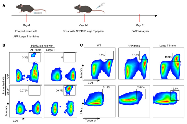
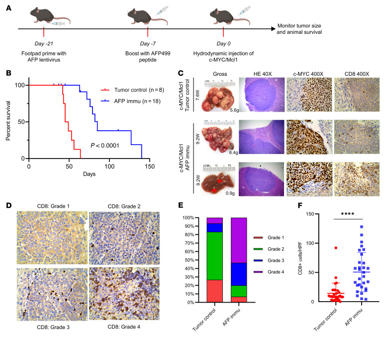
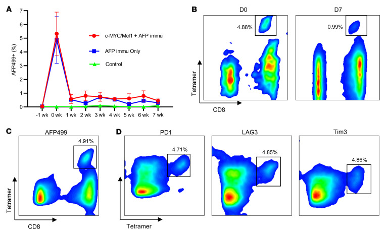
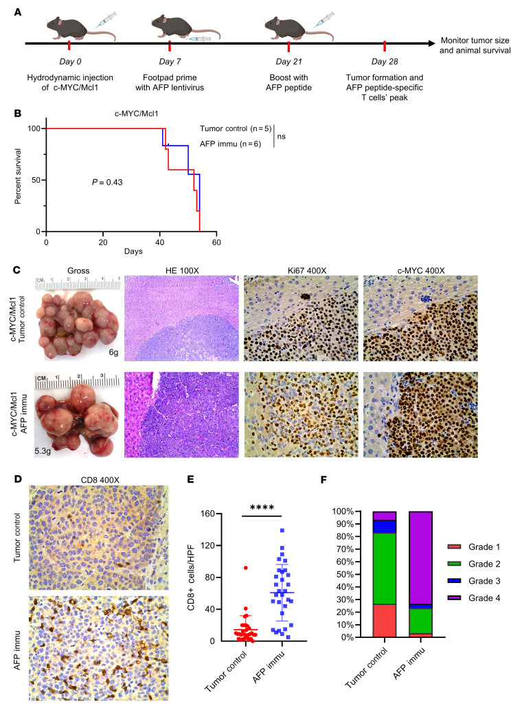
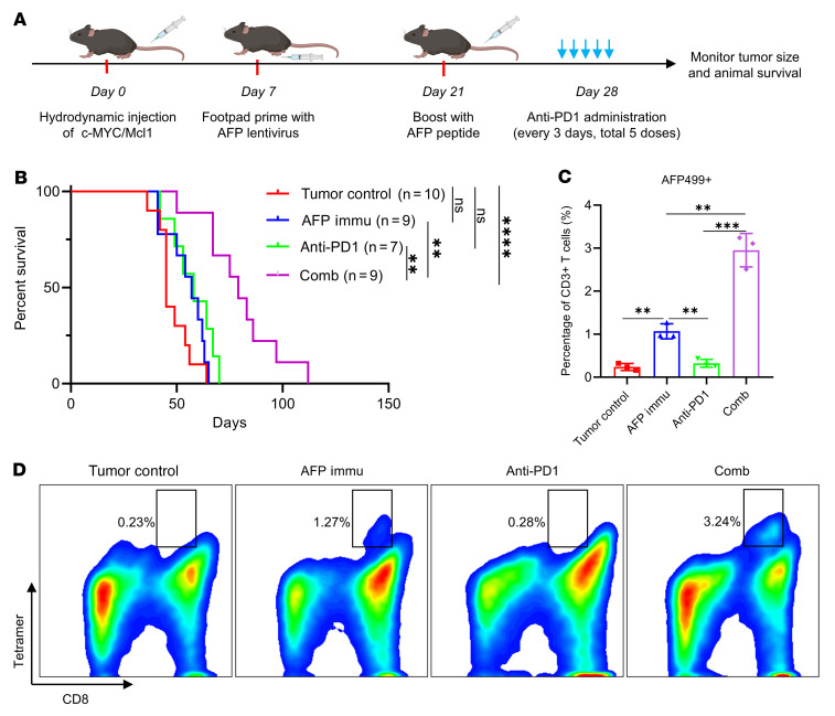
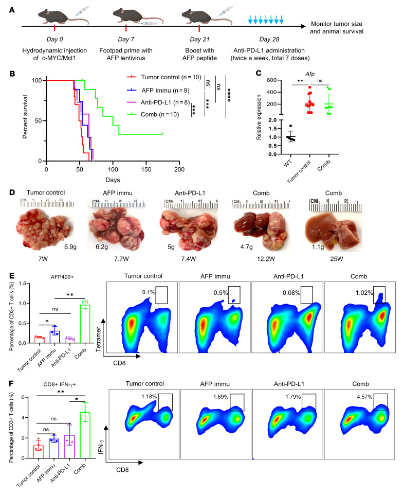
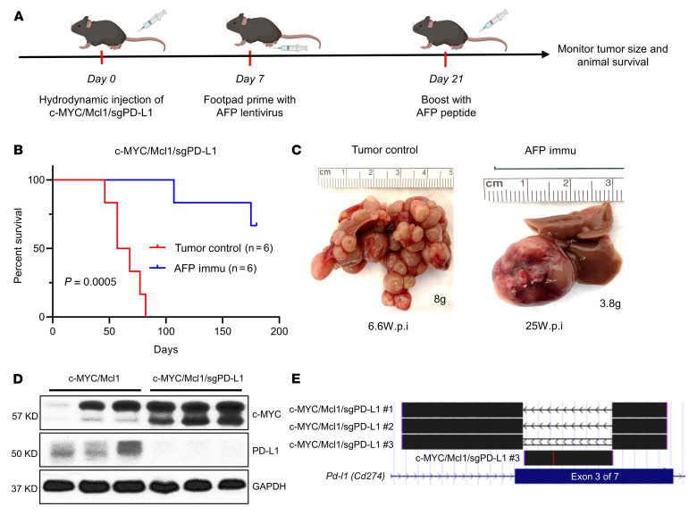
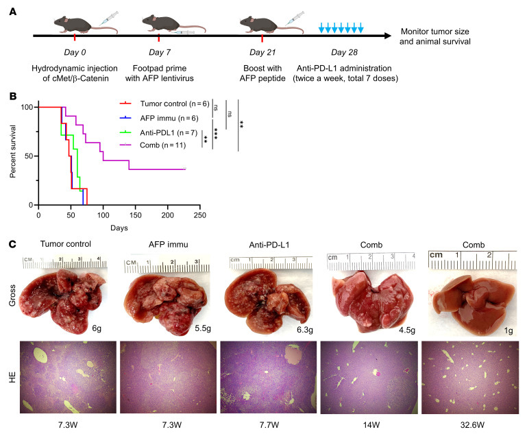
Many patients with hepatocellular carcinoma (HCC) do not respond to the first-line immune checkpoint inhibitor treatment. Immunization with effective cancer vaccines is an attractive alternative approach to immunotherapy. However, its efficacy remains insufficiently evaluated in preclinical studies. Here, we investigated HCC-associated self/tumor antigen, α-fetoprotein-based (AFP-based) vaccine immunization for treating AFP (+) HCC mouse models. We found that AFP immunization effectively induced AFP-specific CD8+ T cells in vivo. However, these CD8+ T cells expressed exhaustion markers, including PD1, LAG3, and Tim3. Furthermore, the AFP vaccine effectively prevented c-MYC/Mcl1 HCC initiation when administered before tumor formation, while it was ineffective against full-blown c-MYC/Mcl1 tumors. Similarly, anti-PD1 and anti-PD-L1 monotherapy showed no efficacy in this murine HCC model. In striking contrast, AFP immunization combined with anti-PD-L1 treatment triggered significant inhibition of HCC progression in most liver tumor nodules, while in combination with anti-PD1, it induced slower tumor progression. Mechanistically, we demonstrated that HCC-intrinsic PD-L1 expression was the primary target of anti-PD-L1 in this combination therapy. Notably, the combination therapy had a similar therapeutic effect in the cMet/β-catenin mouse HCC model. These findings suggest that combining the AFP vaccine and immune checkpoint inhibitors may be effective for AFP (+) HCC treatment.

Keywords: Hepatology; Immunology; Immunotherapy; Liver cancer; T cells.


Figures

Figure 1
Figure 1. AFP or Large T antigen immunization elicits functional peptide-specific CD8+ T cells.
(A) Overview of AFP and Large T peptide immunization procedure. (B) Representative results of AFP and Large T peptide-specific CD8+ T cells induced by the corresponding peptides. (C) Both AFP and Large T peptide-specific CD8+ T cells exhibit robust IFN-γ expression. Immu, immunization.
Figure 2
Figure 2. AFP immunization prevents the autologous HCC formation induced by c-MYC/Mcl1 hydrodynamic injection.
(A) Study design of AFP immunization and c-MYC/Mcl1 hydrodynamic injection. (B) Survival curve of the tumor control and AFP immunization group in c-MYC/Mcl1 mouse model. (C) Representative gross images and histopathological features of HE, c-MYC, and CD8 staining in the tumor control and AFP immunized group. The numbers indicate the liver weight and weeks from plasmid injections to the sacrifice date for that particular mouse. Original magnification, 40 × and 400 ×, as indicated in the column head. (D) The intensity of CD8 staining in C was graded from 1–4 based on the defined infiltrated CD8+ T cell number; Grade 1: 0–5 cells; Grade 2: 6–20 cells; Grade 3: 21–40 cells; Grade 4: > 40 cells. Original magnification, 400 ×. (E) The infiltrated CD8+ T cells were quantified in the tumor control and AFP-immunized groups (n = 30 high power fields). Kaplan-Meier test was used for survival analysis in B, unpaired 2-tailed Student’s t test was used in (F). Data are presented as mean ± SD. ****P < 0.0001. W, week(s); HPF, high power field; Immu, immunization.
Figure 3
Figure 3. Coexpression of exhaustion markers PD1, LAG3, and Tim3 cooperates for the rapid decrease of AFP499+ CTLs.
(A) AFP499+ CTLs decreased rapidly in the AFP immunization only or combined with the c-MYC/Mcl1 hydrodynamically injected mice. (B) Representative results of AFP499+ CTLs at their peak (D0) or 1 week later (D7) in an AFP-immunized mouse. (C and D) The AFP499+ CTLs (C) are observed to concomitantly express exhaustion markers, including PD1, LAG3, and Tim3. C and D are gated on the same population of CD3+CD8+ T cells. W, week(s); D, day(s); AFP499+, AFP499 peptidespecific CD8+ T cells; PD1, programmed cell death protein 1; LAG3, lymphocyte-activation gene 3; Tim3, T cell immunoglobulin and mucin protein 3; Immu, immunization.
Figure 4
Figure 4. AFP immunization has no therapeutic benefits to already-existing HCC tumors.
(A) Study design. (B) Survival curve of the control and AFP Immunization group mouse models. (C) Representative gross image and histopathological features of HE, Ki67, and c-MYC staining in the tumor control and AFP immunized group. The numbers indicate the liver weight of that particular mouse. (D) Representative CD8 staining of the tumor control and AFP Immunization group. (E and F) The infiltrated CD8+ T cells were quantified (E, n = 30 high power fields) and then graded from 1–4 based on the defined criteria; Grade 1: 0–5 cells; Grade 2: 6–20 cells; Grade 3: 21–40 cells; Grade 4: > 40 cells (F). Kaplan-Meier test was used for survival analysis in B, unpaired 2-tailed Student’s t test was used in (E). Data are presented as mean ± SD. Original magnification: 100 × and 400 ×, as indicated in column heads. HPF, high power field; Immu, immunization. ****P < 0.0001.
Figure 5
Figure 5. AFP immunization synergizes with anti-PD1 to inhibit c-MYC/Mcl1–induced HCC development.
(A) Study design. (B) Survival curve of the various groups tested. (C and D) Quantification of the AFP499+ CTLs and their corresponding representative flow cytometry results in each group. Kaplan-Meier test was used for survival analysis in (B), 1-way ANOVA analysis was used in C. Data are presented as mean ± SD. **P < 0.01, ***P < 0.001. AFP499+, AFP499 peptidespecific CD8+ T cells; Comb, combination therapy; Immu, immunization.
Figure 6
Figure 6. AFP immunization synergizes with anti–PD-L1 to inhibit c-MYC/Mcl1–induced HCC progression.
(A) Study design. (B) Survival curve of the various groups tested. (C) qPCR quantification of Afp expression in the tumor control and combination therapy group in B; WT is used as normal control. (D) Pictures of representative livers from B, the numbers indicate the liver weight and weeks from injection to sacrifice date for that particular mouse. (E) Quantification of the AFP499+ CTLs and their corresponding representative flow cytometry results in each group. (F) Quantification of the CD8+IFN-γ+ T cells and the corresponding representative flow cytometry results in each group. Kaplan-Meier test was used for survival analysis in B, 1-way ANOVA analysis was used in (C, E, and F). Data are presented as mean ± SD. *P < 0.05, **P < 0.01. W, week(s); AFP499+, AFP499 peptide-specific CD8+ T cells; Comb, combination therapy; Immu, immunization.
Figure 7
Figure 7. Deletion of PD-L1 does not affect tumor growth.
(A) Study design. (B) Survival curve of c-MYC/Mcl1/sgPD-L1 with or without AFP immunization. Kaplan-Meier test was used for survival analysis. (C) Pictures of representative livers from B, the numbers indicate the liver weight and weeks from hydrodynamic injection to sacrifice date for that particular mouse. (D and E) Western blot (D) and TA cloning sequencing results (E) from the c-MYC/Mcl1/sgPD-L1 tumors also confirm the deletion of PD-L1 in the tumors. The TA cloning sequencing reads are aligned with mouse GRCm38/mm10 genome at the UCSC genome browser (https://genome.ucsc.edu/). PD-L1 is also called CD274, and the gRNAs are designed to target the third exon of Pd-l1. c-MYC/Mcl1/sgPD-L1 no. 1 and c-MYC/Mcl1/sgPD-L1 no. 2 indicate the depletion of the whole sequence between the 2 sgPD-L1 gRNAs, c-MYC/Mcl1/sgPD-L1 no. 3 indicate the reversion sequence between the 2 sgPD-L1 gRNAs. All these sequence edits cause PD-L1 gene sequence frame-shift and early stop. wpi, weeks after injection; Immu, immunization.
Figure 8
Figure 8. AFP immunization synergizes with anti–PD-L1 administration to inhibit cMet/β-catenin–induced HCC progression.
(A) Study design. (B) Survival curve of the various groups tested. (C) Representative liver gross images and HE results (original magnification: 100 ×) of the investigated groups from B, the numbers indicate the liver weight and weeks from injection to sacrifice date for that particular mouse. Kaplan-Meier test was used for survival analysis in B. **P < 0.01, ***P < 0.001. W, week(s); Comb, combination therapy; Immu, immunization.

References

    1. Llovet JM, et al. Hepatocellular carcinoma. Nat Rev Dis Primers. 2021;7(1):6. doi: 10.1038/s41572-020-00240-3. - DOI - PubMed
    1. Kudo M, et al. Lenvatinib versus sorafenib in first-line treatment of patients with unresectable hepatocellular carcinoma: a randomised phase 3 non-inferiority trial. Lancet. 2018;391(10126):1163–1173. doi: 10.1016/S0140-6736(18)30207-1. - DOI - PubMed
    1. Finn RS, et al. Atezolizumab plus bevacizumab in unresectable hepatocellular carcinoma. N Engl J Med. 2020;382(20):1894–1905. doi: 10.1056/NEJMoa1915745. - DOI - PubMed
    1. Abou-Alfa GK, et al. Safety, efficacy, and pharmacodynamics of tremelimumab plus durvalumab for patients with unresectable hepatocellular carcinoma: randomized expansion of a phase I/II study. J Clin Oncol. 2022;1(27):2991–3001. - PMC - PubMed
    1. Liu C, et al. Value of α-fetoprotein in association with clinicopathological features of hepatocellular carcinoma. World J Gastroenterol. 2013;19(11):1811–1819. doi: 10.3748/wjg.v19.i11.1811. - DOI - PMC - PubMed

Publication types

MeSH terms

Substances