. 2023 May 15;6(5):1960-1969.
doi: 10.1021/acsabm.3c00155. Epub 2023 Apr 11.

Evaluation of Nonmodified Wireframe DNA Origami for Acute Toxicity and Biodistribution in Mice

Affiliations

Evaluation of Nonmodified Wireframe DNA Origami for Acute Toxicity and Biodistribution in Mice

Eike-Christian Wamhoff et al. ACS Appl Bio Mater. .

Abstract

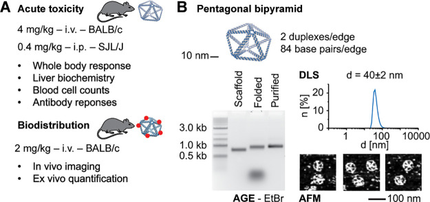
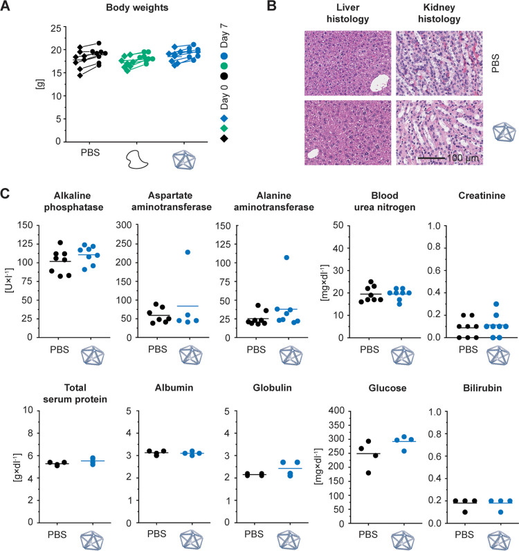
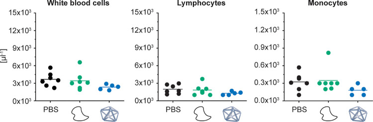
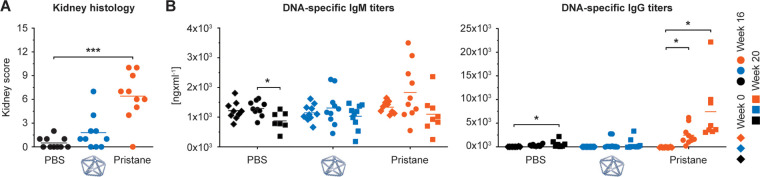
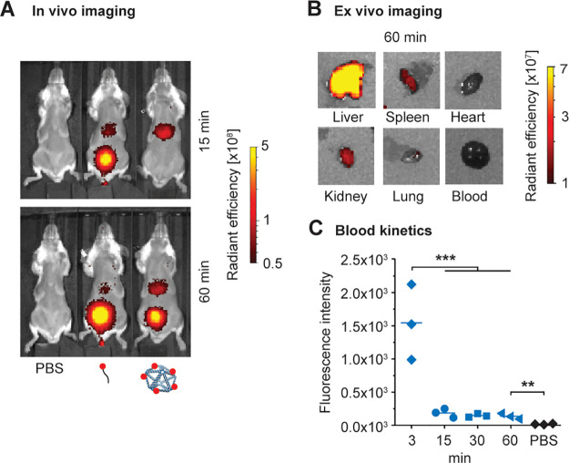
Wireframe DNA origami can be used to fabricate virus-like particles for a range of biomedical applications, including the delivery of nucleic acid therapeutics. However, the acute toxicity and biodistribution of these wireframe nucleic acid nanoparticles (NANPs) have not been previously characterized in animal models. In the present study, we observed no indications of toxicity in BALB/c mice following a therapeutically relevant dosage of nonmodified DNA-based NANPs via intravenous administration, based on liver and kidney histology, liver and kidney biochemistry, and body weight. Further, the immunotoxicity of these NANPs was minimal, as indicated by blood cell counts and type-I interferon and pro-inflammatory cytokines. In an SJL/J model of autoimmunity, we observed no indications of NANP-mediated DNA-specific antibody response or immune-mediated kidney pathology following the intraperitoneal administration of NANPs. Finally, biodistribution studies revealed that these NANPs accumulate in the liver within one hour, concomitant with substantial renal clearance. Our observations support the continued development of wireframe DNA-based NANPs as next-generation nucleic acid therapeutic delivery platforms.

Keywords: DNA origami; biodistribution; immunotoxicity; nanotechnology; therapeutic delivery; toxicity.


Conflict of interest statement

The authors declare no competing financial interest.

Figures

Figure 1
Figure 1
Characterizing the acute toxicity and biodistribution of wireframe DNA origami. (a) The experimental design for this study investigated the acute toxicity, immunotoxicity, and biodistribution of wireframe NANPs across two different animal models via two different administration routes. (b) The model NANP, a pentagonal bipyramid with two duplexes and 84 base pairs per edge (PB84), used in this study. PB84 was characterized by agarose gel electrophoresis (AGE), dynamic light scattering (DLS), and atomic force microscopy (AFM).
Figure 2
Figure 2
Effect of wireframe DNA origami on body weight as well as liver and kidney histology and function in BALB/c mice. Intravenous administration of PB84 (4 mg/kg), PBS control, and an unstructured ssDNA control (4 mg/kg). (a) Body weights were monitored before and 7 days after administration. (b) Histological sections of kidney and liver were visualized by hematoxylin and eosin (H&E) staining and observed under a light microscope at 40× magnification. (c) A panel of 10 biomarkers for liver and kidney function shows no phenotype when NANPs were administered, consistent with a PBS control. General toxicity was assessed from n ≥ 4 biological replicates per group. Representative histology images are shown. Student’s t test was performed for the body weights. One-way ANOVA was performed for the liver and kidney biochemistry panel followed by Tukey’s multiple comparison test. Significant differences are denoted as * - p < 0.050, ** - p < 0.010, and *** - p < 0.001.
Figure 3
Figure 3
Characterizing blood cell counts in BALB/c mice after i.v. administration. BALB/c mice were intravenously administrated through tail-vein injection with 4 mg/kg of PB84 per animal. Total white blood cell, lymphocyte, and monocyte cell counts were not elevated when NANPs were administered, comparable to aPBS control and an unstructured ssDNA control. Blood cell counts were assessed from n ≥ 5 biological replicates per group. One-way ANOVA was performed for the blood cell counts followed by Tukey’s multiple comparison test. Significant differences are denoted as * - p < 0.050, ** - p < 0.010, and *** - p < 0.001.
Figure 4
Figure 4
Characterizing autoimmunity induction in SJL/J mice after i.p. administration. SJL/J mice were intraperitoneally administered with 0.4 mg/kg of PB84 per animal. (a) Kidney scores were assessed at 20 weeks post treatment and (b) DNA-specific IgM and IgG antibody titers were assessed before treatment (time 0 or baseline) and after 16 and 20 weeks post treatment from ten animals (n = 10) per group. One-way ANOVA was performed followed by Tukey’s multiple comparison test. Significant differences are denoted as * - p < 0.050, ** - p < 0.010, and *** - p < 0.001.
Figure 5
Figure 5
Biodistribution in BALB/c mice after i.v. administration. In vivo optical imaging for analysis of pharmacokinetics and biodistribution. BALB/c mice were injected with PBS, AlexaFluor750-labeled oligonucleotide, or PB84–5xAF750 and imaged at multiple time points postinjection. (a) Representative fluorescence images at 15 and 60 min postinjection (n = 3 per group). (b) Representative fluorescence images of ex vivo organs harvested 60 min postinjection of PB84–5xAF750. PB84–5xAF750 was mostly accumulated in the liver, with minor accumulation in the kidney, spleen, and lung after 60 min. (c) Fluorescence intensity measurements of drawn blood for up to 60 min postinjection. One-way ANOVA was performed for blood kinetics measurements followed by Tukey’s multiple comparison test. Significant differences are denoted as * - p < 0.050, ** - p < 0.010, and *** - p < 0.001.

Update of

References

    1. Bujold K. E.; Lacroix A.; Sleiman H. F. DNA Nanostructures at the Interface with Biology. Chem. 2018, 4 (3), 495–521. 10.1016/j.chempr.2018.02.005. - DOI
    1. He L.; Mu J.; Gang O.; Chen X. Rationally Programming Nanomaterials with DNA for Biomedical Applications. Adv. Sci. 2021, 8 (8), 2003775.10.1002/advs.202003775. - DOI - PMC - PubMed
    1. Afonin K. A.; Dobrovolskaia M. A.; Church G.; Bathe M. Opportunities, Barriers, and a Strategy for Overcoming Translational Challenges to Therapeutic Nucleic Acid Nanotechnology. ACS Nano 2020, 14 (8), 9221–9227. 10.1021/acsnano.0c04753. - DOI - PMC - PubMed
    1. Liu S.; Jiang Q.; Zhao X.; Zhao R.; Wang Y.; Wang Y.; Liu J.; Shang Y.; Zhao S.; Wu T.; Zhang Y.; Nie G.; Ding B. A DNA nanodevice-based vaccine for cancer immunotherapy. Nat. Mater. 2021, 20 (3), 421–430. 10.1038/s41563-020-0793-6. - DOI - PubMed
    1. Veneziano R.; Moyer T. J.; Stone M. B.; Wamhoff E. C.; Read B. J.; Mukherjee S.; Shepherd T. R.; Das J.; Schief W. R.; Irvine D. J.; Bathe M. Role of nanoscale antigen organization on B-cell activation probed using DNA origami. Nat. Nanotechnol. 2020, 15 (8), 716–723. 10.1038/s41565-020-0719-0. - DOI - PMC - PubMed

Publication types