. 2023:4:0006.
doi: 10.34133/cbsystems.0006. Epub 2023 Jan 10.

An On-Chip Viscoelasticity Sensor for Biological Fluids

Affiliations

An On-Chip Viscoelasticity Sensor for Biological Fluids

Qianbin Zhao et al. Cyborg Bionic Syst. 2023.

Abstract

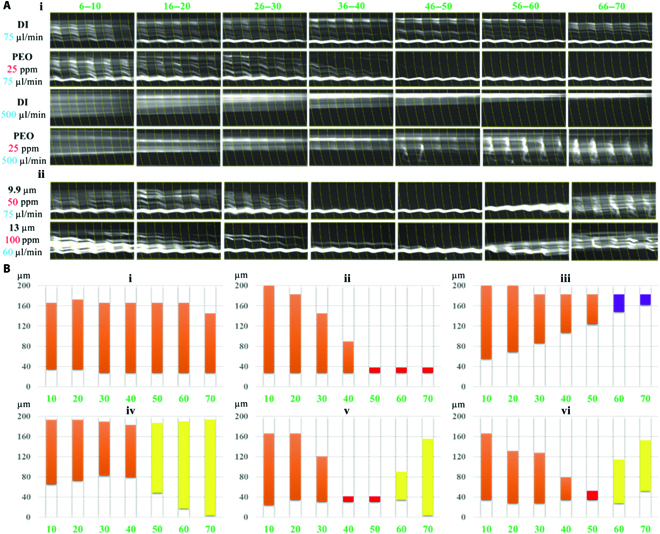
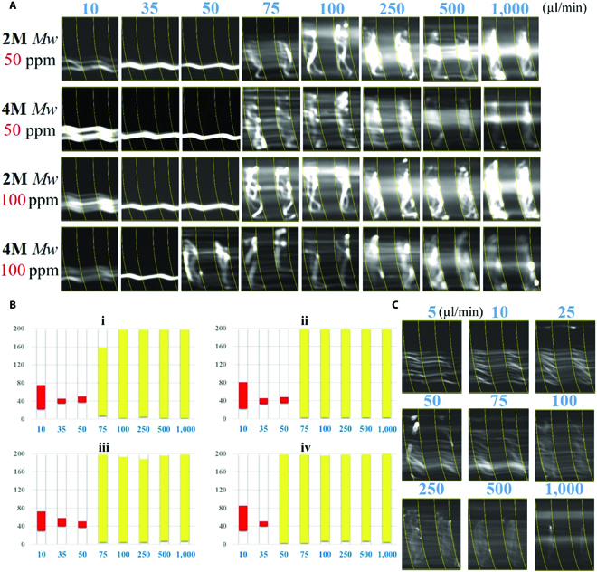
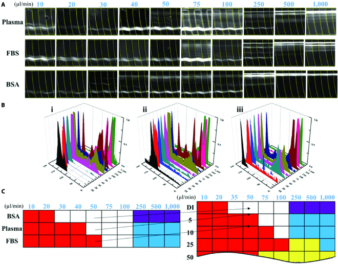
There are so many non-Newtonian fluids in our daily life, such as milk, blood, cytoplasm, and mucus, most of which are viscoelastic heterogeneous liquid containing cells, inorganic ion, metabolites, and hormones. In microfluidic microparticle-manipulating applications, the target particles are practically distributed within the biological fluids like blood and urine. The viscoelasticity of biological fluid is constantly ignored for simplicity especially when the fluid is substantially diluted and contains rather complex components. However, even the fluid's ultraweak viscoelasticity actually affects the microparticle migration and may bring a completely different behavior compared with the Newtonian fluids. As a result, a robust and easy operated on-chip viscoelasticity sensor is potential and desired in many research and industrial fields, including assay sample preparation, clinical diagnostics, and on-chip sensor. In this work, we employed stable non-Newtonian fluid-polyethylene oxide (PEO) solutions with various concentrations to investigate and calibrate effects of the weak fluidic viscoelasticity on microparticle behaviors in a double-layered microfluidic channel. An analogy-based database of fluidic patterns for viscoelasticity sensing and relaxation time measurement was established. Then, we tested different biological fluids including blood plasma and fetal bovine serum and proved that they exhibited similar viscoelasticity effects to the PEO solutions with the corresponding concentration, which reached a good agreement with available results by references. The detection limitation of relaxation time can reach 1 ms. It promised a robust and integrated on-chip microfluidic viscoelasticity sensor for different biological fluids without complicated calculations.


Figures

Fig. 1.
Fig. 1.
(A) Schematic figure of the 2-layer microchannel and the microparticle trajectory in non-Newtonian fluids. (B) Microparticle distribution of 9.9 μm at an outlet under flow rates of 5 to 1000 μl/min, with 25-ppm concentration of PEO. (C) Top view of the shear rate square contour (γ·2). The contour bar could be founded in the Supplementary Materials.
Fig. 2.
Fig. 2.
(A) Particle trajectories of 9.9-μm beads at the outlet the microchannel. The red arrow indicates the hydrophoretic focusing. The purple arrow indicates the secondary flow focusing. The weak viscoelasticity of PEO solutions deteriorated the secondary flow focusing, as indicated by the blue arrow. The green line indicates the disturbance of elastic lift force enhancement on the microbead focusing. In the yellow region, the elastic turbulence and deterioration of hydrophoretic focusing simultaneously existed. Scale bar was 100 μm. (B) Flow field distribution and bead migration at different flow conditions. i, Elastic-assisting hydrophoretic focusing (red square); ii, secondary flow focusing (purple square); iii, transitional section (white square); iv, elastic-deteriorating secondary flow focusing (blue square); v, elastic turbulence region (yellow square). (C) Microbead migration mappings. The color of units represented the corresponding microbead focusing regimes.
Fig. 3.
Fig. 3.
(A) The trajectories of microbeads in the grooved channel. The channel has 70 grooves, indicated by the green numbers. i. The microbeads migration in DI water and PEO solutions. ii. Intriguing microbead “dispersion–focusing–dispersion” phenomenon of 9.9- and 13-μm microbeads migrated in the microchannel. (B) Microbead distributions along microchannel according to the normalized fluorescent intensity, under different PEO concentrations and flow rates: (i) DI water, 75 μl min−1; (ii) 25-ppm PEO, 75 μl min−1; (iii) DI water, 500 μl min−1; (iv) 25-ppm PEO, 500 μl min−1; (v) 50-ppm PEO, 75 μl min−1; and (vi) 100-ppm PEO, 60 μl min−1. The colored columns represent the microbead distribution across the microchannel, and the colors suggest the focusing patterns consistent with the color mapping identified in Fig. 2C.
Fig. 4.
Fig. 4.
(A) The 9.9-μm particle trajectories in the PEO solutions with the Mw of 2M and 4M Mw at flow rates from 10 to 1000 μl min−1. (B) Particle distribution obtained from the normalized fluorescent intensity. (C) Particle trajectories of 4.8-μm microbeads in the 25-ppm PEO solutions.
Fig. 5.
Fig. 5.
(A) Particle trajectories of 9.9-μm microbeads in blood plasma, FBS, and BSA solutions. (B) The obtained fluorescent intensity profiles indicating the microbead distribution across the microchannel at the flow rates from 10 to 1000 μl min−1. (C) Color mappings of the 3 biological fluids.

References

    1. Yan C, Pochan DJ. Rheological properties of peptide-based hydrogels for biomedical and other applications. Chem Soc Rev. 2010;39:3528–3540. - PMC - PubMed
    1. Dickinson E. Hydrocolloids at interfaces and the influence on the properties of dispersed systems. Food Hydrocoll. 2003;17:25–39.
    1. Del Giudice F. Simultaneous measurement of rheological properties in a microfluidic rheometer. Phys Fluids. 2020;32:052001.
    1. Heinrich J, Balleisen L, Schulte H, Assmann G, Loo J. Fibrinogen and factor VII in the prediction of coronary risk. Results from the PROCAM study in healthy men. Arterioscler Thromb. 1994;14:54–59. - PubMed
    1. Yang S, Kim JY, Lee SJ, Lee SS, Kim JM. Sheathless elasto-inertial particle focusing and continuous separation in a straight rectangular microchannel. Lab Chip. 2011;11:266–273. - PubMed