. 2023:6:0034.
doi: 10.34133/research.0034. Epub 2023 Jan 16.

Bioinspired Jellyfish Microparticles from Microfluidics

Affiliations

Bioinspired Jellyfish Microparticles from Microfluidics

Chaoyu Yang et al. Research (Wash D C). 2023.

Abstract

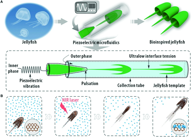
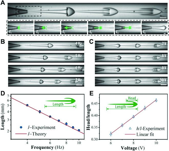
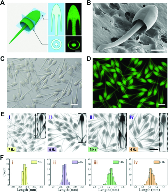
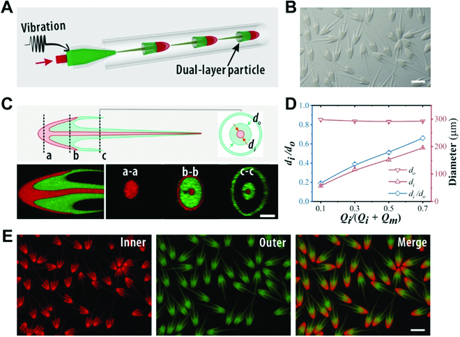
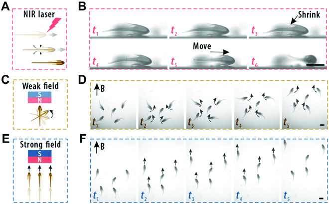
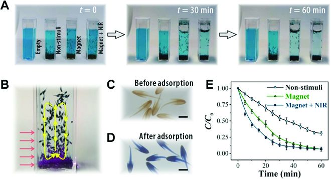
Nonspherical particles have attracted increasing interest because of their shape anisotropy. However, the current methods to prepare anisotropic particles suffer from complex generation processes and limited shape diversity. Here, we develop a piezoelectric microfluidic system to generate complex flow configurations and fabricate jellyfish-like microparticles. In this delicate system, the piezoelectric vibration could evolve a jellyfish-like flow configuration in the microchannel and the in situ photopolymerization could instantly capture the flow architecture. The sizes and morphologies of the particles are precisely controlled by tuning the piezoelectric and microfluidic parameters. Furthermore, multi-compartmental microparticles with a dual-layer structure are achieved by modifying the injecting channel geometry. Moreover, such unique a shape endows the particles with flexible motion ability especially when stimuli-responsive materials are incorporated. On the basis of that, we demonstrate the capability of the jellyfish-like microparticles in highly efficient adsorption of organic pollutants under external control. Thus, it is believed that such jellyfish-like microparticles are highly versatile in potential applications and the piezoelectric-integrated microfluidic strategy could open an avenue for the creation of such anisotropic particles.


Figures

Fig. 1.
Fig. 1.
Schematic illustrations of the generation and adsorption process of the jellyfish particles. (A) Schematic diagram of jellyfish, the piezoelectric microfluidic setup, and the bioinspired jellyfish microparticles. The bottom panel demonstrates the preparation process of the bioinspired jellyfish. (B) Schematic diagram of the movement of a stimuli-responsive microparticle under NIR laser irradiation and the adsorption process for water contaminants.
Fig. 2.
Fig. 2.
Generation dynamics and morphology control of the jellyfish-like ligament template in microfluidics. (A) High-speed real-time images of the generation of jellyfish ligament template in the microfluidic collection tube. The bottom panels show an entire process of fluid retraction and advancement. (B and C) High-speed real-time images of the dynamic behaviors of the jellyfish ligament template under different (B) piezoelectric frequencies and (C) actuation amplitudes. (D) Plot of the length of the jellyfish-like ligament template as a function of the piezoelectric frequency. (E) Plot of the ratio of the head to the length of the ligament template as a function of the piezoelectric voltage amplitude at the position of the third jellyfish in (C). The scale bars are 500 μm in the upper panel of (A), 250 μm in the bottom panels of (A), and 1000 μm in (B) and (C).
Fig. 3.
Fig. 3.
Formation of the single-layered jellyfish-like particles in microfluidics. (A) Schematic illustrations and cross-sectional confocal laser scanning microscope (CLSM) images of the resultant particle. (B) SEM image of one jellyfish particle. (C and D) Microscopy images of a batch of bioinspired particles with fluorescent polystyrene nanoparticles (E) in bright field and (D) fluorescent field. (E) Jellyfish microparticles produced under different piezoelectric frequencies. (F) The corresponding length distributions of the particles. The scale bars are 100 μm in (A), 200 μm in (B), and 1000 μm in (C) to (E).
Fig. 4.
Fig. 4.
Formation of the dual-layered jellyfish-like particles in microfluidics. (A) Schematic illustration of the 2-stage nested capillary microfluidic device for the generation of dual-layered jellyfish-like microparticles. (B) Microscopy images of a batch of bioinspired dual-layered particles. (C) Schematic illustrations of the dual-layered particles with longitudinal and cross-sectional CLSM images. (D) The ratio of the internal thread diameter di to the outer thread diameter do as a function of φ. (E) Fluorescent images of a batch of dual-layered particles with red and green fluorescent polystyrene nanoparticles added to the inner and outer layer, respectively. The scale bars are 1000 μm in (B) and (E) and 200 μm in (C).
Fig. 5.
Fig. 5.
Stimuli-triggered motion of the dual-layered particles. (A and B) Schematic illustration and real-time images showing the movement of a single dual-layered jellyfish microparticle under NIR laser irradiation. (C and D) Schematic illustration and real-time images showing the reorientation of the magnetic-responsive dual-layered jellyfish microparticles under a weak magnetic field. (E and F) Schematic illustration and real-time images showing the directional migration of the magnetic-responsive dual-layered jellyfish microparticles under a strong magnetic field. The scale bars are 1000 μm.
Fig. 6.
Fig. 6.
Adsorption processes of the dual-layered jellyfish particles under different conditions. (A) The adsorption process of MB using magnetic GO/NIPAM dual-layered jellyfish particles under different stimulation modes. (B) The circulation of a swarm of particles under NIR stimulation. (C and D) Microscopic images of the particles (C) before and (D) after MB adsorption. (E) The MB adsorption kinetics of the dual-layered jellyfish particles. C was the real-time concentration, and C0 was the initial concentration of MB. The scale bars are 1000 μm.

Similar articles

Cited by

References

    1. Pearce AK, Wilks TR, Arno MC, O’Reilly RK. Synthesis and applications of anisotropic nanoparticles with precisely defined dimensions. Nat Rev Chem. 2021;5:21–45. - PubMed
    1. Daly AC, Riley L, Segura T, Burdick JA. Hydrogel microparticles for biomedical applications. Nat Rev Mater. 2020;5(1):20–43. - PMC - PubMed
    1. Liang Q, Bie N, Yong T, Tang K, Shi X, Wei Z, Jia H, Zhang X, Zhao H, Huang W, et al. . The softness of tumour-cell-derived microparticles regulates their drug-delivery efficiency. Nat Biomed Eng. 2019;3(9):729–740. - PubMed
    1. Mage PL, Csordas AT, Brown T, Klinger D, Eisenstein M, Mitragotri S, Hawker C, Tom Soh H. Shape-based separation of synthetic microparticles. Nat Mater. 2019;18:82–89. - PubMed
    1. Cai L, Bian F, Chen H, Guo J, Wang Y, Zhao Y. Anisotropic microparticles from microfluidics. Chem. 2021;7:93–136.

LinkOut - more resources