. 2023 Apr 18;120(16):e2214430120.
doi: 10.1073/pnas.2214430120. Epub 2023 Apr 11.

A mutagenesis study of autoantigen optimization for potential T1D vaccine design

Affiliations

A mutagenesis study of autoantigen optimization for potential T1D vaccine design

Yi Song et al. Proc Natl Acad Sci U S A. .

Abstract

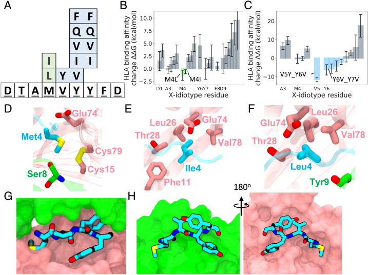
A previously reported autoreactive antigen, termed the X-idiotype, isolated from a unique cell population in Type 1 diabetes (T1D) patients, was found to stimulate their CD4+ T cells. This antigen was previously determined to bind more favorably than insulin and its mimic (insulin superagonist) to HLA-DQ8, supporting its strong role in CD4+ T cell activation. In this work, we probed HLA-X-idiotype-TCR binding and designed enhanced-reactive pHLA-TCR antigens using an in silico mutagenesis approach which we functionally validated by cell proliferation assays and flow cytometry. From a combination of single, double, and swap mutations, we identified antigen-binding sites p4 and p6 as potential mutation sites for HLA binding affinity enhancement. Site p6 is revealed to favor smaller but more hydrophobic residues than the native tyrosine, such as valine (Y6V) and isoleucine (Y6I), indicating a steric mechanism in binding affinity improvement. Meanwhile, site p4 methionine mutation to hydrophobic residues isoleucine (M4I) or leucine (M4L) modestly increases HLA binding affinity. Select p6 mutations to cysteine (Y6C) or isoleucine (Y6I) exhibit favorable TCR binding affinities, while a swap p5-p6 tyrosine-valine double mutant (V5Y_Y6V) and a p6-p7 glutamine-glutamine double mutant (Y6Q_Y7Q) exhibit enhanced HLA binding affinity but weakened TCR affinity. This work holds relevance to potential T1D antigen-based vaccine design and optimization.

Keywords: autoantigen design; free energy perturbation; molecular dynamics; mutagenesis; type 1 diabetes.


Conflict of interest statement

The authors declare no competing interest.

Figures

Fig. 1.
Fig. 1.
HLA-bound X-idiotype structure. (A) HLA-bound X-idiotype structure I. side view and II. top view. X-idiotype epitope is shown in cyan with nonepitope residues in gray sticks and HLA in gray shadow. (B) HLA-bound X-idiotype residue solvent exposure. X-idiotype core epitope DTAMVYYFD is labeled sites 1 to 9, while the X-idiotype antigen (CARQEDTAMVYYFDYW) is labeled sites 1′, 2′, 3′..., 16′. Residue exposure reveals sites bound to HLA (R3′, Y7, and anchor residues D1, M4, Y6). (C) HLA interactions with X-idiotype residue Tyr7. Tyr7 is a nonanchor residue with disproportionately high contact area with the HLA as seen from (B). Tyr7 interacts with a mixture of polar, hydrophobic, and aromatic residues from HLA-β (pink). No Tyr7–HLA-α interactions are observed.
Fig. 2.
Fig. 2.
X-idiotype mutation binding free energy results. (A) Favorable point mutations (green), swap and double mutations (blue) overlaid onto X-idiotype sequence. (B) The change in HLA binding free energy, ΔΔ G for point mutations. Favorable point mutations shown in green. (C) The change in HLA binding free energy ΔΔ G for swap and double mutations, with favorable mutations shown in blue. (B and C) Error bar is ±95% CI. (D) HLA interactions with native X-idiotype anchor residue p4 methionine. HLA-α residues shown in green, HLA-β in pink. (E and F) HLA interactions with mutated X-idiotype anchor residue p4 isoleucine (E) and leucine (F). (G and H) HLA interactions with native X-idiotype residues p4 to p7 MVYY. HLA-α residues shown in green, HLA-β in pink. (G) Top and (H) side views.
Fig. 3.
Fig. 3.
X-idiotype double mutation decomposition and Tyr6 mutation free energy results. (A) Favorable double mutations of Tyr6 and decomposition to individual residues, errors are ±95% CI. (B) X-idiotype Tyr6 mutation binding affinity results in relation to residue volume and residue hydrophobicity. Volume relative to Tyr6 is computed as Vmut/VTyr with volumes from ref. . Eisenberg hydrophobicity scores from refs. , . Error bars are for affinity and represent ±95% CI. The R2 Pearson’s correlation shown at top left is correlation between binding affinity and relative volume. (C) Tyr6–HLA residue interactions with (D) Val6 mutant and (E) Ile6 mutant interactions. Native Tyr6 and mutants interact with same residue set.
Fig. 4.
Fig. 4.
X-idiotype Tyr6 TCR mutation free energy results. (A) HLA-X-idiotype-TCR structure with Tyr6 of X-idiotype in cyan. Only TCR variable domains were included for free energy analysis. (B) Favorable mutations of Tyr6 for HLA and TCR binding, error bars are ±95% CI. (C) TCR structure for the native Tyr6 with CDR3 loops highlighted. (D) TCR contact surface of HLA-X-idiotype assembly for the native Tyr6. (EH) Mutational TCR contact surfaces for Tyr5_Val6, Cys6, and Ile6. TCRα shown in orange, TCRβ in yellow. The X-idiotype antigen is in gray sticks, except for enumerated residues in cyan. Note: contact surface represents HLA-TCR and X-idiotype-TCR contacts combined.
Fig. 5.
Fig. 5.
(A) Representative dot plots show CFSE dilution by gated CD4 T cells among PBMCs from DQ8+ T1D subject that were used at readout (see Table 1 for X-idiotype mutants). Numbers indicate percentages of gated CFSElow CD4 T cells. Uns represents unstimulated CD4 T cells. (B) Percentage of CFSElow CD4 T cells in response to different antigens. (C) CD4 T cell expansion response to different peptides using the response of autologous unstimulated as the denominator. (D) Upregulation of CD69 by gated CFSElow CD4 T cells when compared with CFSEhigh CD4 T cells. (E) Fold change in cell surface CD69 by proliferated CD4 T cells compared with nonproliferated cells on stimulation with indicated peptides.

References

    1. Garyu J. W., Meffre E., Cotsapas C., Herold K. C., Progress and challenges for treating Type 1 diabetes. J. Autoimmunity 71, 1–9 (2016). - PMC - PubMed
    1. Marrack P., Kappler J. W., Do MHCII-presented neoantigens drive type 1 diabetes and other autoimmune diseases? Cold Spring Harb. Perspect Med. 2, a007765–a007765 (2012). - PMC - PubMed
    1. Delong T., et al. , Pathogenic CD4 T cells in type 1 diabetes recognize epitopes formed by peptide fusion. Science 351, 711–714 (2016). - PMC - PubMed
    1. Han S., Donelan W., Wang H., Reeves W., Yang L.-J., Novel autoantigens in type 1 diabetes. Am. J. Transl. Res. 5, 379–392 (2013). - PMC - PubMed
    1. Roep B. O., Peakman M., Antigen targets of type 1 diabetes autoimmunity. Cold Spring Harb. Perspect Med. 2, a007781–a007781 (2012). - PMC - PubMed

Publication types