. 2023 Apr 18;120(16):e2210418120.
doi: 10.1073/pnas.2210418120. Epub 2023 Apr 11.

ABL kinases regulate the stabilization of HIF-1α and MYC through CPSF1

Affiliations

ABL kinases regulate the stabilization of HIF-1α and MYC through CPSF1

Benjamin Mayro et al. Proc Natl Acad Sci U S A. .

Abstract

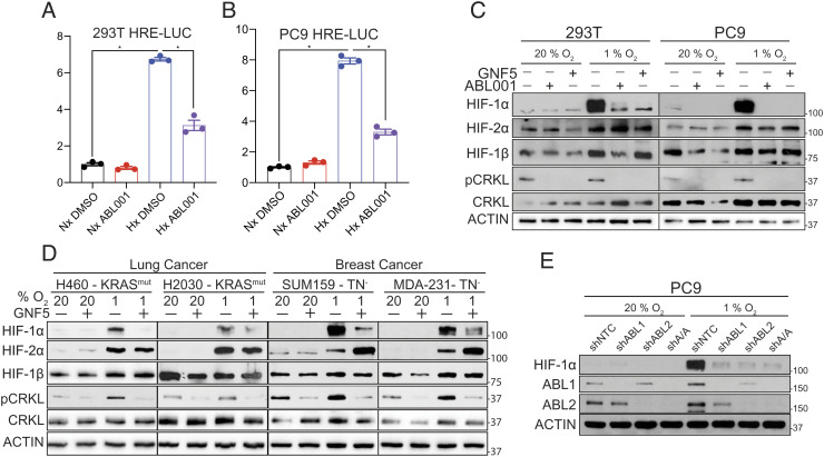
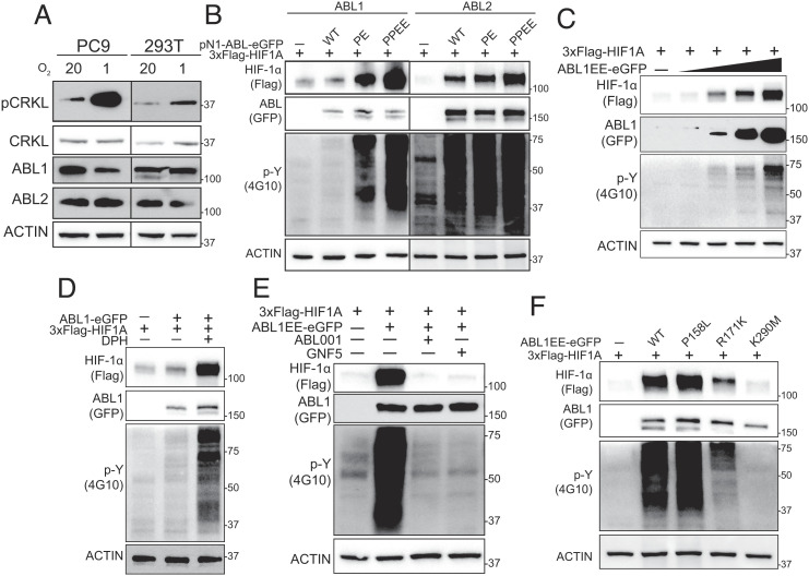
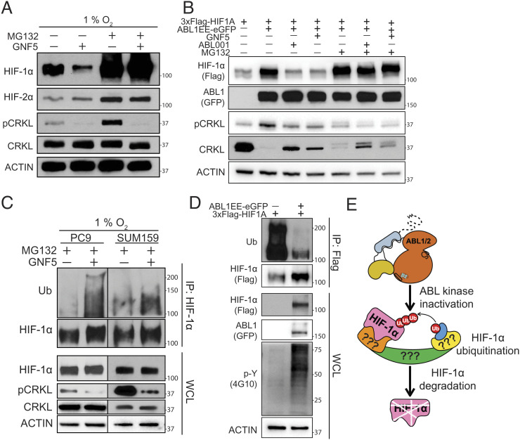
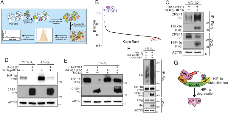
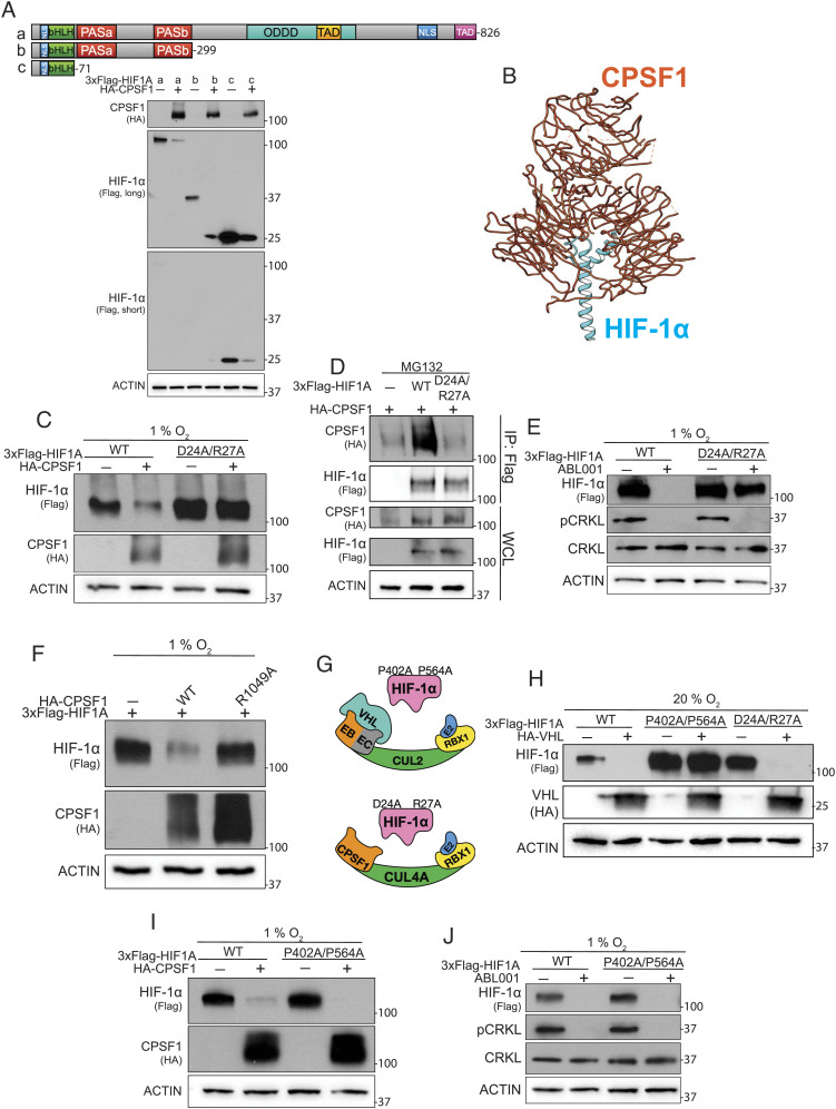
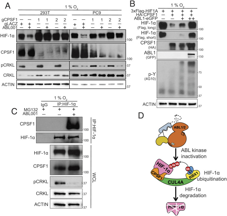
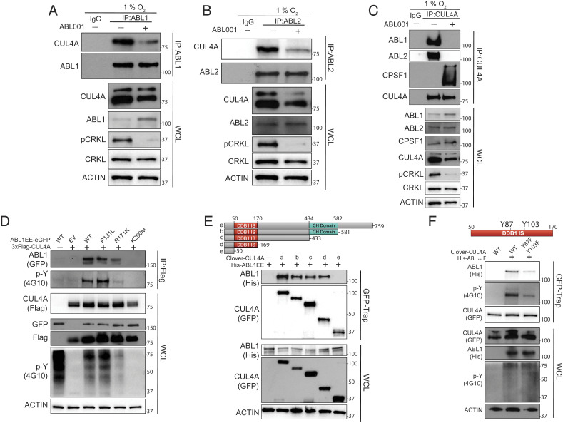
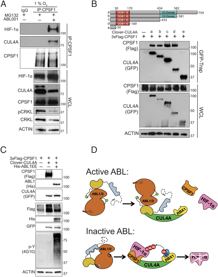
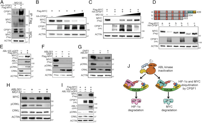
The hypoxia-inducible factor 1-α (HIF-1α) enables cells to adapt and respond to hypoxia (Hx), and the activity of this transcription factor is regulated by several oncogenic signals and cellular stressors. While the pathways controlling normoxic degradation of HIF-1α are well understood, the mechanisms supporting the sustained stabilization and activity of HIF-1α under Hx are less clear. We report that ABL kinase activity protects HIF-1α from proteasomal degradation during Hx. Using a fluorescence-activated cell sorting (FACS)-based CRISPR/Cas9 screen, we identified HIF-1α as a substrate of the cleavage and polyadenylation specificity factor-1 (CPSF1), an E3-ligase which targets HIF-1α for degradation in the presence of an ABL kinase inhibitor in Hx. We show that ABL kinases phosphorylate and interact with CUL4A, a cullin ring ligase adaptor, and compete with CPSF1 for CUL4A binding, leading to increased HIF-1α protein levels. Further, we identified the MYC proto-oncogene protein as a second CPSF1 substrate and show that active ABL kinase protects MYC from CPSF1-mediated degradation. These studies uncover a role for CPSF1 in cancer pathobiology as an E3-ligase antagonizing the expression of the oncogenic transcription factors, HIF-1α and MYC.

Keywords: ABL kinases; CPSF1; E3-ligase; HIF-1α; MYC.


Conflict of interest statement

A.M.P. is a consultant and advisory board member for The Pew Charitable Trusts. All other authors declare that they have no competing interests.

Figures

Fig. 1.
Fig. 1.
ABL kinases are necessary for hypoxic accumulation of HIF-1α. (A and B) Bioluminescence levels of PC9 and 293T cells transduced with a hypoxia response element reporter and treated with ABL001 (20 µM) during normoxia (Nx) or hypoxia (Hx) (1% O2) for 24 h. (C) Immunoblot (IB) analysis of whole-cell lysates (WCL) derived from HEK293T and PC9 cells treated with ABL001 (20 µM) or GNF5 (20 µM) during normoxia or hypoxia (1% O2) for 24 h. (D) IB analysis of WCL derived from KRAS-mutant lung cancer cells (H460, H2030) and triple-negative (TN−) breast cancer cells (SUM159, MDA-MB-231) treated with GNF5 during normoxia or hypoxia (1% O2) for 24 h. (E) IB analysis of WCL derived from PC9 cells transduced with indicated short hairpin RNA (shRNA) lentiviruses and cultured under normoxia or hypoxia for 24 h.
Fig. 2.
Fig. 2.
ABL kinase activation induces HIF-1α protein stabilization. (A) IB analysis of WCL derived from PC9 and 293T exposed to normoxia or hypoxia (1% O2) for 24 h. (B and C) IB analysis of WCL derived from HEK293T cells transfected with 3xFlag-HIF1A and indicated ABL-eGFP constructs for 48 h. ABL PE has a moderate increase in ABL kinase activity and ABL PPEE elicits a higher increase in ABL kinase activity. (D) IB analysis of WCL derived from HEK293T cells transfected with 3xFlag-HIF1A and ABL1-eGFP constructs for 24 h and then treated with the ABL kinase allosteric activator DPH (2.5 µM) as indicated for 24 h before harvesting. (E) IB analysis of WCL derived from HEK293T cells transfected with 3xFlag-HIF1A and ABL1-eGFP constructs for 24 h and then treated with ABL001 and GNF5 as indicated for 24 h before harvesting. (F) IB analysis of WCL derived from HEK293T cells transfected with 3xFlag-HIF1A and indicated ABL1-eGFP constructs for 48 h.
Fig. 3.
Fig. 3.
The ABL kinases regulate the proteasomal degradation of HIF-1α. (A) IB analysis of WCL derived from PC9 cells treated with ABL001 and MG132 (4 µM) as indicated during hypoxia for 24 h. (B) IB analysis of WCL derived from HEK293T cells transfected with 3xFlag-HIF1A and ABL1-eGFP constructs for 24 h and then treated with ABL001, GNF5, and MG132 as indicated for 24 h before harvesting. (C) IB analysis of IP and WCL derived from PC9 and SUM159 cells treated with ABL001 and MG132 as indicated during hypoxia for 24 h. (D) IB analysis of IP and WCL derived from HEK293T cells transfected with 3xFlag-HIF1A and ABL1-eGFP constructs for 24 h. (E) Proposed model depicting that loss of ABL kinase activity leads to HIF-1α ubiquitination and proteasome-mediated protein degradation.
Fig. 4.
Fig. 4.
CPSF1 is a HIF-1α-targeting E3-ligase. (A) Scheme of the FACS-based CRISPR E3 ligase screen performed. (B) Snake plot depicting the β scores across 593 E3-ligase-related gene identified in PC9 lung cancer cells treated with ABL kinase inhibitor under hypoxia. (C) IB analysis of IP- and WCL-derived HEK293T cells transfected with 3xFlag-HIF1A and HA-CPSF1 constructs for 24 h and then treated with MG132 during hypoxia for 24 h before harvesting. (D) IB analysis of WCL derived from HEK293T cells transfected with 3xFlag-HIF1A and HA-CPS1 constructs for 24 h and then exposed to normoxia or hypoxia for 24 h. (E) IB analysis of WCL derived from HEK293T cells transfected with 3xFlag-HIF1A and HA-CPSF1 constructs for 24 h and then treated with or without MG132 during hypoxia for 24 h before harvesting. (F) IB analysis of IP derived from HEK293T cells transfected with 3xFlag-HIF1A and HA-CSPF1 constructs for 24 h and then treated with MG132 for the final 24 h. (G) Proposed model depicting CPSF1-dependent HIF-1α ubiquitination and proteasome-mediated protein degradation.
Fig. 5.
Fig. 5.
CPSF1 is an E3-ligase targeting the HIF-1α DNA-binding domain. (A, Top) Structural diagram of full-length HIF-1α and truncation mutants. (A, Bottom) IB analysis of WCL derived from HEK293T cells transfected with HA-CPSF1 and indicated 3xFlag-HIF1A truncation constructs for 24 h and exposed to hypoxia for 24 h. (B) HDOCK protein–protein docking server prediction of the interaction of full-length CPSF1 (PDB: 6F9N) with HIF-1α AA 1-71 (PDB:4ZPR). CPSF1 is shown in orange and HIF-1α is shown in blue. (C) IB analysis of WCL derived from HEK293T cells transfected with indicated 3xFlag-HIF1A and HA-CPSF1 constructs for 24 h and exposed to hypoxia for 24 h. (D) IB and IP analysis of WCL derived from HEK293T cells transfected with HA-CPSF1 and indicated 3xFlag-HIF1A constructs for 24 h and exposed to hypoxia for 24 h. (E) IB analysis of WCL-derived 293T cells transfected with indicated 3xFlag-HIF1A constructs for 24 h and then treated with or without ABL001 during hypoxia for 24 h. (F) IB analysis of WCL derived from HEK293T cells transfected with 3xFlag-HIF1A and indicated HA-CPSF1 constructs for 24 h and exposed to hypoxia for 24 h. (G) Scheme of CPSF1- and VHL-resistant HIF-1α mutants. (H) IB analysis of WCL derived from HEK293T cells transfected with indicated 3xFlag-HIF1A and HA-VHL constructs for 24 h and exposed to hypoxia for 24 h. (I) IB analysis of WCL derived from HEK293T cells transfected with indicated 3xFlag-HIF1A and HA-CPSF1 constructs for 24 h and exposed to hypoxia for 24 h. (J) IB analysis of WCL-derived 293T cells transfected with indicated 3xFlag-HIF1A constructs for 24 h and then treated with or without ABL001 during hypoxia for 24 h.
Fig. 6.
Fig. 6.
ABL kinases protect HIF-1α from CPSF1-dependent degradation. (A) IB analysis of WCL derived from HEK293T and PC9 cells transduced with indicated gRNA/CRISPR-Cas9 constructs and then treated with ABL001 during hypoxia for 24 h. (B) IB analysis of WCL derived from HEK293T cells transfected with 3xFlag-HIF1A, ABL1-eGFP, and HA-CPSF1 constructs for 24 h and then exposed to hypoxia for 24 h. (C) IB and IP analysis of WCL derived from PC9 cells treated with ABL001 (24 h) and MG132 (final 6 h) during hypoxia for 24 h. IgG sample added to the vehicle and MG132-treated lysate. (D) Proposed model depicting loss of ABL kinase activity leading to CPSF1-dependent HIF-1α ubiquitination and proteasome-mediated protein degradation.
Fig. 7.
Fig. 7.
ABL kinases interact with and phosphorylate CUL4A. (AC) IB and IP analysis of WCL of indicated endogenous ABL and CUL4A proteins from PC9 cells treated with or without ABL001 (24 h) during hypoxia for 24 h. Cell lysates were incubated with IgG control or indicated antibodies. (DF) IB analysis of IP- and WCL-derived HEK293T cells transfected with indicated epitope-tagged CUL4A and ABL1 constructs for 24 h and then exposed to hypoxia for 24 h before harvesting. DDB1 IS = DDB1 interaction site; CH domain = Cullin homology domain.
Fig. 8.
Fig. 8.
The ABL kinases and CPSF1 form mutually exclusive complexes with CUL4A. (A) IB and IP analysis of WCL derived from PC9 cells treated with ABL001 (24 h) and MG132 (final 6 h) during hypoxia for 24 h. IgG sample added to the vehicle and MG132-treated lysate. (B) IB analysis of IP- and WCL-derived HEK293T cells transfected with 3xFlag-CPSF1 and indicated Clover-CUL4A constructs for 24 h and exposed to hypoxia for 24 h before harvesting. (C) IB analysis of IP and WCL-derived HEK293T cells transfected with 3xFlag-CPSF1, His-ABl1EE, Clover-CUL4A constructs as indicated for 24 h and exposed to hypoxia for 24 h before harvesting. DDB1 IS = DDB1 interaction site, CH domain = Cullin homology domain. (D) Model for the ABL kinases regulation of HIF-1α through CPSF1.
Fig. 9.
Fig. 9.
ABL kinase protects MYC from CPSF1-dependent degradation. (A) IB analysis of IP- and WCL-derived HEK293T cells transfected with Flag-MYC and HA-CPSF1 constructs for 48 h. (B) IB analysis of WCL derived from HEK293T cells transfected with Flag-MYC and HA-CPSF1 constructs for 48 h. (C) IB analysis of WCL derived from HEK293T cells transfected with Flag-MYC and HA-CPSF1 constructs for 24 h and then treated with MG132 for 24 h before harvesting during normoxia. (D, Top) Structural diagram of full-length MYC and truncation mutants. (A, Bottom) IB analysis of WCL derived from HEK293T cells transfected with HA-CPSF1 and indicated Flag-MYC truncation constructs for 24 h and exposed to hypoxia for 24 h. (E) IB analysis of WCL derived from HEK293T cells transfected with Flag-MYC and ABL1-eGFP constructs for 48 h during normoxia. (F) IB analysis of WCL derived from HEK293T cells transfected with Flag-MYC for 24 h and then treated with ABL001 and GNF5 as indicated for 24 h during normoxia. (G and H) IB analysis of WCL derived from PC9 cells treated with ABL001, GNF5, and MG132 as indicated for 24 h during normoxia. (I) IB analysis of WCL derived from HEK293T cells transfected with Flag-MYC, ABL1-eGFP, and HA-CPSF1 constructs for 48 h. (J) Model for the ABL kinase regulation of HIF-1α and MYC through CPSF1.

Similar articles

Cited by

References

    1. Kaelin W. G. Jr., Ratcliffe P. J., Oxygen sensing by metazoans: The central role of the HIF hydroxylase pathway. Mol. Cell 30, 393–402 (2008). - PubMed
    1. Kaelin W. G. Jr., The von Hippel-Lindau tumour suppressor protein: O2 sensing and cancer. Nat. Rev. Cancer 8, 865–873 (2008). - PubMed
    1. Ivan M., et al. , HIFalpha targeted for VHL-mediated destruction by proline hydroxylation: Implications for O2 sensing. Science 292, 464–468 (2001). - PubMed
    1. Jaakkola P., et al. , Targeting of HIF-alpha to the von Hippel-Lindau ubiquitylation complex by O2-regulated prolyl hydroxylation. Science 292, 468–472 (2001). - PubMed
    1. Masson N., Willam C., Maxwell P. H., Pugh C. W., Ratcliffe P. J., Independent function of two destruction domains in hypoxia-inducible factor-alpha chains activated by prolyl hydroxylation. EMBO J. 20, 5197–5206 (2001). - PMC - PubMed

Publication types

MeSH terms

Substances