. 2023 Apr 11;16(780):eade2438.
doi: 10.1126/scisignal.ade2438. Epub 2023 Apr 11.

HSP90 inhibition in the mouse spinal cord enhances opioid signaling by suppressing an AMPK-mediated negative feedback loop

Affiliations

HSP90 inhibition in the mouse spinal cord enhances opioid signaling by suppressing an AMPK-mediated negative feedback loop

Katherin A Gabriel et al. Sci Signal. .

Abstract

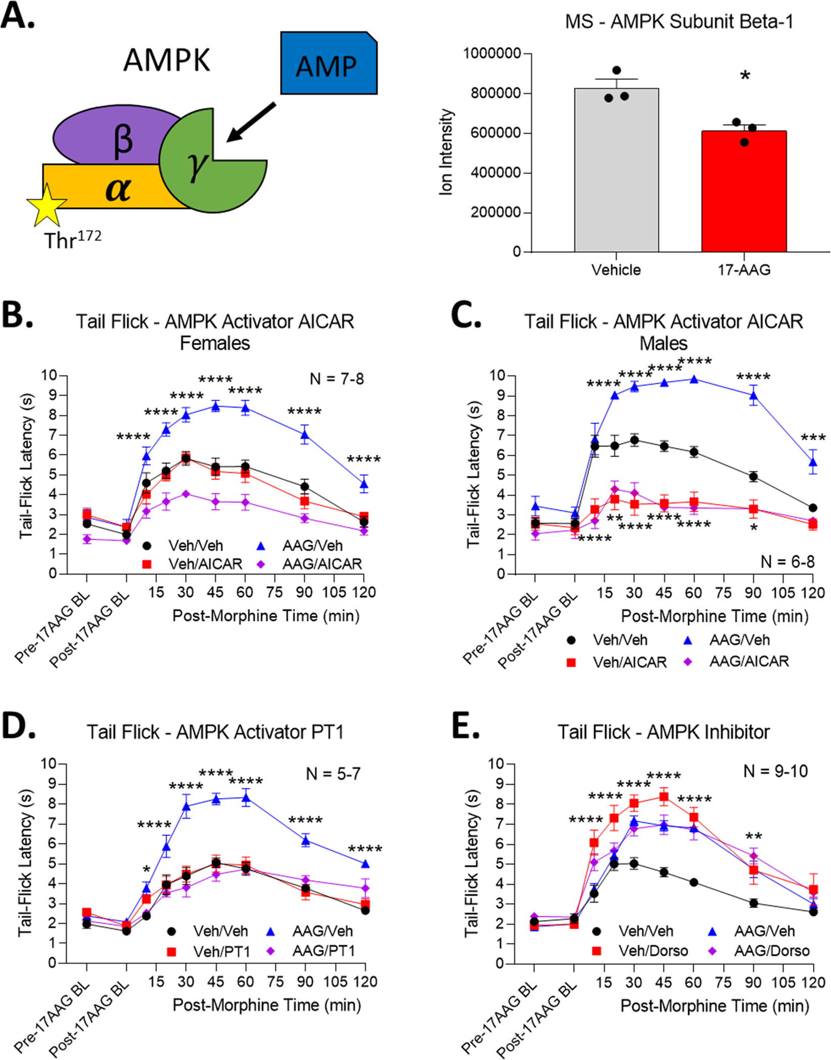
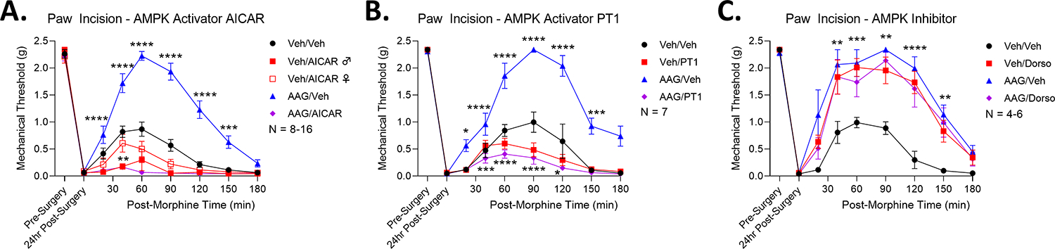
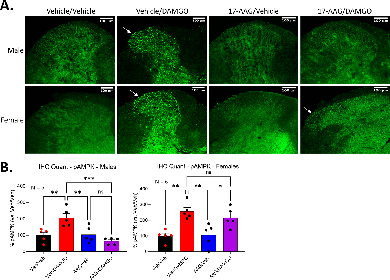
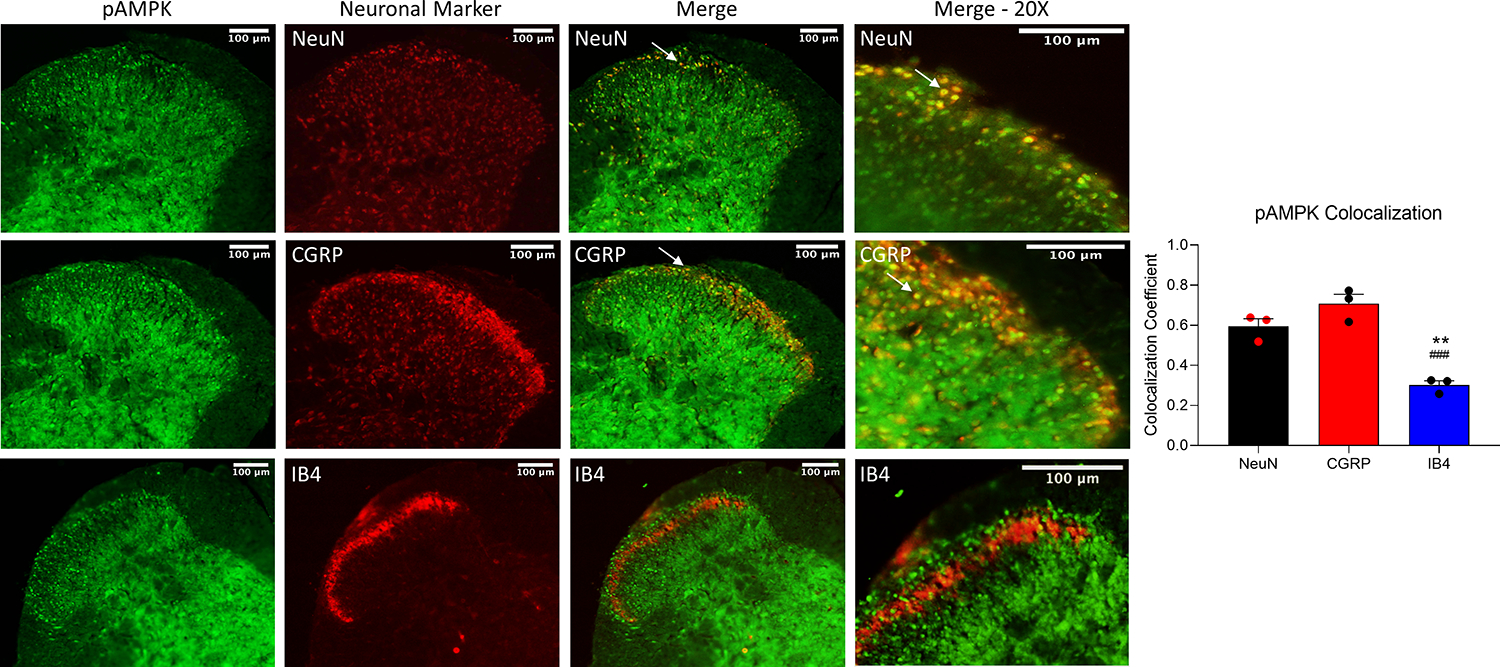
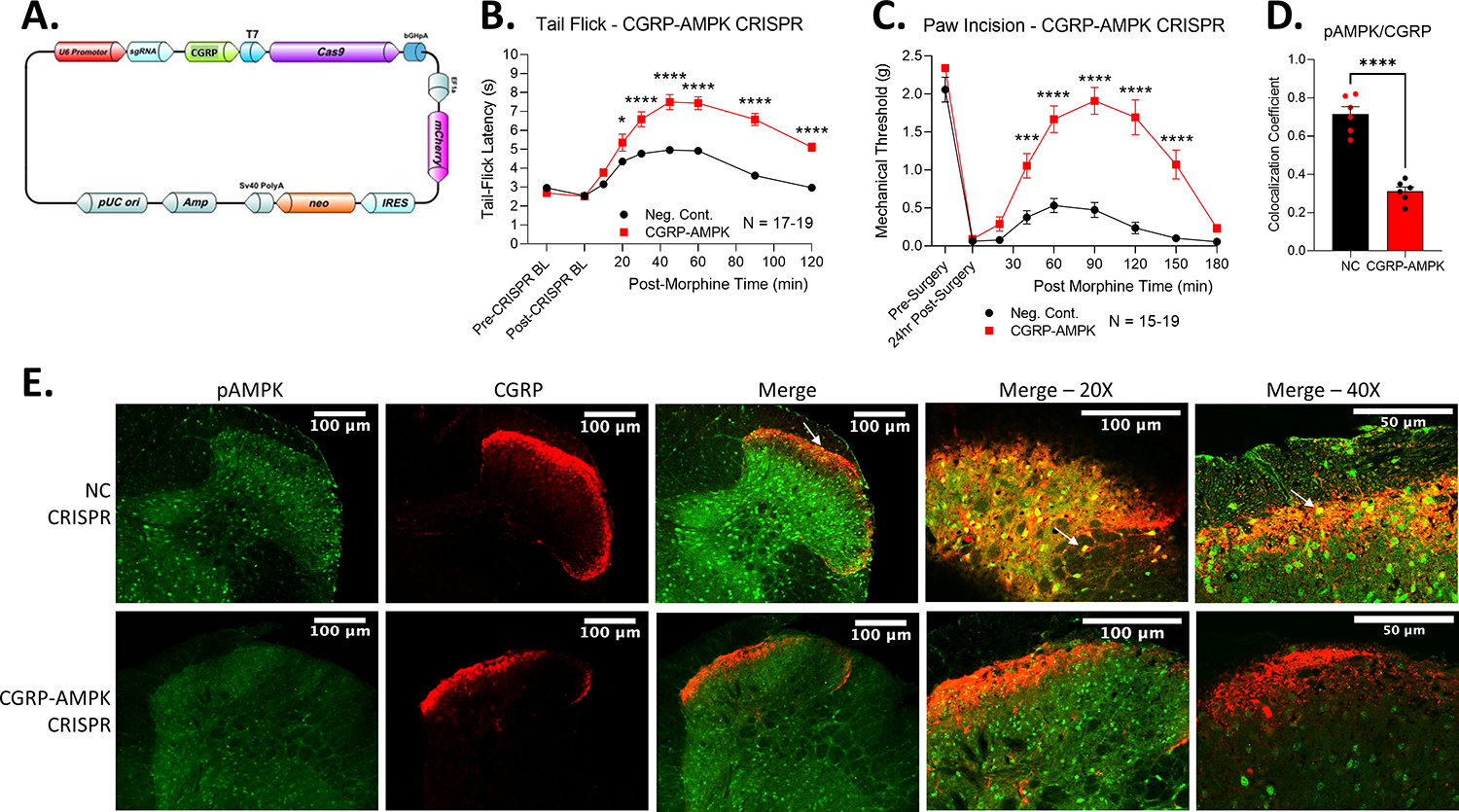
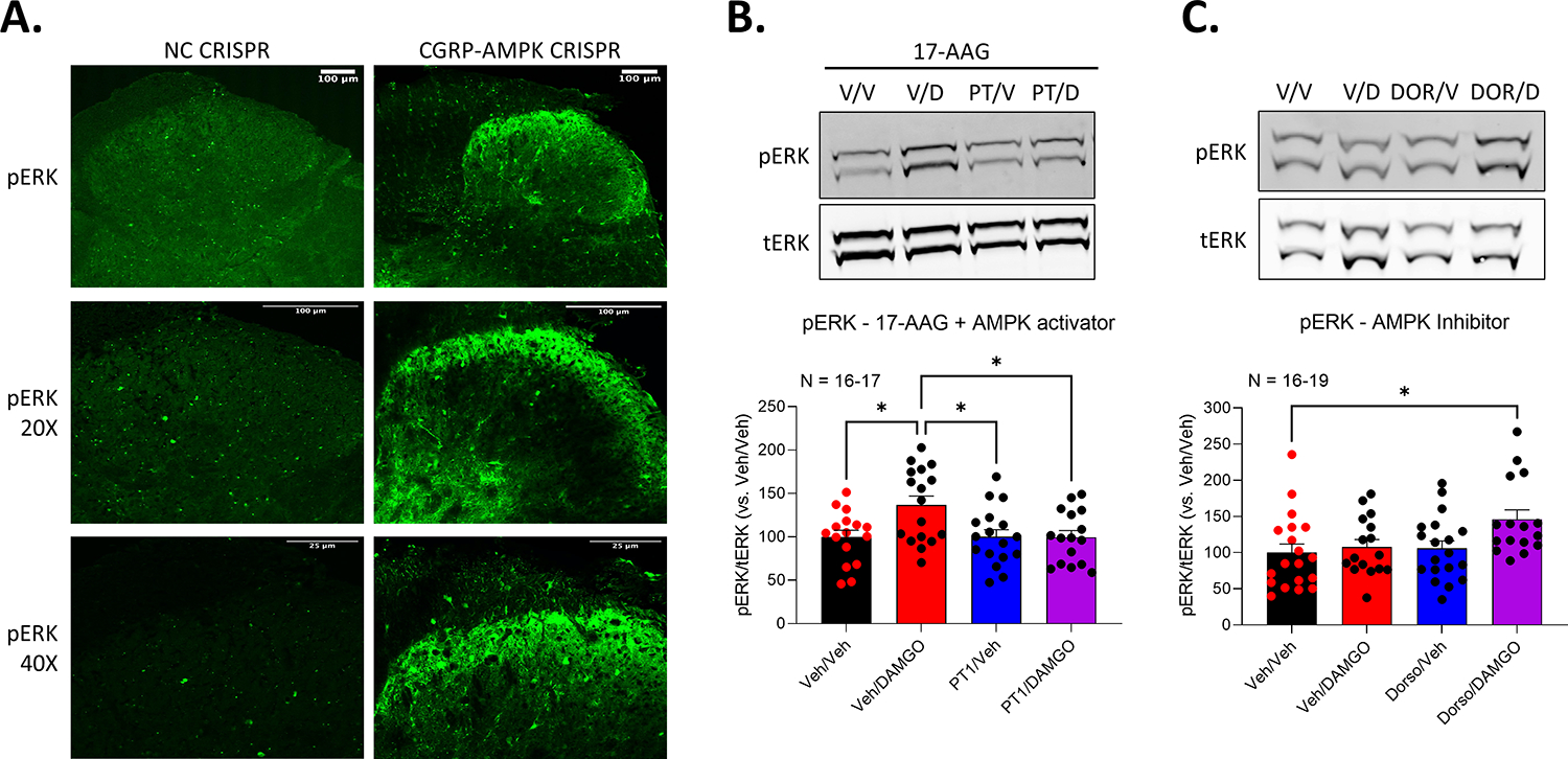
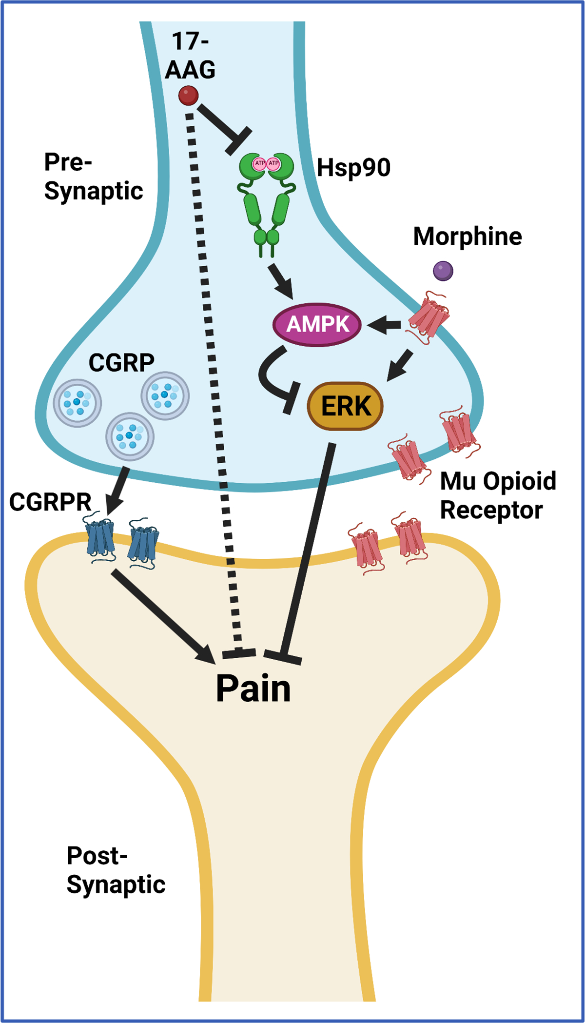
Opioids and other agonists of the μ-opioid receptor are effective at managing acute pain, but their chronic use can lead to tolerance that limits their efficacy. We previously reported that inhibiting the chaperone protein HSP90 in the spinal cords of mice promotes the antinociceptive effects of opioids in a manner that involved increased activation of the kinase ERK. Here, we found that the underlying mechanism involves the relief of a negative feedback loop mediated by the kinase AMPK. Intrathecal treatment of male and female mice with the HSP90 inhibitor 17-AAG decreased the abundance of the β1 subunit of AMPK in the spinal cord. The antinociceptive effects of 17-AAG with morphine were suppressed by intrathecal administration of AMPK activators and enhanced by an AMPK inhibitor. Opioid treatment increased the abundance of phosphorylated AMPK in the dorsal horn of the spinal cord, where it colocalized with a neuronal marker and the neuropeptide CGRP. Knocking down AMPK in CGRP-positive neurons enhanced the antinociceptive effects of morphine and demonstrated that AMPK mediated the signal transduction between HSP90 inhibition and ERK activation. These data suggest that AMPK mediates an opioid-induced negative feedback loop in CGRP neurons of the spinal cord and that this loop can be disabled by HSP90 inhibition to enhance the efficacy of opioids.


Conflict of interest statement

Competing interests: JMS is an equity holder in Teleport Pharmaceuticals, LLC and Botanical Results, LLC, but these companies are not Hsp90-related. The authors have no other relevant conflicts of interest to declare.

Figures

Figure 1:
Figure 1:. AMPK signaling suppresses opioid anti-nociception, which is reversed by HSP90 inhibition.
(A) Left, diagram of the active AMPK protein complex, showing the α/β/γ ternary complex. The protein is activated by AMP binding to the γ subunit, leading to phosphorylation of Thr172 in the α subunit and subsequent activation. Right, proteomic quantification of the β1 subunit of AMPK in spinal cord of female CD-1 mice 24 hours after treatment with vehicle or 0.5 nmol 17-AAG (HSP90 inhibitor) intrathecally (i.t.). N=3 mice in each group. Ion intensity mass spectrometry counts for each group are reported and compared. * = p < 0.05 by unpaired 2-tailed t test. This suggests AMPK downregulation after HSP90 inhibitor treatment. Data taken from proteomic data set reported in (11). (B to E) Tail flick time course analysis in male and female CD-1 mice treated i.t. with vehicle or 0.5 nmol 17-AAG, followed 24 hours later by i.t. treatment with vehicle or AMPK activator (100 nmol, B and C; 10 nmol, D) or inhibitor (20 nmol, E) for 10 min, then subcutaneously with 3.2 mg/kg morphine. BL, baseline. Data are mean ± SEM, N (animals per group) is noted in each graph. Four technical replicates were performed for each experiment. *P < 0.05, **P < 0.01, ***P < 0.001, and **** P < 0.0001 vs. same time point in the Veh/Veh group by RM 2-Way ANOVA with Dunnett’s post hoc test.
Figure 2:
Figure 2:. AMPK signaling suppresses opioid anti-nociception in post-surgical pain.
(A to C) Mechanical allodynia was measured in male and female CD-1 mice after paw incision surgery was performed along with intrathecal injection of vehicle or 17-AAG (0.5 nmol) and, after 24 hours recovery, injected intrathecally with either AMPK activator AICAR (100 nmol, A) or PT1 (10 nmol, B) or AMPK inhibitor dorsomorphin (20 nmol, C), and 10 min later subcutaneously with morphine (3.2 mg/kg). Data are the mean ± SEM; N mice per group noted in the graphs. The experiments were performed with 3 technical replicates. *P < 0.05, **P < 0.01, ***P < 0.001, and **** P < 0.0001 vs. same time point Veh/Veh group by RM 2-Way ANOVA with Dunnett’s post hoc test.
Figure 3:
Figure 3:. AMPK is activated in the dorsal horn of the spinal cord by opioid treatment, which is blocked by HSP90 inhibition in males.
(A and B) AMPK phosphorylation (pAMPK, green) analyzed by IHC in dorsal horn tissue slices from male and female CD-1 mice injected intrathecally with vehicle or 0.5 nmol 17-AAG then 24 hours later with vehicle again or 0.1 nmol DAMGO and sacrificed 10 min later. Representative images are shown (A); arrows note punctate staining. Scale bars, 100 μm. Data for the signal intensity of pAMPK (B) are mean ± SEM from N=5 mice/group with 3 slices analyzed per mouse. *P < 0.05, **P < 0.01, and ***P < 0.001 vs. indicated group by one-way ANOVA with Tukey’s post hoc test.
Figure 4:
Figure 4:. Localization of activated AMPK signaling to CGRP-expressing neurons in the dorsal horn of the spinal cord.
IHC for phosphorylated AMPK (pAMPK), NeuN, CGRP, and IB4 in dorsal horn tissue from male and female CD-1 mice injected intrathecally with vehicle followed 24 hours later with 0.1 nmol DAMGO, sacrificed 10 min later. Representative images are shown, and Pearson correlation coefficients for colocalization (such as indicated by arrows) are mean ± SEM from N=3 mice per group and 3 slices per mouse. Scale bars, 100 μm. **P < 0.01 vs. NeuN group; ###P < 0.001 vs. CGRP group; both by one-way ANOVA with Tukey’s post hoc test.
Figure 5:
Figure 5:. Selective CRISPR gene editing of AMPK in CGRP-expressing neurons enhances opioid anti-nociception.
(A) Diagram of the CGRP-AMPK CRISPR construct. The AMPK sgRNA is driven by a universal promoter U6, but the Cas9 protein is driven by the CGRP promoter (Calca). (B) Tail flick time course analysis in male and female CD-1 mice injected intrathecally with negative control (NC) CRISPR or CGRP-AMPK CRISPR constructs and, 10 days later, injected subcutaneously with 3.2 mg/kg morphine. BL, baseline. Data are mean ± SEM; N mice per group noted in the graph. Experiments were performed with 4 technical replicates. *P < 0.05 and **** P < 0.0001 vs. same time point NC group by RM two-way ANOVA with Sidak’s post hoc test. (C) Mechanical allodynia was measured in the CRISPR mice described in (B) with paw incision surgery one day before morphine treatment. Data are presented and analyzed as in (B), ***P < 0.001 and **** P < 0.0001. (D and E) IHC analysis of pAMPK and CGRP colocalization in male and female CRISPR CD-1 mice as described in (B), with intrathecal 0.1 nmol DAMGO on day 10, and sacrificed 10 min later. Pearson correlation coefficients are mean ± SEM of N=3 mice per group, with 2 sections per mouse analyzed. ****P < 0.0001 by unpaired two-tailed t test. Representative images are shown (E). Scale bars, 100 μm and (far right) 50 μm.
Figure 6:
Figure 6:. Placement of AMPK signaling upstream of ERK MAPK signaling in the MOR cascade.
(A) IHC of phosphorylated ERK (pERK) in the spinal cord from male and female CD-1 mice injected intrathecally with negative control (NC) CRISPR or CGRP-AMPK CRISPR constructs and, 10 days later, with 0.1 nmol DAMGO and sacrificed 10 min later. Images are representative of N=3 mice in each group, with 3 slices analyzed per mouse. Scale bars, 100 μm and, bottom 25 μm. (B) Western blotting for phosphorylated and total ERK in spinal cord tissue from male and female CD-1 mice injected intrathecally with vehicle or 0.5 nmol 17-AAG followed 24 hours later with vehicle or 10 nmol PT1 (AMPK activator), followed 10 min later with vehicle or 0.1 nmol DAMGO and sacrificed 10 min later. Analysis of pERK, normalized to total ERK in each sample then further normalized to the Veh/Veh group (set to 100), are mean ± SEM; N mice per group noted in the graphs. *P < 0.05 by one-way ANOVA with Tukey’s post hoc test. (C) Western blotting for phosphorylated and total ERK in spinal cord tissue from male and female CD-1 mice injected intrathecally with vehicle or 20 nmol dorsomorphin (AMPK inhibitor) followed 10 min later with vehicle again or 0.1 nmol DAMGO, then sacrificed 10 min later. Analysis as in (B).
Figure 7:
Figure 7:. Summary model of MOR-HSP90-AMPK-ERK signal transduction cascade in spinal cord CGRP neurons.
Activation of the MOR by morphine and other opioids stimulates AMPK signaling, which suppresses ERK activation by the MOR. This limits opioid anti-nociception. HSP90 maintains and promotes AMPK signaling, so when HSP90 is inhibited by 17-AAG, AMPK signaling is lost, and ERK stimulation by the MOR is enabled and opioid anti-nociception is increased. This cascade takes place in the context of CGRP neurons.

Similar articles

Cited by

References

    1. Berthelot JM, Darrieutort-Lafitte C, Le Goff B, Maugars Y, Strong opioids for noncancer pain due to musculoskeletal diseases: Not more effective than acetaminophen or NSAIDs. Joint Bone Spine 82, 397–401 (2015). - PubMed
    1. Zöllner C, Stein C, Opioids. analgesia, 31–63 (2006).
    1. Al-Hasani R, Bruchas MR, Molecular mechanisms of opioid receptor-dependent signaling and behavior. The Journal of the American Society of Anesthesiologists 115, 1363–1381 (2011). - PMC - PubMed
    1. Schmid CL et al., Bias Factor and Therapeutic Window Correlate to Predict Safer Opioid Analgesics. Cell 171, 1165–1175 e1113 (2017). - PMC - PubMed
    1. Varga BR, Streicher JM, Majumdar S, Strategies towards safer opioid analgesics—A review of old and upcoming targets. British journal of pharmacology, (2021). - PMC - PubMed

Publication types