. 2023 May 22;58(10):885-897.e4.
doi: 10.1016/j.devcel.2023.03.013. Epub 2023 Apr 10.

Beclin-1-dependent autophagy, but not apoptosis, is critical for stem-cell-mediated endometrial programming and the establishment of pregnancy

Affiliations

Beclin-1-dependent autophagy, but not apoptosis, is critical for stem-cell-mediated endometrial programming and the establishment of pregnancy

Pooja Popli et al. Dev Cell. .

Abstract

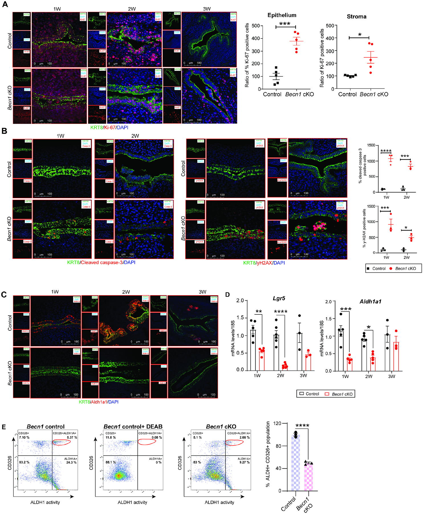
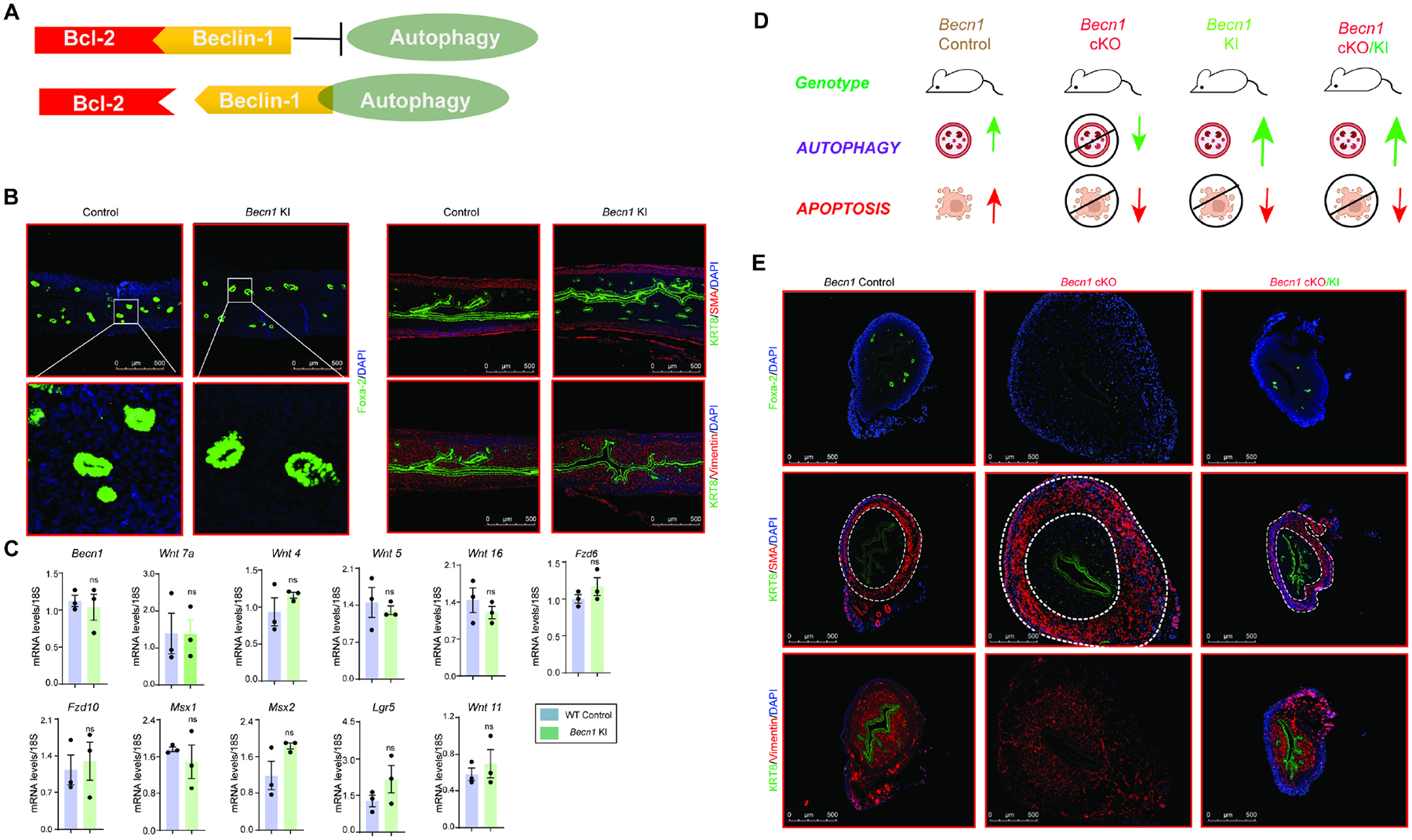
The human endometrium shows a remarkable regenerative capacity that enables cyclical regeneration and remodeling throughout a woman's reproductive life. Although early postnatal uterine developmental cues direct this regeneration, the vital factors that govern early endometrial programming are largely unknown. We report that Beclin-1, an essential autophagy-associated protein, plays an integral role in uterine morphogenesis during the early postnatal period. We show that conditional depletion of Beclin-1 in the uterus triggers apoptosis and causes progressive loss of Lgr5+/Aldh1a1+ endometrial progenitor stem cells, with concomitant loss of Wnt signaling, which is crucial for stem cell renewal and epithelial gland development. Beclin-1 knockin (Becn1 KI) mice with disabled apoptosis exhibit normal uterine development. Importantly, the restoration of Beclin-1-driven autophagy, but not apoptosis, promotes normal uterine adenogenesis and morphogenesis. Together, the data suggest that Beclin-1-mediated autophagy acts as a molecular switch that governs the early uterine morphogenetic program by maintaining the endometrial progenitor stem cells.

Keywords: Beclin-1; autophagy; endometrium; morphogenesis; stem cells.


Conflict of interest statement

Declaration of interests The authors declare no competing interests.

Figures

Figure 1:
Figure 1:. Conditional ablation of Becn1 results in female infertility with intact ovarian function.
(A) Mouse breeding strategy employed to generate control (Becn1f/f) mice or Becn1f/f;PRcre/+ (Becn1 cKO) mice. (B) Relative transcript levels of Becn1 in 8-week-old virgin control and cKO uteri (whole uterus including epithelial, stromal, and myometrial compartment), ovary, and liver (n=6). mRNA levels are normalized to levels of 18S m-RNA. Data are presented as mean ± SEM; ***P<0.001, P>0.05, ns=non-significant. (C & D) Left panel, Western blotting to show protein levels of Beclin-1 in uteri from 8-week-old virgin control and cKO uteri. GAPDH is used as a loading control. Right panel, Immunohistological analysis of Beclin-1 expression in Control and Becn1 cKO mice. Scale bar: 40 μm (E & F) Relative number of littermates and uterine weight of Becn1 control and cKO mice sacrificed after breeding trial. Data are presented as mean ± SEM; P>0.05, ns=non-significant. (G) H&E staining of ovaries from 8-week-old control and Becn1 cKO mice; scale bar: 2mm (upper panel), 500 μm (lower panel) (H) Levels of reproductive hormones estradiol and progesterone from serum collected during euthanasia of 8-week-old virgin mice. (I) Super-ovulated oocytes retrieved from 4-week-old Becn1 control and cKO mice (left). Number of super-ovulated oocytes in 4-week-old control and Becn1 cKO mice (Right); scale bar: 200 μm
Figure 2:
Figure 2:. Becn1 is critical for embryo implantation and uterine receptivity.
(A) Images of 5.0 dpc uteri of control and Becn1 cKO mice injected with Chicago Sky Blue dye to visualize implantation sites (denoted by black arrows) (Left). Quantification of implantation sites in control (n=6) and Becn1 cKO (n=6) mice on day 5 of pregnancy (Right). (B) H&E-stained cross-sections of 5.0 dpc uteri of control and Becn1 cKO mice to visualize embryo implantation. LE, luminal epithelium; S, stroma. Asterisk denotes the embryo. Scale bar: 200 μm, 50 μm (C) Immunofluorescence analysis of uterine tissues from Control and Becn1 cKO mice, stained with Muc1. scale bar: 500 μm, 100 μm. (D) Experimental protocol for hormonal induction of uterine receptivity in ovariectomized mice. (E) Representative immunofluorescence images of uteri from Control and Becn1 cKO mice stained for Ki-67 following Oil or E2 or E2+P4 treatment (n = 5 mice/group); scale bar: 100 μm. (F) Transcript levels of Becn1 and Igf1, in uteri from Control and Becn1 cKO mice in the indicated treatment groups. Data are presented as mean ± SEM (n = 5), P>0.05, ns=non-significant.
Figure 3:
Figure 3:. Becn1 is indispensable for uterine glandular development and morphogenesis.
(A) Graphical illustration to show gland development events in uterus (B) Immunofluorescence analysis to show Beclin-1 expression in 1 to 3-week (W)-old control and Becn1 cKO uteri; scale bar: 500 μm, 100 μm (C & D) Immunofluorescence analysis to show Foxa2-positive glandular structures in uteri from Control and Becn1 cKO at 2W of age (lower panel showing magnified images) and different PNDs (3 to 12W) of development (left); scale bar: 500 μm. Quantitative analysis of Foxa2-positive glands on indicated PNDs. Data represents the number of glands counted from four different areas of uterine cross-sections; images taken at 100X magnification and the average number of glands from n=3 mice was presented. Data shown represent the mean± S.E.M., *P>0.05, ns=non-significant.
Figure 4:
Figure 4:. Loss of Becn1 leads to undifferentiated stroma and myometrium resulting in hyper-muscular uteri.
(A) Immunolocalization of KRT8 (green) and Vimentin (red) in the uteri of Control and Becn1 cKO at different PNDs; scale bar: 100 μm (B) Immunofluorescence of KRT8 (green) and smooth muscle α-actin (red) in the uteri of Control and Becn1 cKO at different PNDs. DAPI (blue) was used to counterstain the nuclei; scale bar: 500 μm (C) Relative mRNA expression of genes implicated in uterine development at different PNDs from Control and Becn1 cKO mice.
Figure 5:
Figure 5:. Endometrial progenitor stem cell maintenance requires Becn1 during early uterine morphogenesis.
(A) Immunofluorescence of KRT8 (green) and Ki67 (red) in uteri of Control and Becn1 cKO mice at 1, 2 and 3W of postnatal stage of uterus development (left); scale bar: 100 μm. Quantification of Ki67-positively stained cells in the luminal epithelia and stromal compartment of uteri at 3W of postnatal age. Data represents the ratio of the number of Ki67-positive cells to the total number of cells counted in four different areas and plotted as percent positive cells from n=6 mice (right). (B) Immunofluorescence of KRT8 (green), cleaved caspase-3 (red), and γH2AX in uteri of Control and Becn1 cKO mice during 1W, and 2W of postnatal age of uterus development (left); scale bar: 100 μm. Quantification of cleaved caspase-3 and γH2AX-positively stained epithelial cells in uteri at 1W, and 2W of postnatal ages. Data represents the ratio of the number of positive cells to the total number of cells counted in four different areas and plotted as percent positive cells from n=3 mice (right). (C) Immunolocalization of endometrial progenitor stem cell marker; Aldh1a1 in uteri at different PNDs (upper panel); scale bar: 100 μm (D) Transcript levels of endometrial progenitor stem cell markers Aldh1a1 and Lgr5 during the critical window of uterine maturation. (E) Representative flow cytometry analysis of ALDH1A1 activity in uterine cells isolated from Becn1 2W old control and cKO uteri. Control cells treated with DEAB was used as negative control for Aldefluor staining.
Figure 6:
Figure 6:. Pharmacological inhibition of autophagy impedes uterine glandular development in mice and human organoids.
(A) (Upper panel) Experiment strategy to inhibit autophagy in neonatal uteri. Mice pups were treated with Vehicle or Spautin on PND5 and euthanized at PND14 time-point for tissue harvesting (Lower panel). Immunofluorescence imaging to show Foxa2 (green)-positive glandular structures in Vehicle or Spautin-treated 2W old WT mice pups. DAPI (blue) was used to counterstain the nuclei; scale bar: 500 μm. (B) Experimental timeline of human endometrial epithelia organoid cultures. Organoids were derived in ExM and then subjected to treatment with Vehicle or E2 or E2+Spautin or Spautin alone. (C) Representative bright-field images showing morphology of endometrial organoids prior to treatment on day 4 or after treatment with Vehicle or E2 (10nM) or E2 (10nM) +Spautin (15 μM) or Spautin (15 μM) alone for two days on day 6; scale bar: 600 μm (D) Relative mRNA levels of PGR, FOXA2, LGR5 and ALDH1A1 in hormone-or Spautin-treated organoids. Data are means ± SEMs. *P ≤ 0.05, **P ≤ 0.01, ***P ≤ 0.001, ****P ≤ 0.0001. (E) (left) Immunolocalization of FOXA2, KRT8 (Green)+ Ki-67 (Red), KRT8 (Green)+ γH2AX (Red) in response to E2 or Spautin inhibitor. Organoids were counterstained with DAPI (blue) to visualize nuclei, scale bar: 40 μm. Quantification of the percentage of FOXA2, Ki-67 and γH2AX positive cells relative to the total number of cells in organoid sections (right). Three independent experiments were performed and for each experiment, 7–10 organoids per treatment group were counted and plotted as percent positive cells. Data are presented as mean ± SE.
Figure 7:
Figure 7:. Becn1-dependent autophagy but not apoptosis is required for postnatal uterine gland development in mice.
(A) Illustration of Beclin-1-Bcl2 interaction (B) Immunolocalization of Foxa2-positive glandular structures in upper panel and magnified images in lower panel; scale bar: 500 μm. Immunofluorescence of KRT8 (Green)+ Vimentin (Red), KRT8 (Green)+ smooth muscle α-actin (Red) in uteri from control wild-type and Becn1 KI mice by 2W of postnatal age of development. DAPI (blue) was used to counterstain the nuclei; scale bar: 500 μm (C) Transcript levels of various candidate genes associated with gland development and uterine maturation. (D) Graphical illustration to demonstrate Beclin-1-dependent autophagy or apoptosis in different genetically engineered mice. (E) Immunolocalization of Foxa2 (Green), KRT8 (Green)+ SM-α-actin (Red), KRT8 (Green)+ Vimentin (Red) in Becn1 control, cKO and cKO/KI mice; scale bar: 500 μm.

References

    1. Matsumoto H (2017). Molecular and cellular events during blastocyst implantation in the receptive uterus: clues from mouse models. J Reprod Dev 63, 445–454. 10.1262/jrd.2017-047. - DOI - PMC - PubMed
    1. Robertshaw I, Bian F, and Das SK (2016). Mechanisms of uterine estrogen signaling during early pregnancy in mice: an update. J Mol Endocrinol 56, R127–138. 10.1530/JME-15-0300. - DOI - PMC - PubMed
    1. Ramathal CY, Bagchi IC, Taylor RN, and Bagchi MK (2010). Endometrial decidualization: of mice and men. Semin Reprod Med 28, 17–26. 10.1055/s-0029-1242989. - DOI - PMC - PubMed
    1. Filant J, and Spencer TE (2014). Uterine glands: biological roles in conceptus implantation, uterine receptivity and decidualization. Int J Dev Biol 58, 107–116. 10.1387/ijdb.130344ts. - DOI - PMC - PubMed
    1. Spencer TE (2014). Biological roles of uterine glands in pregnancy. Semin Reprod Med 32, 346–357. 10.1055/s-0034-1376354. - DOI - PMC - PubMed

Publication types

LinkOut - more resources