. 2023 Apr 11;14(1):2026.
doi: 10.1038/s41467-023-37703-6.

FOXI3 pathogenic variants cause one form of craniofacial microsomia

Affiliations

FOXI3 pathogenic variants cause one form of craniofacial microsomia

Ke Mao et al. Nat Commun. .

Abstract

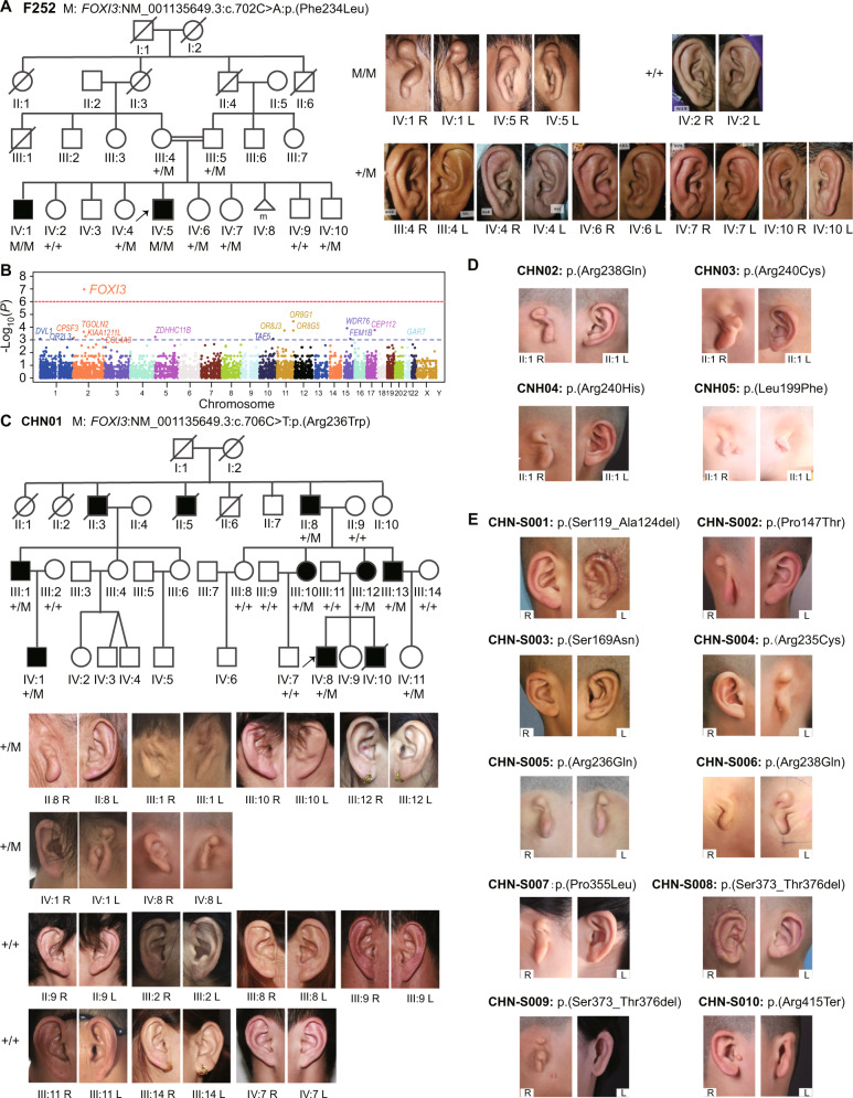
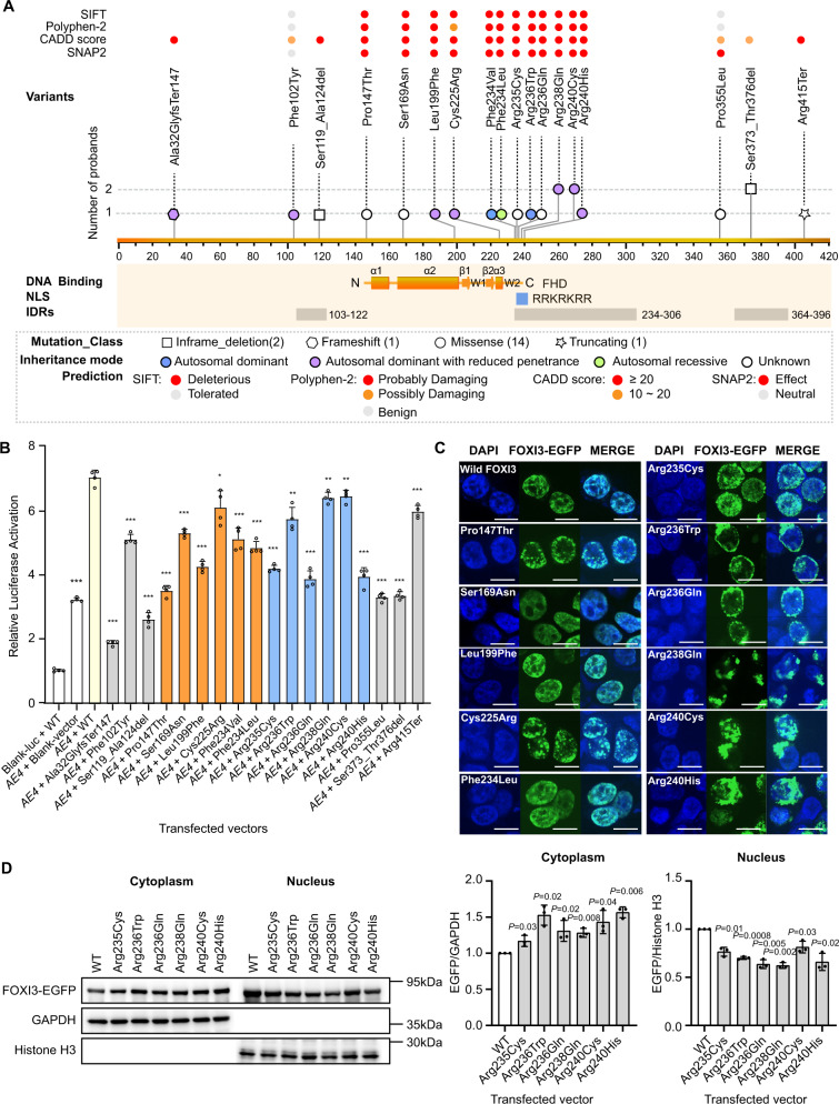
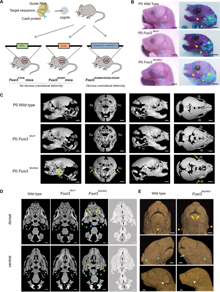
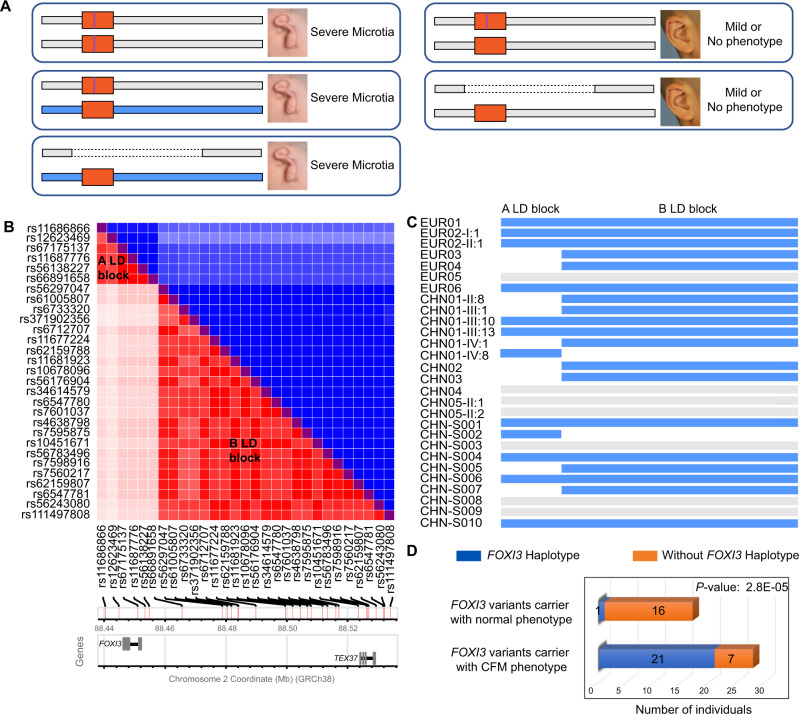
Craniofacial microsomia (CFM; also known as Goldenhar syndrome), is a craniofacial developmental disorder of variable expressivity and severity with a recognizable set of abnormalities. These birth defects are associated with structures derived from the first and second pharyngeal arches, can occur unilaterally and include ear dysplasia, microtia, preauricular tags and pits, facial asymmetry and other malformations. The inheritance pattern is controversial, and the molecular etiology of this syndrome is largely unknown. A total of 670 patients belonging to unrelated pedigrees with European and Chinese ancestry with CFM, are investigated. We identify 18 likely pathogenic variants in 21 probands (3.1%) in FOXI3. Biochemical experiments on transcriptional activity and subcellular localization of the likely pathogenic FOXI3 variants, and knock-in mouse studies strongly support the involvement of FOXI3 in CFM. Our findings indicate autosomal dominant inheritance with reduced penetrance, and/or autosomal recessive inheritance. The phenotypic expression of the FOXI3 variants is variable. The penetrance of the likely pathogenic variants in the seemingly dominant form is reduced, since a considerable number of such variants in affected individuals were inherited from non-affected parents. Here we provide suggestive evidence that common variation in the FOXI3 allele in trans with the pathogenic variant could modify the phenotypic severity and accounts for the incomplete penetrance.


Conflict of interest statement

S.E.A. is a cofounder and CEO of Medigenome, and serves on the Scientific Advisory Board of the “Imaging” institute in Paris; J.R.L. has stock ownership in 23andMe, is a paid consultant for Regeneron Genetics Center, is a co-inventor on multiple United States and European patents related to molecular diagnostics for inherited neuropathies, eye diseases, and bacterial genomic fingerprinting, and serves on the Scientific Advisory Board of BG. The Department of Molecular and Human Genetics at Baylor College of Medicine receives revenue from clinical genetic and genomic testing conducted at Baylor Genetics (BG) Laboratories. The remaining authors declare no competing interests.

Figures

Fig. 1
Fig. 1. Identification of FOXI3 as the pathogenic gene for CFM.
A The Pakistani consanguineous pedigree F252 with the likely pathogenic FOXI3: p.(Phe234Leu) variant. Photographs of the microtia type III of both ears of affected individuals IV:1 and IV:5. The bottom panel depicts the ears of additional members of the F252 family. The Sanger sequences are shown in Supplementary Fig. 1. M depicts the mutant allele, and + the normal allele. B Family-based gene-level association on rare LoF (Loss-of-Function) variants from 48 CFM Chinese families. The blue dashed line indicates the significance threshold of 1E-03, and the red dashed line indicates the Bonferroni correction threshold of 1.03E-06. Source data are provided as a Source Data file. C The pedigree, genotype, and external ear phenotype for the Chinese CFM family CHN01. M depicts the mutant allele, and + indicates the normal allele. D The genotype and external auricular phenotype for the four Chinese CFM families. E The genotype and external auricular phenotype of the sporadic Chinese patients.
Fig. 2
Fig. 2. The effect of FOXI3 variants with in silico prediction and in vitro experiments.
A The 18 identified variants on FOXI3. The predictions of the variants are classified by bins and shown on the top of each variant. The variants are classified by the type of inframe_deletion (square), frameshift (hexagon), missense (circle), and truncating (star), and the inheritance mode of autosomal dominant (blue) or with reduced penetrance (purple), autosomal recessive (green), and unknown (white). The variants of p.(Arg238Gln), p.(Arg240Cys), and p.(Ser373_Thr376del) are detected in two probands and the rest are in one proband. B Effects of the 18 variants on the transactivation of FOXI3 in HEK293T cells (n = 4/group). Gray Bars: intrinsically disordered regions (IDRs), orange bars: forkhead domain (FHD), blue bar: nuclear localization signal (NLS). *P < 0.05, **P < 0.01, ***P < 0.001. Each experiment was repeated at least three times. C The localization and distribution of wild type or mutant FOXI3-EGFP fusion proteins in HEK-293T cells. DAPI counterstain (blue) shows the location of the nucleus. EGFP (green fluorescent protein) shows the subcellular localization of the FOXI3-EGFP fusion protein. Wild-type FOXI3 is almost completely transferred into the nucleus and uniformly distributed. A majority of FOXI3 with mutant NLS is blocked outside the nucleus; FOXI3 with mutant FHD tends to form intranuclear aggregation; most of FOXI3 with mutant IDR is similar to Wild-type FOXI3. Scale bar = 10 μm. Each experiment was performed in triplicate and repeated at least three times. D Representative western blots of FOXI3-EGFP with mutant NLS. Immunoprecipitation on the EGFP-tagged FOXI3 using antibody of EGFP and detect the amount of FOXI3-EGFP in the nucleus and cytoplasmic fractions of HEK293T cells (n = 3 biologically independent experiments). Compared to the wild type, the mutant FOXI3-EGFP fusion protein is significantly trapped in the cytoplasm and the amount of mutant FOXI3-EGFP is significantly reduced in the nucleus. Significant differences between two groups (wild type and each mutant) were determined by unpaired Student’s t-test (two-tailed). Data are presented as the means ± s.d. Source data are provided as a Source Data file.
Fig. 3
Fig. 3. Generation and characterization of FOXI3 mutant mice.
A A schematic illustration of the generation of Foxi3 mutant mice. Three mouse lines are created including Foxi3F218L, Foxi3R224H, and Foxi3R220W-R222Q-R224H. B Craniofacial differences of wild-type, heterozygous, and homozygous Foxi3R220W-R222Q-R224H neonatal mice (n = 3 for each genotype). Comparing with wild type or heterozygotes, the cyan arrows indicate homozygotes with small pinnae, and various overt cranioskeletal defects, including abnormal middle and inner ear bones, syngnathia (mandible and maxilla fusion), and anomalous squamosal development (yellow arrows). The left panel was photographed with bright field; the right panel of mice were stained with Alizarin Red and Alcian Blue. Scale bar = 2 mm. C Asymmetric presentation of cranioskeletal features in the neonatal Foxi3R220W-R222Q-R224H homozygous mice (n = 4 for each genotype). Micro-CT images show variable development of the jugal bone, and almost complete loss of the squamous bone in one or both sides in homozygous neonates. The blue arrow indicates the fusion of the mandible (Md) to the maxilla (Mx) in a homozygote. The yellow arrow indicates the poorly developed squamosal (Sq), jugal (Jg), palatine (Pt), and condylar process (Cp). The red asterisk (*) indicates the retrotympanic process (Rtp) and the tympanic ring (Tr) that are absent or severely hypoplastic in homozygotes. D Dorsal and ventral views of the cranial base show the absent or hypoplastic bony ear structures (noted in (C); asterisked) as well as hypoplasia of the lateral pterygoids of the basisphenoid (cyan arrows) and cleft palate (yellow arrows, dorsal view), with ventrally protruding palatine bones (yellow arrows, ventral view) of newborn triple-mutant mice. Dashed black line denotes the asymmetry of the skull centered on the cranial base. E Surface rendering of the facial soft tissue from the same micro-CT scans highlights the severe left-side microtia in the homozygote (white and cyan arrowheads) and asymmetry in positions of the pinnae (yellow arrowheads). Homozygotes exhibit microstomia that is asymmetric in presentation (yellow arrows) and, variably, a row of whisker follicles on the ‘mandible’ (blue arrow) for the newborn triple-mutant mice. Scale bar = 1 mm in (C), (D), and (E). P0: Postnatal day 0.
Fig. 4
Fig. 4. Presumed FOXI3 haplotype that modifies the clinical severity of pathogenic FOXI3 variants.
A The hypothesis of FOXI3 haplotype for the reduced penetrance of CFM with microtia types II or III. The graph shows three possible genotype combination for CFM with microtia. The orange rectangular box indicates FOXI3 genes in the human genome, the purple line means a pathogenic variant allele. The blue rectangular depicts the presumed modifier trans FOXI3 haplotype while the gray rectangular is the common haplotype. The dashed line indicates the missing haplotype. B Linkage disequilibrium (LD) blocks A and B (see text) of polymorphic sites shown in the y- and x-axis (data from Europeans and Chinese). The x-axis also depicts a schematic representation of the map position of the polymorphic sites along chromosome 2 (GRCh38). The blue part is the D’ and the red is the r2 statistics. C Presence of the presumed modifier haplotype of A LD block and/or B or both in the DNA of individuals with microtia type III or preauricular tags described in this study. D Chi-square test for the trans FOXI3 haplotype in CFM and normal individuals. The blue column square indicates the individual who carried FOXI3 variants, as well as trans FOXI3 haplotype, and the orange column square, indicates the individual who carried FOXI3 variants but without trans FOXI3 haplotype.

References

    1. Klein D. Living history–autobiography: genetics and environment from a personal perspective. Am. J. Med. Genet. 1990;37:325–335. doi: 10.1002/ajmg.1320370307. - DOI - PubMed
    1. Goldenhar M. Associations malformatives de l’oeil et de l’oreille, en particulier le svndrome dermoide epibulbaire- appendices auriculaires-fistula auris congenita et ses relations avec la dysostose mandibulo-faciale. J. Genet. Hum. 1952;1:243–282.
    1. Cohen MM, Jr., Rollnick BR, Kaye CI. Oculoauriculovertebral spectrum: an updated critique. Cleft Palate J. 1989;26:276–286. - PubMed
    1. Beleza-Meireles A, Clayton-Smith J, Saraiva JM, Tassabehji M. Oculo-auriculo-vertebral spectrum: a review of the literature and genetic update. J. Med. Genet. 2014;51:635–645. doi: 10.1136/jmedgenet-2014-102476. - DOI - PubMed
    1. Mastroiacovo P, et al. Epidemiology and genetics of microtia-anotia: a registry based study on over one million births. J. Med. Genet. 1995;32:453–457. doi: 10.1136/jmg.32.6.453. - DOI - PMC - PubMed

Publication types

Substances