. 2023 Apr 12;14(4):264.
doi: 10.1038/s41419-023-05777-1.

USP1 modulates hepatocellular carcinoma progression via the Hippo/TAZ axis

Affiliations

USP1 modulates hepatocellular carcinoma progression via the Hippo/TAZ axis

Dongyi Liu et al. Cell Death Dis. .

Abstract

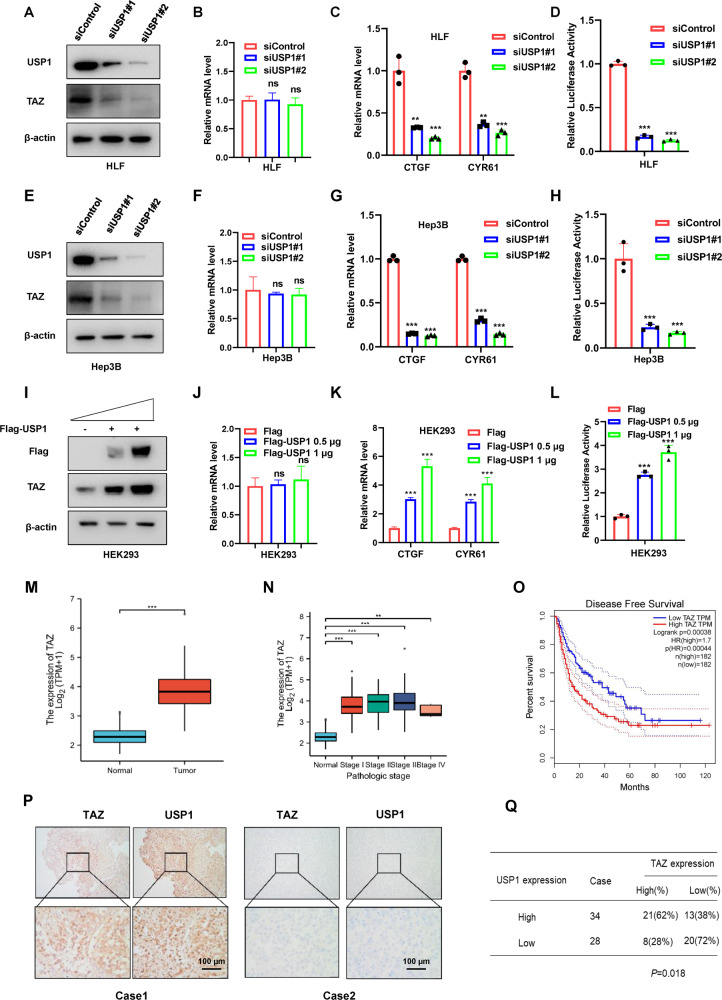
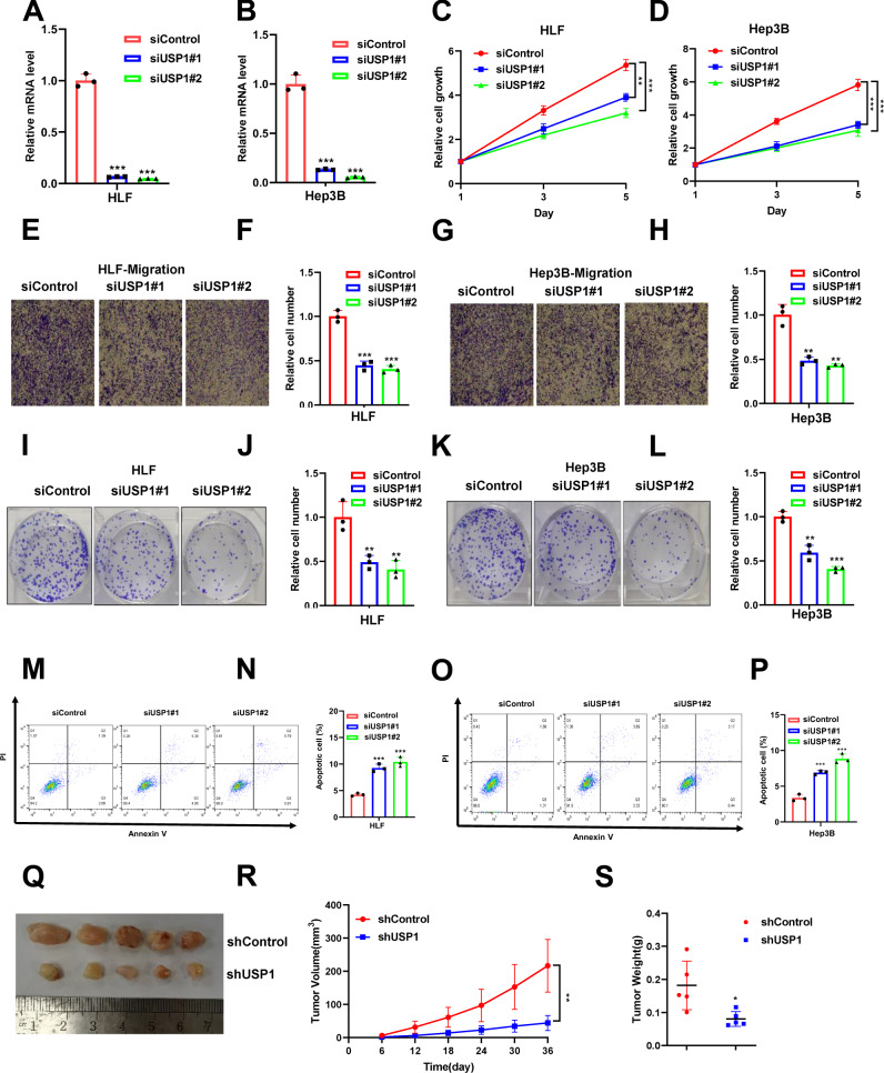
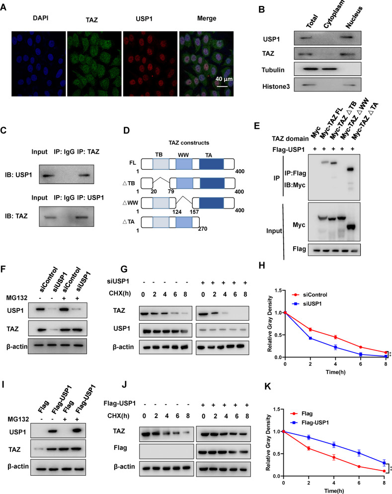
Hepatocellular carcinoma (HCC) is one of the most lethal malignancies worldwide. The Hippo signaling pathway has emerged as a significant suppressive pathway for hepatocellular carcinogenesis. The core components of the Hippo pathway constitute a kinase cascade, which inhibits the functional activation of YAP/TAZ. Interestingly, the overactivation of YAP/TAZ is commonly observed in hepatocellular carcinoma, although the inhibitory kinase cascade of the Hippo pathway is still functional. Recent studies have indicated that the ubiquitin‒proteasome system also plays important roles in modulating Hippo signaling activity. Our DUB (deubiquitinase) siRNA screen showed that USP1 is a critical regulator of Hippo signaling activity. Analysis of TCGA data demonstrated that USP1 expression is elevated in HCC and associated with poor survival in HCC patients. RNA sequencing analysis revealed that USP1 depletion affects Hippo signaling activity in HCC cell lines. Mechanistic assays revealed that USP1 is required for Hippo/TAZ axis activity and HCC progression. USP1 interacted with the WW domain of TAZ, which subsequently enhanced TAZ stability by suppressing K11-linked polyubiquitination of TAZ. Our study identifies a novel mechanism linking USP1 and TAZ in regulating the Hippo pathway and one possible therapeutic target for HCC.


Conflict of interest statement

The authors declare no competing interests.

Figures

Fig. 1
Fig. 1. USP1 is an important regulator of Hippo signaling activity, and its expression is elevated in human hepatocellular carcinoma.
A The flowchart shows the siRNA screening procedure for identifying novel deubiquitinases involved in modulating Hippo signaling. HEK293 cells were seeded in 24-well plates. Each well was transfected with a siRNA against one deubiquitinase and incubated for 48 h. RNA was extracted, and CTGF expression was assessed by qPCR. B In the siRNA screen of deubiquitinases, relative CTGF mRNA levels were determined. The red columns represent relative mRNA level of CTGF after depletion of USP1. C The relative RNA level of USP1 in HCC tumor samples (n = 371) versus normal samples (n = 50) in the TCGA database (https://portal.gdc.cancer.gov/). D The relative RNA level of USP1 in HCC tumors of different stages (Stag I, n = 168; Stag II, n = 84; Stag III, n = 82; Stag IV, n = 6) was compared with that in normal liver tissue (n = 50). Data were obtained from the TCGA database (https://portal.gdc.cancer.gov/). E Comparing with lower USP1 expression (n = 275), higher USP1 expression (n = 90) was associated with poorer overall survival in HCC patients based on Kaplan‒Meier analysis in TCGA. P < 0.001, log-rank test. F TCGA analysis of the correlation of USP1 expression with that of classical downstream genes of the Hippo signaling pathway in HCC (n = 371), with p < 0.001 as the threshold. G USP1 expression was significantly correlated with that of CCN1 (CYR61) and CCN2 (CTGF) in HCC (n = 371). H Volcano plot of RNA-seq data in HLF cell lines treated with siControl or siUSP1. The volcano plot revealed a significant increase in the expression of CTGF and CYR61 downstream of YAP as a result of USP1 depletion. Threshold values of P < 0.05 and fold change>2 was set as screening criteria. I These are the top 10 KEGG pathways that are significantly depleted (top) or enriched (bottom) in HLF cells treated with siUSP1. The pathway enrichment analysis consisted of differentially regulated genes identified with threshold criteria of P < 0.001 and fold change >2. The cells were treated for 48 h with 50 nM siUSP1 or vehicle. Total mRNA was extracted for RNA sequencing analysis. n = 3. J HLF cells treated with USP1 siRNA had depletion of Hippo pathway signature genes, according to gene set enrichment analysis (GSEA).
Fig. 2
Fig. 2. USP1 depletion inhibits Hippo/TAZ axis activity in HCC cells.
A, E, I Western blot analysis of USP1 and TAZ expression in HLF, Hep3B, and HEK293 cells exposed as indicated. In this study, actin was employed as an internal reference. B, F, J RT–qPCR results of TAZ mRNA expression in HLF, Hep3B, and HEK293 cell lines exposed as indicated. C, G, K RT–qPCR results of CTGF and CYR61 mRNA expression in HLF, Hep3B, and HEK293 cell lines exposed as indicated. D, H, L In HLF, Hep3B, and HEK293 cells exposed as indicated, transcriptional activity of TEAD response elements was measured by a luciferase assay using a reporter containing tandem TEAD-binding sites. M In the TCGA database, the relative level of TAZ RNA in HCC tumor samples (n = 371) was compared to that in normal samples (n = 50) (https://www.genome.gov/). N In HCC tumors of various stages (Stag I, n = 168; Stag II, n = 84; Stag III, n = 82; Stag IV, n = 6), the relative RNA level of TAZ was compared with that in normal liver tissue (n = 50). Data were obtained from the TCGA database. (https://www.genome.gov/). O Comparing with lower TAZ expression (n = 275), higher TAZ expression (n = 90) was associated with poorer progression-free survival in HCC patients based on Kaplan‒Meier analysis in TCGA. P = 0.00044, log-rank test. P, Q Immunohistochemical (IHC) staining to evaluate USP1 and TAZ expression in HCC tissues. In Panels AL the results are representative of three independent experiments. All the data are presented as the means ± SDs. **P < 0.01, ***P < 0.001 (Student’s t test).
Fig. 3
Fig. 3. USP1 depletion inhibits hepatocellular carcinoma progression in vivo and in vitro.
A, B RT‒PCR was performed to determine USP1 mRNA levels in HLF and Hep3B cells following USP1 siRNA treatment. C, D HLF and Hep3B cells transfected with siControl or siUSP1 were tested for viability using the CCK-8 assay at the indicated time points. EH HLF and Hep3B cells were tested for their migration ability using Transwell assays. F and H show the quantitative analysis of the colony formation assay results. IL Colony formation (left panel) of HLF and Hep3B cells transfected with scrambled siRNA or one of two independent USP1 siRNAs. J and L show the quantitative analysis of the colony formation assay results. MP The percentage of apoptotic cells was determined by FACS analysis after HLF and Hep3B cells were treated with USP1 siRNA. PI and Annexin V staining were performed on the cells. Q A representative image of a tumor derived from a nude mouse injected with stably transfected shControl or shUSP1 HLF cells is shown. R, S The tumor volume (R) and weight (S) in nude mice subcutaneously inoculated with stably transfected shControl or shUSP1 HLF cells. Three independent experiments were conducted to obtain the results shown in Panels AP. All the data are presented as the means ± SDs. **P < 0.01, ***P < 0.001 (Student’s t test).
Fig. 4
Fig. 4. Hepatocellular carcinomas with TAZ overexpression display partial reversal of the antitumor effects of USP1 depletion.
A Western blot analysis of TAZ and USP1 expression in HLF cells treated with siControl or siUSP1 for 24 h, then transfected with Myc or Myc-TAZ for another 24 h. In this study, β-Actin was used as the internal reference. B Results of RT‒qPCR to measure CTGF and CYR61 mRNA levels in HLF cells treated as indicated. C TEAD response element transcriptional activity was measured via luciferase reporter assays with tandem TEAD-binding sites in HLF cells treated as indicated. D At the indicated time points, a CCK-8 assay was performed to determine the viability of HLF cells treated as indicated. E, F Colony formation (left panel) of HLF cells treated as indicated. F shows the quantitative analysis of the colony formation assay results. G, H FACS analysis (left panel) was performed on HLF cells treated as indicated. I, J Transwell assays was performed on HLF cells transfected treated as indicated. K As indicated, images of tumors derived from nude mice injected with shControl, shUSP1- or shUSP1+TAZ-transfected HLF cells are shown. L, M Tumor volume (L) and weight (M) in nude mice injected with stably transfected shControl, shUSP1, or shUSP1+TAZ HLF cells. In Panels AJ, the results are representative of three independent experiments. In Panels KM, the results are representative of five independent experiments. The data are presented as the means ± SDs. **P < 0.01, ***P < 0.001 (Student’s t test).
Fig. 5
Fig. 5. In hepatocellular carcinoma cells, USP1 interacts with TAZ and modulates its stability.
A Immunofluorescence imaging of TAZ (green), USP1 (red) and DAPI (blue) in HLF cells, scale bar 40 µm. B Western blot analysis of USP1 and TAZ protein localization in HLF cells. In this study, the cytoplasmic and nuclear fractions were separated using a subcellular protein fractionation kit (Thermo Scientific 78840). The control cytoplasmic and nuclear fractions were analyzed using antibodies specific for tubulin and histone3, respectively. C USP1 and TAZ proteins were found to be associated in HLF cells by a coimmunoprecipitation (Co-IP) assay. D The wild-type and truncated TAZ constructs are shown in a schematic diagram. E An immunoblot demonstrating the interaction between USP1 and WT TAZ or truncated TAZ, as assessed by Co-IP with USP1 (with an anti-Flag antibody). F, I Western blotting were used to measure TAZ and USP1 protein levels in HLF and HEK293 cells treated as indicated. MG132 (10 μM) was applied to the cells for 8 h before western blot analysis was performed. G, H USP1 depletion decreased the TAZ protein half-life in HLF cells. Cells were treated with 100 μmol/L CHX for the indicated time periods before being collected for western blot analysis. H Quantitative analysis of the half-life of the TAZ protein. J, K USP1 prolonged the TAZ protein half-life in HEK293 cells. Cells were treated with 100 μmol/L CHX for the indicated time periods before being collected for western blot analysis. K Quantitative analysis of the half-life of the TAZ protein. All the results are representative of 3 independent experiments. The data are presented as the means ± SDs. **P < 0.01 (Student’s t test).
Fig. 6
Fig. 6. USP1 regulates TAZ protein stability by inhibiting K11-linked polyubiquitination of TAZ.
A Western blot analysis of polyubiquitinated TAZ was performed after coimmunoprecipitation (Co-IP) in HLF cells treated as indicated. B, C Western blot analysis of polyubiquitinated TAZ was performed after coimmunoprecipitation in HEK293 cells treated as indicated. D, E Western blot analysis of K11 linkage-specific polyubiquitinated TAZ was performed after coimmunoprecipitation in HLF or HEK293 cells treated as indicated. F Western blot analysis of K11R linkage-specific polyubiquitinated TAZ was performed after coimmunoprecipitation in HEK293 cells treated as indicated. G The hypothetical mechanistic model of USP1-regulated Hippo signaling in HCC progression. The USP1 associates with TAZ and decreases TAZ protein degradation via K11-linked polyubiquitination of TAZ, which increases Hippo/TAZ axis activation and enhances malignant progression in HCC cells. All the results are representative of three independent experiments.

References

    1. Siegel RL, Miller KD, Jemal A. Cancer statistics, 2019. CA Cancer J Clin. 2019;69:7–34. doi: 10.3322/caac.21551. - DOI - PubMed
    1. Sung H, Ferlay J, Siegel RL, Laversanne M, Soerjomataram I, Jemal A, et al. Global Cancer Statistics 2020: GLOBOCAN estimates of incidence and mortality worldwide for 36 cancers in 185 countries. CA Cancer J Clin. 2021;71:209–49. - PubMed
    1. Llovet JM, Kelley RK, Villanueva A, Singal AG, Pikarsky E, Roayaie S, et al. Hepatocellular carcinoma. Nat Rev Dis Primers. 2021;7:6. doi: 10.1038/s41572-020-00240-3. - DOI - PubMed
    1. Garcia-Lezana T, Lopez-Canovas JL, Villanueva A. Signaling pathways in hepatocellular carcinoma. Adv Cancer Res. 2021;149:63–101. doi: 10.1016/bs.acr.2020.10.002. - DOI - PubMed
    1. Misra JR, Irvine KD. The hippo signaling network and its biological functions. Annu Rev Genet. 2018;52:65–87. doi: 10.1146/annurev-genet-120417-031621. - DOI - PMC - PubMed

Publication types

MeSH terms

Substances