. 2023 Apr 11;14(1):2025.
doi: 10.1038/s41467-023-37779-0.

Cryo-electron microscopy structures of capsids and in situ portals of DNA-devoid capsids of human cytomegalovirus

Affiliations

Cryo-electron microscopy structures of capsids and in situ portals of DNA-devoid capsids of human cytomegalovirus

Zhihai Li et al. Nat Commun. .

Abstract

The portal-scaffold complex is believed to nucleate the assembly of herpesvirus procapsids. During capsid maturation, two events occur: scaffold expulsion and DNA incorporation. The portal-scaffold interaction and the conformational changes that occur to the portal during the different stages of capsid formation have yet to be elucidated structurally. Here we present high-resolution structures of the A- and B-capsids and in-situ portals of human cytomegalovirus. We show that scaffolds bind to the hydrophobic cavities formed by the dimerization and Johnson-fold domains of the major capsid proteins. We further show that 12 loop-helix-loop fragments-presumably from the scaffold domain-insert into the hydrophobic pocket of the portal crown domain. The portal also undergoes significant changes both positionally and conformationally as it accompanies DNA packaging. These findings unravel the mechanism by which the portal interacts with the scaffold to nucleate capsid assembly and further our understanding of scaffold expulsion and DNA incorporation.


Conflict of interest statement

The authors declare no competing interests.

Figures

Fig. 1
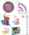
Fig. 1. Asymmetric reconstructions of capsids and in situ portals of the human cytomegalovirus (HCMV) A- and B-capsids.
Composite structures of the HCMV B-capsid (a) and A-capsid (b), respectively. The front-halves of both capsid shells (light blue) are removed to show their internal components. The front-right quarter of the scaffold in (a) is removed to show the inner three-layer scaffold (the inner core and middle band are radially colored from dark to medium purple and the outer shell is highlighted in red). The portal vertices are colored as indicated below in (c). c Close-up views of the portal vertex regions in B- (left), A- (middle) and virion (right) capsids. The capsid shell components of the portal vertex are colored by protein. The portal is colored by domain. For both B- (left) and A-capsid (middle), the composite cryoEM maps were assembled from reconstructions of C1 capsid (light blue), C5 portal vertex (CVSC, salmon; Ta, white; Tc, cyan; portal turret, magenta; portal 10-helix anchor, violet) and C12 portal main body (yellow). The portal-bound scaffold fragments from C12 reconstruction of portal in B-capsid are highlighted in red. For virion capsid (right), the composite cryoEM map were assembled from reconstructions of C1 capsid (light blue, EMD-31292), C1 portal vertex (pDNA, orange. EMD-31290), C5 portal vertex (CVSC, salmon; Ta, white; Tc, cyan; portal 10-helix anchor, violet. EMD-31297), C6 portal (portal turret: magenta. EMD-31299) and C12 portal main body (yellow. EMD-31295). d Top-views of in situ portals of B- (left), A- (middle), and virion (right) capsids. Portal turrets are highlighted to show the symmetry difference in arrangement between DNA-devoid and DNA-filled capsids.
Fig. 2
Fig. 2. Interactions between scaffolds and MCPs.
a Asymmetric reconstruction of the HCMV B-capsid with the front half of the capsid shell (gray) removed. The portal is in yellow and the three layers of scaffolds are colored as in Fig. 1a. b Cross-section of one-quarter of the B-capsid map, showing the radial distribution of the three layers of the scaffolds. Density map (c) and schematic (d) of one asymmetric unit of the icosahedral B-capsid, showing scaffold densities contacting with the capsid shell. The MCPs in center- (C-hexon), peripenton- (P-hexon), edge-hexons (E-hexon) and penton are in light blue, pink, medium purple and gold, respectively. The triplexes Ta to Te are in wheat, yellow, purple, cyan and magenta, respectively. e Enlarged view of the boxed region in (c), exemplifying that each of the nine patches of the outer shell scaffolds interacts with the capsid floor at the hydrophobic groove (inset. hydrophobic, gold; hydrophilic, cyan) formed by two neighboring MCPs. f Summary of the regions on MCPs associated with the nine patches of the outer shell scaffolds.
Fig. 3
Fig. 3. In situ portal structure of the DNA-devoid capsid.
a Superposition of the C1 density map of the portal vertex region (transparent gray) with the segments of the C5 portal turret (magenta), C5 portal 10-helix anchor (violet), and the C12 portal main body (colored by domain: Clip, cyan; Stem, pink; Wing, yellow; β-hairpin, red; Crown, blue). b Pipe-and-plank depiction of the portal. The C5 turret, the C12 main body, and the C5 10-helix anchor are in magenta, yellow and violet, respectively. Structural comparison of the dodecameric (c) and monomeric (d) portal main bodies between the A-capsid (colored) and the virion capsid (gray). The portal in the A-capsid is colored by domain, as indicated in (a). Insets are zoomed-in views of the boxed regions in (d), showing significant structural differences between the A-capsid and the virion capsid. e Summary of the RMSDs of the different domains of the portal main body between the A-capsid and the virion capsid.
Fig. 4
Fig. 4. Structure of the portal-scaffold complex in the B-capsid.
a Superposition of the C1 density map of the portal vertex region (transparent gray) with the composite cryoEM map of the portal and the portal-bound scaffold in the B-capsid that was assembled from reconstructions as in Fig. 1. b Atomic model of the portal main body (yellow) and the portal-bound scaffold fragments (red). Insets are zoomed-in views of the boxed regions on the left, showing the CryoEM map and model of residues 167–180 (sharpened map, contour level: 2.5σ), residues 535–550 (sharpened map, contour level: 3.0σ) of portal and the scaffold-binding region of portal-scaffold complex (unsharpened map, contour level: 2.0σ), respectively. c, d Zoomed-in view of the contact region between portal and one of the 12 scaffold fragments. c Ribbon model of the portal (colored by molecule) and the scaffold fragment (red). Inset highlights the two conserved tryptophan residues (Trp118 and Trp142) in the portal at the portal-scaffold interface. d Hydrophobic surface (portal, hydrophobic, gold; hydrophilic, cyan) and ribbon model (scaffold), showing the hydrophobic interaction between the scaffold fragment and the portal protein.
Fig. 5
Fig. 5. CVSC structure in DNA-devoid capsids.
a Unsharpened maps of the portal vertex (left) and the segmentation of the portal, CVSC, Ta and Tc (right). The color codes for different molecules are indicated. b Unsharpened maps of the one CVSC-binding penton vertex (left) and the segmentation of the penton, CVSC, Ta and Tc (right). The color codes for different molecules are indicated. c Sharpened map the CVSC, colored by protein. Side (d) and back (e) views of the CVSC model. The structural motifs in (e) (helices 346–382 and 399–416, and strand 385–387) of the pUL93 protein that interact with pUL77 molecules are highlighted.
Fig. 6
Fig. 6. Asymmetric reconstructions of capsids and in situ portal of NIEP capsids.
a Composite map of the asymmetric structure of the NIEP capsid. Upper-front quarter of the capsid and the outer scaffold shell are removed to show the portal vertex and inner capsid materials. The composite map is assembled from reconstructions of C1 capsid (capsid, light blue; pp150, pink), C5 portal vertex (CVSC, salmon; Ta, white; Tc, cyan; portal turret, magenta and 10-helix anchor of portal, violet) and C12 portal (portal main body, yellow and scaffold, red). b Zoomed-in view of the boxed region in (a), showing the structural architecture of the portal vertex in the NIEP capsid. c Structural comparison of the portal vertex between NIEP- (left), B- (middle) and virion capsid (right). Insets, cross-sections of the portal cap of the NIEP- and virion capsid, respectively. The composite maps of the B- and virion capsids are assembled using the same components as in Fig. 1.

Similar articles

Cited by

References

    1. Adler SP. Congenital cytomegalovirus screening. Pediatr. Infect. Dis. J. 2005;24:1105–1106. doi: 10.1097/00006454-200512000-00016. - DOI - PubMed
    1. Ramanan P, Razonable RR. Cytomegalovirus infections in solid organ transplantation: a review. Infect. Chemother. 2013;45:260–271. doi: 10.3947/ic.2013.45.3.260. - DOI - PMC - PubMed
    1. Lerner CW, Tapper ML. Opportunistic infection complicating acquired immune deficiency syndrome. Clinical features of 25 cases. Medicine. 1984;63:155–164. doi: 10.1097/00005792-198405000-00002. - DOI - PubMed
    1. Hoover DR, et al. Clinical manifestations of AIDS in the era of pneumocystis prophylaxis. Multicenter AIDS Cohort Study. N. Engl. J. Med. 1993;329:1922–1926. doi: 10.1056/NEJM199312233292604. - DOI - PubMed
    1. van der Bij W, Speich R. Management of cytomegalovirus infection and disease after solid-organ transplantation. Clin. Infect. Dis. 2001;33:S32–S37. doi: 10.1086/320902. - DOI - PubMed

Publication types