. 2023 Apr 11;13(1):5909.
doi: 10.1038/s41598-023-32235-x.

Neuronal growth on high-aspect-ratio diamond nanopillar arrays for biosensing applications

Affiliations

Neuronal growth on high-aspect-ratio diamond nanopillar arrays for biosensing applications

Elena Losero et al. Sci Rep. .

Abstract

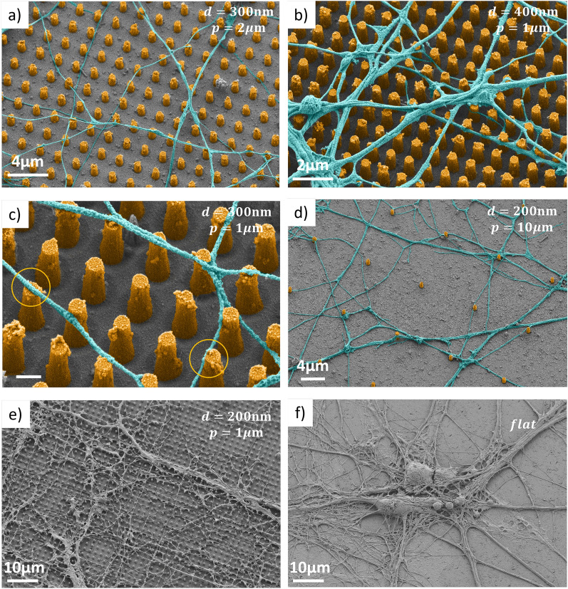
Monitoring neuronal activity with simultaneously high spatial and temporal resolution in living cell cultures is crucial to advance understanding of the development and functioning of our brain, and to gain further insights in the origin of brain disorders. While it has been demonstrated that the quantum sensing capabilities of nitrogen-vacancy (NV) centers in diamond allow real time detection of action potentials from large neurons in marine invertebrates, quantum monitoring of mammalian neurons (presenting much smaller dimensions and thus producing much lower signal and requiring higher spatial resolution) has hitherto remained elusive. In this context, diamond nanostructuring can offer the opportunity to boost the diamond platform sensitivity to the required level. However, a comprehensive analysis of the impact of a nanostructured diamond surface on the neuronal viability and growth was lacking. Here, we pattern a single crystal diamond surface with large-scale nanopillar arrays and we successfully demonstrate growth of a network of living and functional primary mouse hippocampal neurons on it. Our study on geometrical parameters reveals preferential growth along the nanopillar grid axes with excellent physical contact between cell membrane and nanopillar apex. Our results suggest that neuron growth can be tailored on diamond nanopillars to realize a nanophotonic quantum sensing platform for wide-field and label-free neuronal activity recording with sub-cellular resolution.


Conflict of interest statement

The authors declare no competing interests.

Figures

Figure 1
Figure 1
(a) Design of 25 nanopillar arrays with different geometrical parameters on the same chip, each array covering an area of 400μm×400μm. They all present the same height h1μm. (b) Optical image of the diamond surface before plating the neurons. The different pillar densities and diameters yield different colors. Pillars with diameters d300 nm and/or pitch p2μm cannot be clearly distinguished.
Figure 2
Figure 2
(a–c) SEM images of adjacent diamond pillar square arrays. They reveal high homogeneity and smooth and clean diamond surface between the pillars. (b) SEM image of an individual diamond pillar, with a diameter of 200 nm and a height of 1 μm. As a result of the optimized etching process, the top is flat and the sidewalls exhibit high verticality.
Figure 3
Figure 3
(a) Photoluminescence xy-map at the diamond surface level (z=0). The pillars (d=500 nm and p=1μm) can be clearly distinguished as brighter spots. (b) Photoluminescence xz-map, demonstrating the pillar waveguiding effect. The dashed line represents the estimated surface level. (c) Schematic of the experimental configuration used for both PL and ODMR measurements. Figure not on scale. MW = Microwave. (d) Continuous-wave ODMR spectrum acquired from one pillar, in presence of a magnetic field (few mT). For better visibility only the lower half of the spectrum is reported. The other four resonances are symmetric with respect to the zero-field resonance at 2.87 GHz. (e) Continuous-wave ODMR spectrum acquired in between pillars, in the same experimental conditions as for the spectrum in (d). All the plots are normalized to the largest PL signal level measured on this area.
Figure 4
Figure 4
Axon-pillar relative positions. SEM images of primary hippocampal neurons plated on different diamond substrates, at magnification up to ×50k. The samples are tilted by 20 with respect to horizontal to better appreciate the three-dimensional morphology. In panels (a–e) the substrate is nanostructured with pillar arrays (height 1μm, pitch p and diameter d reported in each figure). In panel (f) the substrate is flat. Images (a–d) have been artificially colored for better visibility.
Figure 5
Figure 5
Preferential growth direction of primary hippocampal neurons neurites on nanopillar arrays. The first row (a–c) refers to a nanostructured substrate (d=300 nm, p=2μm), the second row (d–f) to a flat surface. (a,d) SEM images at ×4.5k magnification. (b,e) Neurites detected using NeuronJ. (c,f) Histograms reporting the axon direction with respect to one of the main axis of the grid, as illustrated in panel (a). Red lines mark the two perpendicular directions along nearest neighbors of the array. Each histogram is obtained analysing 15 images.
Figure 6
Figure 6
(a) Elicited action potentials from primary hippocampal neurons plated on the nanostructured diamond substrate. The curves are obtained by injecting discrete, incremental, depolarizing current pulses close to threshold. Action potentials present a clear overshoot and hyperpolarization phase. (b) Excitatory post synaptic potentials (EPSPs) produced at −65 mV membrane potential by spontaneous synaptic activation. In the inset a single ESPS peak is fit giving a time decay constant τ25 ms.
Figure 7
Figure 7
Fabrication process for large-scale diamond nanopillar arrays. (I) The single-crystal diamond substrate is cleaned, polished (see main text) and die-attached on a Si carrier wafer. (II) Sputtering of Ti hard mask (200 nm). (III) Spin-coating of HSQ negative resist (150 nm), electron beam lithography for pillars, and development in TMAH. (IV) Cl2-based reactive ion etching patterning of the Ti layer. (V) Highly directional O2-plasma etch of diamond substrate. (VI) Stripping of the hard mask in diluted hydrofluoric acid (HF).

References

    1. Adolphs R. The unsolved problems of neuroscience. Trends Cognit. Sci. 2015;19:173–175. doi: 10.1016/j.tics.2015.01.007. - DOI - PMC - PubMed
    1. Lee S-J, Lim H-S, Masliah E, Lee H-J. Protein aggregate spreading in neurodegenerative diseases: Problems and perspectives. Neurosci. Res. 2011;70:339–348. doi: 10.1016/j.neures.2011.05.008. - DOI - PMC - PubMed
    1. Neher E, Sakmann B. Single-channel currents recorded from membrane of denervated frog muscle fibres. Nature. 1976;260:799–802. doi: 10.1038/260799a0. - DOI - PubMed
    1. Hamill OP, Marty A, Neher E, Sakmann B, Sigworth FJ. Improved patch-clamp techniques for high-resolution current recording from cells and cell-free membrane patches. Pflügers Arch. 1981;391:85–100. doi: 10.1007/BF00656997. - DOI - PubMed
    1. Neher E, Sakmann B. The patch clamp technique. Sci. Am. 1992;266:44–51. doi: 10.1038/scientificamerican0392-44. - DOI - PubMed

Publication types