

A study on the differential of solid lung adenocarcinoma and tuberculous granuloma nodules in CT images by Radiomics machine learning
- PMID: 37041262
- PMCID: PMC10090156
- DOI: 10.1038/s41598-023-32979-6
A study on the differential of solid lung adenocarcinoma and tuberculous granuloma nodules in CT images by Radiomics machine learning
Abstract
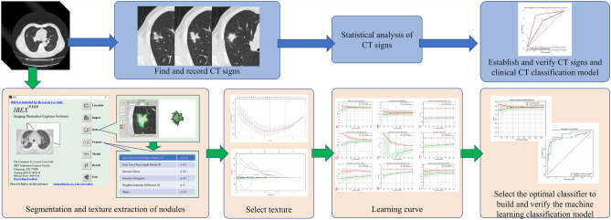
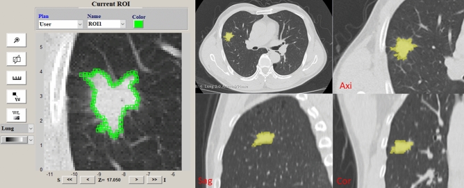
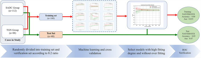
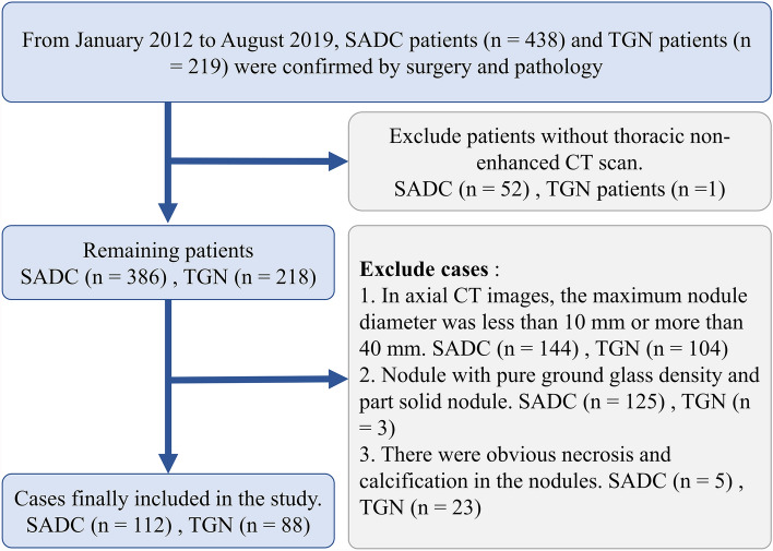
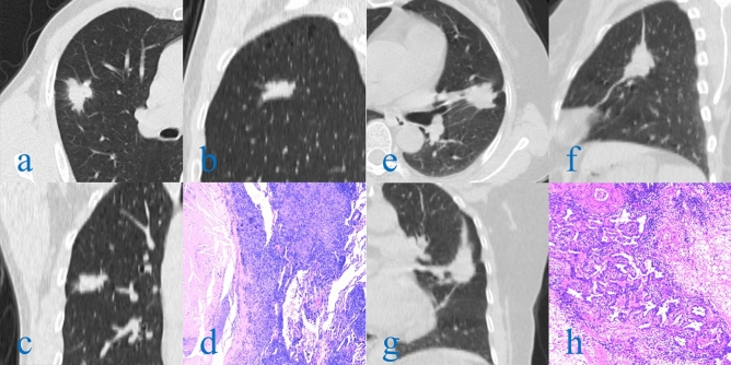
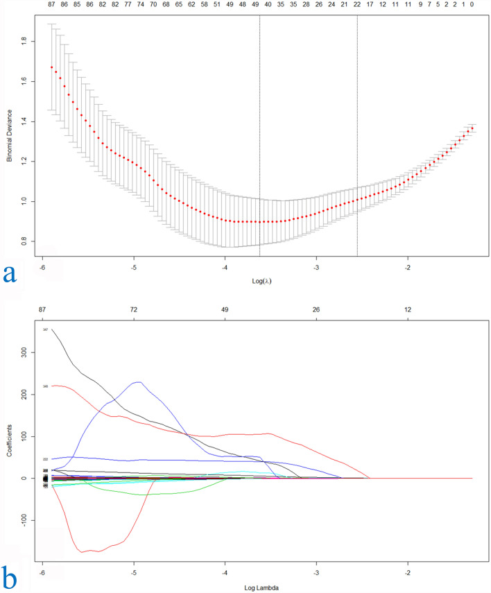
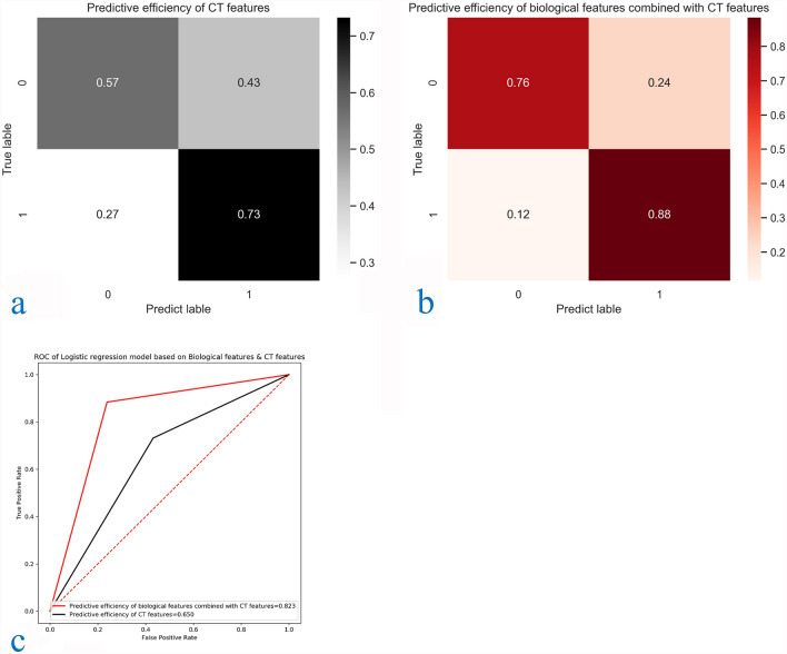
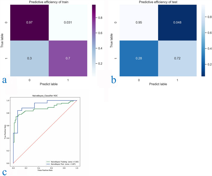
To study the classification efficiency of using texture feature machine learning method in distinguishing solid lung adenocarcinoma (SADC) and tuberculous granulomatous nodules (TGN) that appear as solid nodules (SN) in non-enhanced CT images. 200 patients with SADC and TGN who underwent thoracic non-enhanced CT examination from January 2012 to October 2019 were included in the study, 490 texture eigenvalues of 6 categories were extracted from the lesions in the non-enhanced CT images of these patients for machine learning, the classification prediction model is established by using relatively the best classifier selected according to the fitting degree of learning curve in the process of machine learning, and the effectiveness of the model was tested and verified. The logistic regression model of clinical data (including demographic data and CT parameters and CT signs of solitary nodules) was used for comparison. The prediction model of clinical data was established by logistic regression, and the classifier was established by machine learning of radiologic texture features. The area under the curve was 0.82 and 0.65 for the prediction model based on clinical CT and only CT parameters and CT signs, and 0.870 based on Radiomics characteristics. The machine learning prediction model developed by us can improve the differentiation efficiency of SADC and TGN with SN, and provide appropriate support for treatment decisions.
© 2023. The Author(s).
Conflict of interest statement
The authors declare no competing interests.
Figures
Similar articles
-
CT-based radiomics signature for differentiating solitary granulomatous nodules from solid lung adenocarcinoma.Lung Cancer. 2018 Nov;125:109-114. doi: 10.1016/j.lungcan.2018.09.013. Epub 2018 Sep 17. Lung Cancer. 2018. PMID: 30429007
-
Development and validation of a preoperative CT‑based radiomics nomogram to differentiate tuberculosis granulomas from lung adenocarcinomas: an external validation study.BMC Cancer. 2024 Jun 1;24(1):670. doi: 10.1186/s12885-024-12422-3. BMC Cancer. 2024. PMID: 38824514 Free PMC article.
-
Solitary solid pulmonary nodules: a CT-based deep learning nomogram helps differentiate tuberculosis granulomas from lung adenocarcinomas.Eur Radiol. 2020 Dec;30(12):6497-6507. doi: 10.1007/s00330-020-07024-z. Epub 2020 Jun 27. Eur Radiol. 2020. PMID: 32594210
-
CT-based radiomics and machine learning to predict spread through air space in lung adenocarcinoma.Eur Radiol. 2020 Jul;30(7):4050-4057. doi: 10.1007/s00330-020-06694-z. Epub 2020 Feb 28. Eur Radiol. 2020. PMID: 32112116
-
A combined non-enhanced CT radiomics and clinical variable machine learning model for differentiating benign and malignant sub-centimeter pulmonary solid nodules.Med Phys. 2023 May;50(5):2835-2843. doi: 10.1002/mp.16316. Epub 2023 Mar 2. Med Phys. 2023. PMID: 36810703
Cited by
-
Establishing predictive models for malignant and inflammatory pulmonary nodules using clinical data and CT imaging features.Quant Imaging Med Surg. 2025 Apr 1;15(4):2957-2970. doi: 10.21037/qims-24-2338. Epub 2025 Mar 17. Quant Imaging Med Surg. 2025. PMID: 40235774 Free PMC article.
-
Tumour-pleura relationship on computed tomography (CT) provides effective risk stratification for peripheral pulmonary nodules with Lung Imaging Reporting and Data System (Lung-RADS) score of 4X.Quant Imaging Med Surg. 2024 Oct 1;14(10):7138-7150. doi: 10.21037/qims-24-530. Epub 2024 Sep 21. Quant Imaging Med Surg. 2024. PMID: 39429614 Free PMC article.
-
Machine learning-driven imaging data for early prediction of lung toxicity in breast cancer radiotherapy.Sci Rep. 2025 May 27;15(1):18473. doi: 10.1038/s41598-025-02617-4. Sci Rep. 2025. PMID: 40425645 Free PMC article.
-
A spatio-temporal image analysis for growth of indeterminate pulmonary nodules detected by CT scan.Radiol Phys Technol. 2024 Mar;17(1):71-82. doi: 10.1007/s12194-023-00750-1. Epub 2023 Oct 27. Radiol Phys Technol. 2024. PMID: 37889460
References
-
- Lung cancer group RS. Chinese Medical Association Expert consensus on diagnosis and treatment of pulmonary nodules (2018 edition) Chin. J. Tubre Respir. Dis. 2018;40(10):9. doi: 10.3760/cma.j.issn.1001-0939.2018.10.004. - DOI
Publication types
MeSH terms
LinkOut - more resources
Full Text Sources
Medical
Miscellaneous
