

Stereology neuron counts correlate with deep learning estimates in the human hippocampal subregions
- PMID: 37041300
- PMCID: PMC10090178
- DOI: 10.1038/s41598-023-32903-y
Stereology neuron counts correlate with deep learning estimates in the human hippocampal subregions
Abstract
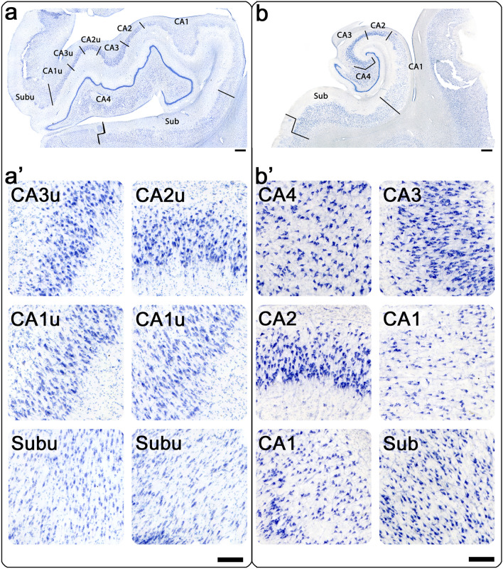
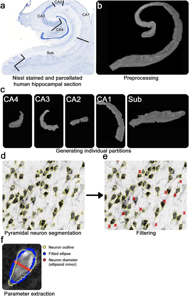
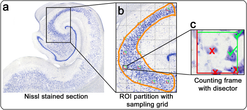
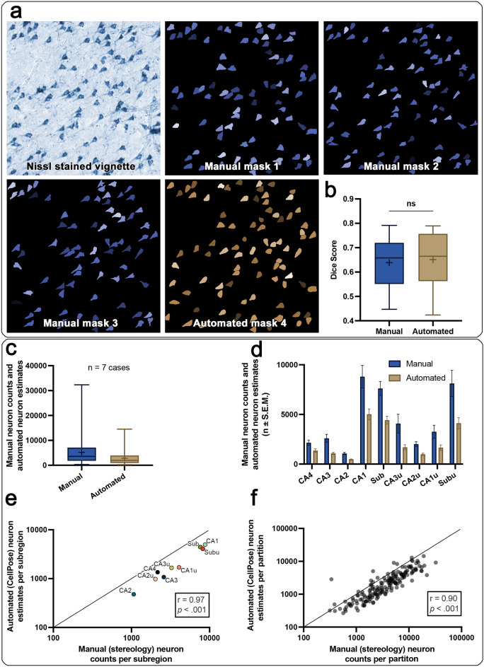
Hippocampal subregions differ in specialization and vulnerability to cell death. Neuron death and hippocampal atrophy have been a marker for the progression of Alzheimer's disease. Relatively few studies have examined neuronal loss in the human brain using stereology. We characterize an automated high-throughput deep learning pipeline to segment hippocampal pyramidal neurons, generate pyramidal neuron estimates within the human hippocampal subfields, and relate our results to stereology neuron counts. Based on seven cases and 168 partitions, we vet deep learning parameters to segment hippocampal pyramidal neurons from the background using the open-source CellPose algorithm, and show the automated removal of false-positive segmentations. There was no difference in Dice scores between neurons segmented by the deep learning pipeline and manual segmentations (Independent Samples t-Test: t(28) = 0.33, p = 0.742). Deep-learning neuron estimates strongly correlate with manual stereological counts per subregion (Spearman's correlation (n = 9): r(7) = 0.97, p < 0.001), and for each partition individually (Spearman's correlation (n = 168): r(166) = 0.90, p <0 .001). The high-throughput deep-learning pipeline provides validation to existing standards. This deep learning approach may benefit future studies in tracking baseline and resilient healthy aging to the earliest disease progression.
© 2023. The Author(s).
Conflict of interest statement
The authors declare no competing interests.
Figures
Similar articles
-
Multi-atlas segmentation of the whole hippocampus and subfields using multiple automatically generated templates.Neuroimage. 2014 Nov 1;101:494-512. doi: 10.1016/j.neuroimage.2014.04.054. Epub 2014 Apr 29. Neuroimage. 2014. PMID: 24784800
-
Automatic ground truth for deep learning stereology of immunostained neurons and microglia in mouse neocortex.J Chem Neuroanat. 2019 Jul;98:1-7. doi: 10.1016/j.jchemneu.2019.02.006. Epub 2019 Mar 2. J Chem Neuroanat. 2019. PMID: 30836126
-
Automated Cell Counts on Tissue Sections by Deep Learning and Unbiased Stereology.J Chem Neuroanat. 2019 Mar;96:94-101. doi: 10.1016/j.jchemneu.2018.12.010. Epub 2018 Dec 27. J Chem Neuroanat. 2019. PMID: 30594529
-
Intrinsic factors in the selective vulnerability of hippocampal pyramidal neurons.Prog Clin Biol Res. 1989;317:333-51. Prog Clin Biol Res. 1989. PMID: 2690106 Review.
-
Hippocampal subfields at ultra high field MRI: An overview of segmentation and measurement methods.Hippocampus. 2017 May;27(5):481-494. doi: 10.1002/hipo.22717. Epub 2017 Feb 23. Hippocampus. 2017. PMID: 28188659 Free PMC article. Review.
Cited by
-
Midbrain cytotoxic T cells as a distinct neuropathological feature of progressive supranuclear palsy.Brain. 2025 Aug 1;148(8):2650-2657. doi: 10.1093/brain/awaf135. Brain. 2025. PMID: 40233181 Free PMC article.
-
Between neurons and networks: investigating mesoscale brain connectivity in neurological and psychiatric disorders.Front Neurosci. 2024 Feb 20;18:1340345. doi: 10.3389/fnins.2024.1340345. eCollection 2024. Front Neurosci. 2024. PMID: 38445254 Free PMC article. Review.
-
Deep learning-based localization algorithms on fluorescence human brain 3D reconstruction: a comparative study using stereology as a reference.Sci Rep. 2024 Jun 25;14(1):14629. doi: 10.1038/s41598-024-65092-3. Sci Rep. 2024. PMID: 38918523 Free PMC article.
-
High-throughput digital quantification of Alzheimer disease pathology and associated infrastructure in large autopsy studies.J Neuropathol Exp Neurol. 2023 Nov 20;82(12):976-986. doi: 10.1093/jnen/nlad086. J Neuropathol Exp Neurol. 2023. PMID: 37944065 Free PMC article.
-
The prosubiculum in the human hippocampus: A rostrocaudal, feature-driven, and systematic approach.J Comp Neurol. 2024 Mar;532(3):e25604. doi: 10.1002/cne.25604. J Comp Neurol. 2024. PMID: 38477395 Free PMC article.
References
Publication types
MeSH terms
Grants and funding
LinkOut - more resources
Full Text Sources
