

Cell death in the lateral geniculate nucleus, and its possible relationship with nicotinic receptors and sudden infant death syndrome (SIDS)
- PMID: 37041306
- PMCID: PMC10224858
- DOI: 10.1007/s12035-023-03332-9
Cell death in the lateral geniculate nucleus, and its possible relationship with nicotinic receptors and sudden infant death syndrome (SIDS)
Abstract
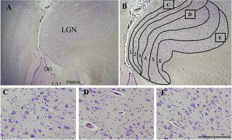
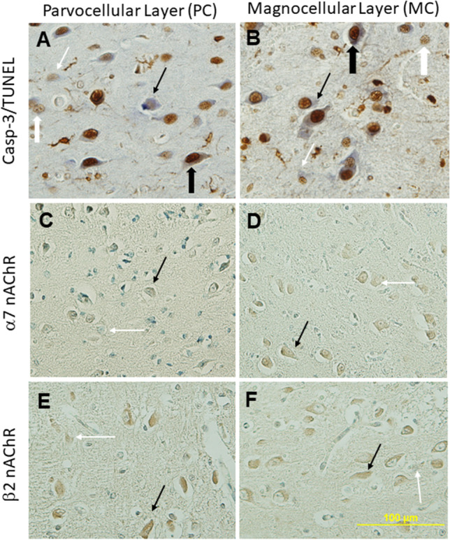
The role of the lateral geniculate nucleus (LGN) in vision has been extensively studied, yet its extraretinal capacities are still being investigated, including its role in arousal from sleep. The β2 nicotinic acetylcholine receptor (nAChR) subunit is involved in the laminal organisation of the LGN with magnocellular (MC) and parvocellular (PC) neurons. Sudden infant death syndrome (SIDS) occurs during a sleep period and, neuropathologically, is associated with increased neuronal cell death and altered nAChRs. A recent qualitative pilot study from our group implicates the possibility of increased neuronal death/apoptosis in the SIDS LGN. The present study used quantitative analysis to report the baseline expression of apoptotic and nAChR subunits α7 and β2 in the PC and MC layers of the LGN, to determine correlations amongst these markers within layers and across layers, and to evaluate changes in the expression of these markers in the LGN of SIDS infants, along with associations with SIDS risk factors, such as age, sex, cigarette smoke exposure, bed-sharing, and presence of an upper respiratory tract infection (URTI). Tissue was immunohistochemically stained for cell death markers of active caspase-3 (Casp-3) and TUNEL, and for the α7 and β2 nAChR subunits. Amongst 43 cases of sudden and unexpected deaths in infancy (SUDI), classifications included explained deaths (eSUDI, n = 9), SIDS I (n = 5) and SIDS II (n = 29). Results indicated a strong correlation of the apoptotic markers and β2 nAChR subunit between the LGN layers, but not across the markers within the layers. Amongst the diagnostic groups, compared to eSUDI, the SIDS II cases had decreased Casp-3 expression while β2 nAChR expression was increased in both PC and MC layers. Amongst the SIDS risk factors, URTI and bed-sharing were associated with changes in neuronal death but not in the α7 and β2 markers. In conclusion, our findings do not support a role for the α7 and β2 nAChRs in apoptotic regulation of the LGN layers during infancy. However, for SIDS victims, an inverse correlation between the changes for markers of apoptosis and the β2 nAChR subunit expression suggests altered LGN function.
Keywords: Acetylcholine; Apoptosis; Cholinergic; LGN; SUDI; Sleep.
© 2023. The Author(s).
Conflict of interest statement
The authors declare they have no conflict of interest.
Figures
Similar articles
-
The α7 and β2 nicotinic acetylcholine receptor subunits regulate apoptosis in the infant hippocampus, and in sudden infant death syndrome (SIDS).Apoptosis. 2020 Aug;25(7-8):574-589. doi: 10.1007/s10495-020-01618-0. Apoptosis. 2020. PMID: 32577853
-
The α3 and α4 nicotinic acetylcholine receptor (nAChR) subunits in the brainstem medulla of sudden infant death syndrome (SIDS).Neurobiol Dis. 2019 May;125:23-30. doi: 10.1016/j.nbd.2019.01.010. Epub 2019 Jan 18. Neurobiol Dis. 2019. PMID: 30665006
-
Effects of cigarette smoke exposure on nicotinic acetylcholine receptor subunits α7 and β2 in the sudden infant death syndrome (SIDS) brainstem.Toxicol Appl Pharmacol. 2011 Dec 15;257(3):396-404. doi: 10.1016/j.taap.2011.09.023. Epub 2011 Oct 6. Toxicol Appl Pharmacol. 2011. PMID: 22000980
-
Diverse strategies targeting α7 homomeric and α6β2* heteromeric nicotinic acetylcholine receptors for smoking cessation.Ann N Y Acad Sci. 2014 Oct;1327(1):27-45. doi: 10.1111/nyas.12421. Epub 2014 Apr 14. Ann N Y Acad Sci. 2014. PMID: 24730978 Free PMC article. Review.
-
The triple risk hypotheses in sudden infant death syndrome.Pediatrics. 2002 Nov;110(5):e64. doi: 10.1542/peds.110.5.e64. Pediatrics. 2002. PMID: 12415070 Review.
Cited by
-
Metabolic acidosis and sudden infant death syndrome: overlooked data provides insight into SIDS pathogenesis.World J Pediatr. 2025 Jan;21(1):29-40. doi: 10.1007/s12519-024-00860-9. Epub 2024 Dec 10. World J Pediatr. 2025. PMID: 39656413 Free PMC article. Review.
-
Academy of Breastfeeding Medicine Clinical Protocol #21: Breastfeeding in the Setting of Substance Use and Substance Use Disorder (Revised 2023).Breastfeed Med. 2023 Oct;18(10):715-733. doi: 10.1089/bfm.2023.29256.abm. Breastfeed Med. 2023. PMID: 37856658 Free PMC article.
-
Prenatal and postnatal factors associated with sudden infant death syndrome: an umbrella review of meta-analyses.World J Pediatr. 2024 May;20(5):451-460. doi: 10.1007/s12519-024-00806-1. Epub 2024 Apr 29. World J Pediatr. 2024. PMID: 38684567
References
-
- Skalicky SE. Ocular and Visual Physiology. Springer Singapore, Singapore; 2016. The Lateral Geniculate Nucleus; pp. 201–206.
-
- Martinovic J. Magno-, Parvo-, Koniocellular Pathways. Springer New York, New York, NY; 2016. pp. 893–896.
MeSH terms
Substances
LinkOut - more resources
Full Text Sources
Medical
Research Materials
