Breast-Conserving Surgery Margin Guidance Using Micro-Computed Tomography: Challenges When Imaging Radiodense Resection Specimens
- PMID: 37041429
- PMCID: PMC10600965
- DOI: 10.1245/s10434-023-13364-z
Breast-Conserving Surgery Margin Guidance Using Micro-Computed Tomography: Challenges When Imaging Radiodense Resection Specimens
Abstract
Background: Breast-conserving surgery (BCS) is an integral component of early-stage breast cancer treatment, but costly reexcision procedures are common due to the high prevalence of cancer-positive margins on primary resections. A need exists to develop and evaluate improved methods of margin assessment to detect positive margins intraoperatively.
Methods: A prospective trial was conducted through which micro-computed tomography (micro-CT) with radiological interpretation by three independent readers was evaluated for BCS margin assessment. Results were compared to standard-of-care intraoperative margin assessment (i.e., specimen palpation and radiography [abbreviated SIA]) for detecting cancer-positive margins.
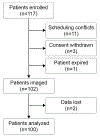
Results: Six hundred margins from 100 patients were analyzed. Twenty-one margins in 14 patients were pathologically positive. On analysis at the specimen-level, SIA yielded a sensitivity, specificity, positive predictive value (PPV), and negative predictive value (NPV) of 42.9%, 76.7%, 23.1%, and 89.2%, respectively. SIA correctly identified six of 14 margin-positive cases with a 23.5% false positive rate (FPR). Micro-CT readers achieved sensitivity, specificity, PPV, and NPV ranges of 35.7-50.0%, 55.8-68.6%, 15.6-15.8%, and 86.8-87.3%, respectively. Micro-CT readers correctly identified five to seven of 14 margin-positive cases with an FPR range of 31.4-44.2%. If micro-CT scanning had been combined with SIA, up to three additional margin-positive specimens would have been identified.
Discussion: Micro-CT identified a similar proportion of margin-positive cases as standard specimen palpation and radiography, but due to difficulty distinguishing between radiodense fibroglandular tissue and cancer, resulted in a higher proportion of false positive margin assessments.
© 2023. Society of Surgical Oncology.
Conflict of interest statement
Figures


Similar articles
-
Micro-computed tomography enables rapid surgical margin assessment during breast conserving surgery (BCS): correlation of whole BCS micro-CT readings to final histopathology.Breast Cancer Res Treat. 2018 Dec;172(3):587-595. doi: 10.1007/s10549-018-4951-3. Epub 2018 Sep 17. Breast Cancer Res Treat. 2018. PMID: 30225621 Free PMC article.
-
Feasibility of Micro-Computed Tomography Imaging for Direct Assessment of Surgical Resection Margins During Breast-Conserving Surgery.J Surg Res. 2019 Sep;241:160-169. doi: 10.1016/j.jss.2019.03.029. Epub 2019 Apr 23. J Surg Res. 2019. PMID: 31026794
-
Micro-computed tomography (micro-CT) for intraoperative surgical margin assessment of breast cancer: A feasibility study in breast conserving surgery.Eur J Surg Oncol. 2018 Nov;44(11):1708-1713. doi: 10.1016/j.ejso.2018.06.022. Epub 2018 Jul 3. Eur J Surg Oncol. 2018. PMID: 30005963
-
Definition and Management of Positive Margins for Invasive Breast Cancer.Surg Clin North Am. 2018 Aug;98(4):761-771. doi: 10.1016/j.suc.2018.03.008. Epub 2018 Apr 24. Surg Clin North Am. 2018. PMID: 30005772 Review.
-
Close/positive margins after breast-conserving therapy: additional resection or no resection?Breast. 2013 Aug;22 Suppl 2:S115-7. doi: 10.1016/j.breast.2013.07.022. Breast. 2013. PMID: 24074771 Review.
Cited by
-
The Value of Micro-CT in the Diagnosis of Lung Carcinoma: A Radio-Histopathological Perspective.Diagnostics (Basel). 2023 Oct 20;13(20):3262. doi: 10.3390/diagnostics13203262. Diagnostics (Basel). 2023. PMID: 37892083 Free PMC article. Review.
-
Assessment of surgical precision and safety in oncoplastic breast-conserving surgery for breast cancer under the guidance of imaging.BMC Surg. 2025 Aug 31;25(1):403. doi: 10.1186/s12893-025-03130-1. BMC Surg. 2025. PMID: 40887662 Free PMC article.
-
Aptamer-based NIR II imaging for breast cancer surgical resection.Npj Imaging. 2025 Aug 19;3(1):38. doi: 10.1038/s44303-025-00095-x. Npj Imaging. 2025. PMID: 40830213 Free PMC article.
References
-
- American Cancer Society. Breast Cancer Facts and Figures 2019–2020. Inc.: American Cancer Society; 2019.
-
- Schnitt SJ, Abner A, Gelman R, et al. The relationship between microscopic margins of resection and the risk of local recurrence in patients with breast cancer treated with breast-conserving surgery and radiation therapy. Cancer. 1994;74(6):1746–51. 10.1002/1097-0142(19940915)74:6<1746::aid-cncr2820740617>3.0.co;2-y. - DOI - PubMed
MeSH terms
Grants and funding
LinkOut - more resources
Full Text Sources
Medical

