

Coronary computed tomography angiography study on the relationship between the Ramus Intermedius and Atherosclerosis in the bifurcation of the left main coronary artery
- PMID: 37041479
- PMCID: PMC10091592
- DOI: 10.1186/s12880-023-01009-2
Coronary computed tomography angiography study on the relationship between the Ramus Intermedius and Atherosclerosis in the bifurcation of the left main coronary artery
Abstract
Objective: This study aimed to explore the relationship between the ramus intermedius (RI) and atherosclerosis in the bifurcation of the left coronary artery (LCA).
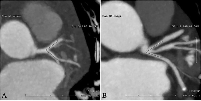
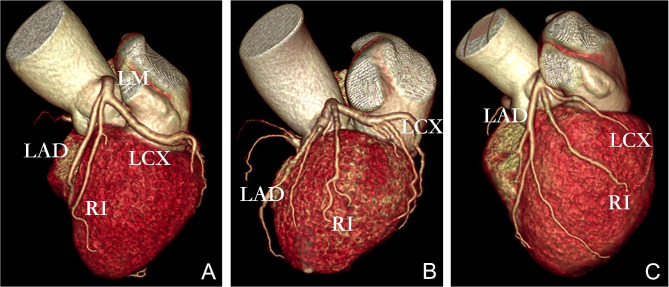
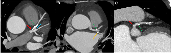
Methods: Screening patients who underwent CCTA from January to September 2021, 100 patients with RI (RI group) and 100 patients without RI (no-RI group) were randomly enrolled, Evaluation of RI distribution characteristics and left main coronary artery(LM),Left anterior descending branch(LAD),left circumflex branch(LCX) proximal segment plaque distribution, measurement of LAD-LCX bifurcation angle(∠LAD-LCX),Comparison of the three distribution characteristics with the incidence of plaques in the left main trunk bifurcation area (LM, LAD, LCX) between groups and within the RI group.
Results: The difference in the incidence of plaques in the proximal LCX and the LM between the RI group and the no-RI group were not statistically significant (P > 0.05). The incidence of plaques in the proximal LAD in the RI group was significantly higher than that in the non-RI group (77% versus 53%, P < 0.05). However, there was no statistically significant difference between the two groups after PSM. A univariate logistic regression analysis revealed that an RI was a risk factor for plaque formation in the proximal LAD (P < 0.001), and a multivariate logistic regression analysis revealed that an RI was not an independent risk factor for plaque formation in the proximal LAD (P > 0.05). When compared within the RI group, the difference in the incidence of plaques in the proximal segment of LAD, the proximal segment of LCX, and the LM among the different distribution groups of RI was not statistically significant, respectively (P > 0.05).
Conclusion: RI is not an independent risk factor for atherosclerosis in the left coronary artery bifurcation zone, but it may indirectly increase the risk of atherosclerosis in the proximal segment of the LAD.
Keywords: Atherosclerosis; Bifurcation angle; Coronary computed tomography angiography; Ramus intermedius.
© 2023. The Author(s).
Conflict of interest statement
The authors declare that they have no competing interests.
Figures
References
-
- OPOLSKI M P, GRODECKI K, STARUCH A D, et al. Accuracy of RESOLVE score derived from coronary computed tomography versus visual angiography to predict side branch occlusion in percutaneous bifurcation intervention. J Cardiovasc Comput Tomogr. 2020;14(3):258–65. doi: 10.1016/j.jcct.2019.11.007. - DOI - PubMed
-
- CHEN X, DAI J, LIN J et al. Image-based morphometric studies of human coronary artery bifurcations with/without coronary artery disease.Comput Methods Biomech Biomed Engin, 2020:1–17. - PubMed
Publication types
MeSH terms
LinkOut - more resources
Full Text Sources
Medical
Research Materials
Miscellaneous
