

METTL3 enhances dentinogenesis differentiation of dental pulp stem cells via increasing GDF6 and STC1 mRNA stability
- PMID: 37041485
- PMCID: PMC10088233
- DOI: 10.1186/s12903-023-02836-z
METTL3 enhances dentinogenesis differentiation of dental pulp stem cells via increasing GDF6 and STC1 mRNA stability
Abstract
Background: The dentinogenesis differentiation of dental pulp stem cells (DPSCs) is controlled by the spatio-temporal expression of differentiation related genes. RNA N6-methyladenosine (m6A) methylation, one of the most abundant internal epigenetic modification in mRNA, influences various events in RNA processing, stem cell pluripotency and differentiation. Methyltransferase like 3 (METTL3), one of the essential regulators, involves in the process of dentin formation and root development, while mechanism of METTL3-mediated RNA m6A methylation in DPSC dentinogenesis differentiation is still unclear.
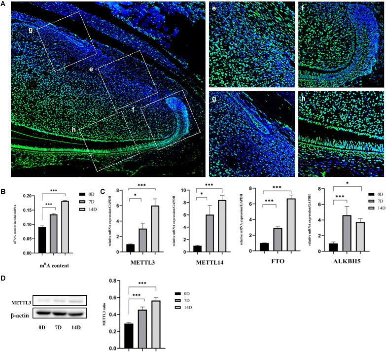
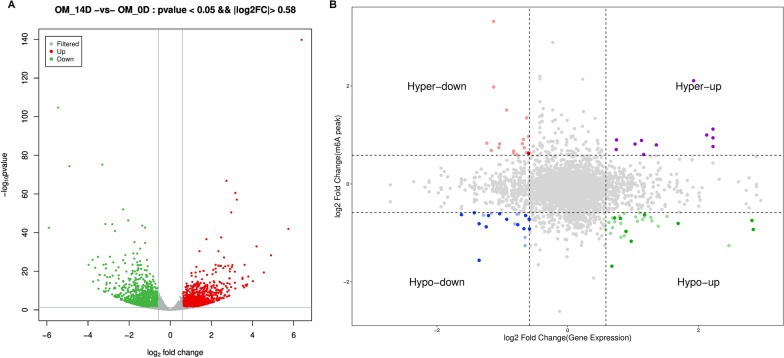
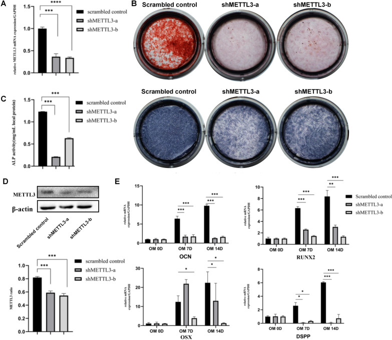
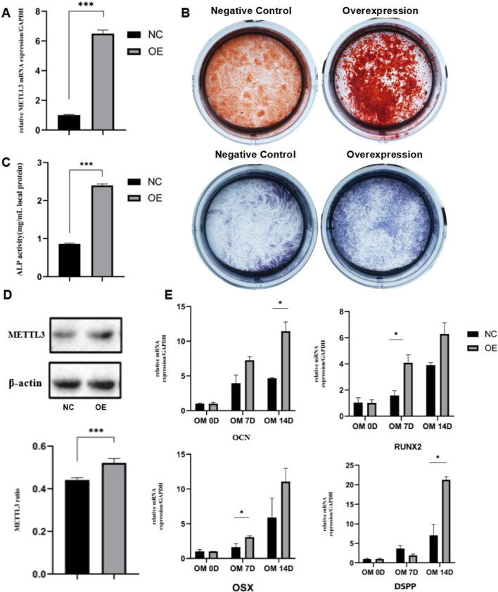
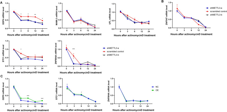
Methods: Immunofluorescence staining and MeRIP-seq were performed to establish m6A modification profile in dentinogenesis differentiation. Lentivirus were used to knockdown or overexpression of METTL3. The dentinogenesis differentiation was analyzed by alkaline phosphatase, alizarin red staining and real time RT-PCR. RNA stability assay was determined by actinomycin D. A direct pulp capping model was established with rat molars to reveal the role of METTL3 in tertiary dentin formation.
Results: Dynamic characteristics of RNA m6A methylation in dentinogenesis differentiation were demonstrated by MeRIP-seq. Methyltransferases (METTL3 and METTL14) and demethylases (FTO and ALKBH5) were gradually up-regulated during dentinogenesis process. Methyltransferase METTL3 was selected for further study. Knockdown of METTL3 impaired the DPSCs dentinogenesis differentiation, and overexpression of METTL3 promoted the differentiation. METTL3-mediated m6A regulated the mRNA stabiliy of GDF6 and STC1. Furthermore, overexpression of METTL3 promoted tertiary dentin formation in direct pulp capping model.
Conclusion: The modification of m6A showed dynamic characteristics during DPSCs dentinogenesis differentiation. METTL3-mediated m6A regulated in dentinogenesis differentiation through affecting the mRNA stability of GDF6 and STC1. METTL3 overexpression promoted tertiary dentin formation in vitro, suggesting its promising application in vital pulp therapy (VPT).
Keywords: Dentinogenesis; Epigenesis; METTL3; Mesenchymal stem cells; RNA stability.
© 2023. The Author(s).
Conflict of interest statement
All authors state that they have no potential competing interests.
Figures


Similar articles
-
METTL3 promotes immature dental pulp stem cells-induced angiogenesis by regulating ETS1 mRNA stability in an m6A-HuR-dependent manner.Odontology. 2025 Jan;113(1):305-317. doi: 10.1007/s10266-024-00977-3. Epub 2024 Jul 5. Odontology. 2025. PMID: 38969870
-
Stage-specific requirement for METTL3-dependent m6A modification during dental pulp stem cell differentiation.J Transl Med. 2022 Dec 16;20(1):605. doi: 10.1186/s12967-022-03814-9. J Transl Med. 2022. PMID: 36527141 Free PMC article.
-
METTL3-mediated m6A modification regulates cell cycle progression of dental pulp stem cells.Stem Cell Res Ther. 2021 Mar 1;12(1):159. doi: 10.1186/s13287-021-02223-x. Stem Cell Res Ther. 2021. PMID: 33648590 Free PMC article.
-
METTL3 plays a crucial function in multiple biological processes.Acta Histochem. 2022 Aug;124(6):151916. doi: 10.1016/j.acthis.2022.151916. Epub 2022 Jun 22. Acta Histochem. 2022. PMID: 35752056 Review.
-
N (6)-Methyladenosine (m(6)A) Methylation in mRNA with A Dynamic and Reversible Epigenetic Modification.Mol Biotechnol. 2016 Jul;58(7):450-9. doi: 10.1007/s12033-016-9947-9. Mol Biotechnol. 2016. PMID: 27179969 Review.
Cited by
-
METTL3 promotes immature dental pulp stem cells-induced angiogenesis by regulating ETS1 mRNA stability in an m6A-HuR-dependent manner.Odontology. 2025 Jan;113(1):305-317. doi: 10.1007/s10266-024-00977-3. Epub 2024 Jul 5. Odontology. 2025. PMID: 38969870
-
m6A-Modified GATA2 Enhances Odontogenic Differentiation in Stem Cells from the Apical Papilla.Int J Mol Sci. 2025 Mar 24;26(7):2920. doi: 10.3390/ijms26072920. Int J Mol Sci. 2025. PMID: 40243520 Free PMC article.
-
Epigenetic control of dental stem cells: progress and prospects in multidirectional differentiation.Epigenetics Chromatin. 2024 Dec 3;17(1):37. doi: 10.1186/s13072-024-00563-5. Epigenetics Chromatin. 2024. PMID: 39623487 Free PMC article. Review.
-
METTL3 Promotes Osteogenic Differentiation of Human Periodontal Ligament Stem Cells through IGF2BP1-Mediated Regulation of Runx2 Stability.Int J Med Sci. 2024 Feb 4;21(4):664-673. doi: 10.7150/ijms.90485. eCollection 2024. Int J Med Sci. 2024. PMID: 38464837 Free PMC article.
-
Dexmedetomidine plays a protective role in sepsis-associated myocardial injury by repressing PRMT5-mediated ferroptosis.Toxicol Res (Camb). 2025 Feb 2;14(1):tfaf010. doi: 10.1093/toxres/tfaf010. eCollection 2025 Feb. Toxicol Res (Camb). 2025. PMID: 39902345
References
Publication types
MeSH terms
Substances
LinkOut - more resources
Full Text Sources
