. 2023 Apr 11;21(1):36.
doi: 10.1186/s12958-023-01084-8.

RA-RAR signaling promotes mouse vaginal opening through increasing β-catenin expression and vaginal epithelial cell apoptosis

Affiliations

RA-RAR signaling promotes mouse vaginal opening through increasing β-catenin expression and vaginal epithelial cell apoptosis

Nana Zheng et al. Reprod Biol Endocrinol. .

Abstract

Background: Retinoic acid (RA) plays important role in the maintenance and differentiation of the Müllerian ducts during the embryonic stage via RA receptors (RARs). However, the function and mechanism of RA-RAR signaling in the vaginal opening are unknown.

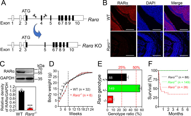
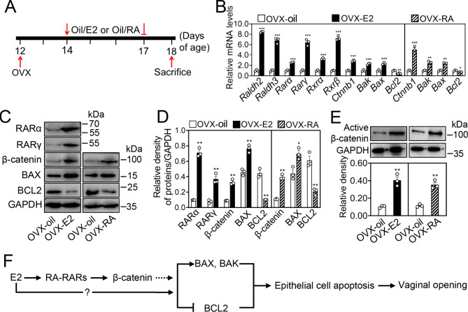
Method: We used the Rarα knockout mouse model and the wild-type ovariectomized mouse models with subcutaneous injection of RA (2.5 mg/kg) or E2 (0.1 µg/kg) to study the role and mechanism of RA-RAR signaling on the vaginal opening. The effects of Rarα deletion on Ctnnb1 mRNA levels and cell apoptosis in the vaginas were analyzed by real-time PCR and immunofluorescence, respectively. The effects of RA on the expression of β-catenin and apoptosis in the vaginas were analyzed by real-time PCR and western blotting. The effects of E2 on RA signaling molecules were analyzed by real-time PCR and western blotting.

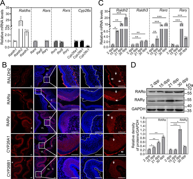
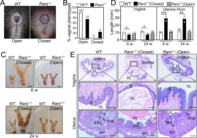
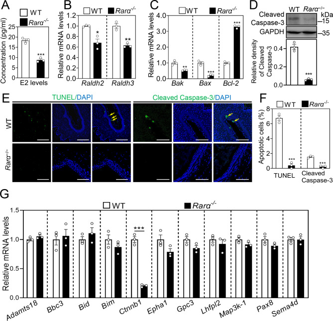
Results: RA signaling molecules were expressed in vaginal epithelial cells, and the mRNA and/or protein levels of RALDH2, RALDH3, RARα and RARγ reached a peak at the time of vaginal opening. The deletion of Rarα resulted in 25.0% of females infertility due to vaginal closure, in which the mRNA (Ctnnb1, Bak and Bax) and protein (Cleaved Caspase-3) levels were significantly decreased, and Bcl2 mRNA levels were significantly increased in the vaginas. The percentage of vaginal epithelium with TUNEL- and Cleaved Caspase-3-positive signals were also significantly decreased in Rarα-/- females with vaginal closure. Furthermore, RA supplementation of ovariectomized wild-type (WT) females significantly increased the expression of β-catenin, active β-catenin, BAK and BAX, and significantly decreased BCL2 expression in the vaginas. Thus, the deletion of Rarα prevents vaginal opening by reducing the vaginal β-catenin expression and epithelial cell apoptosis. The deletion of Rarα also resulted in significant decreases in serum estradiol (E2) and vagina Raldh2/3 mRNA levels. E2 supplementation of ovariectomized WT females significantly increased the expression of RA signaling molecules in the vaginas, suggesting that the up-regulation of RA signaling molecules in the vaginas is dependent on E2 stimulation.

Conclusion: Taken together, we propose that RA-RAR signaling in the vaginas promotes vaginal opening through increasing β-catenin expression and vaginal epithelial cell apoptosis.

Keywords: Apoptosis; Mouse; Retinoic acid receptor; Vaginal opening; β-catenin.


Conflict of interest statement

The authors declare no conflict of interest in this paper.

Figures

Fig. 1
Fig. 1
The expression pattern of RA signaling molecules in the vaginas. (A) The mRNA levels of Raldhs, Rars, Rxrs and Cyp26s in mouse vaginas at 5 weeks. The mRNA values of Raldh1, Rarα, Rxrα and Cyp26a1 were set as 1, and those of other gens were normalized accordingly. (n = 3 independent experiments). Bars indicate the mean ± SEM. (B) Immunofluorescence staining for RALDH2, RARα, RARγ, CYP26A1 and CYP26B1 (red) in the vaginas at 5 weeks. The small white boxes indicate the enlarged areas as shown in the following images. Downwards arrows indicate epithelial cells of spinous layers, while leftwards and rightwards arrows indicate the epithelial cells of cornified layers and the stromal region near basement membrane layers, respectively. Asterisks (*) indicate endothelial cells. The nuclei were counterstained by DAPI (blue). The cells in the dashed white line box are vaginal epithelial cells. Scale bar, 100 μm. VL, vaginal lumen; E, epithelium. (C) The mRNA levels of Raldh2, Raldh3, Rarα and Rarγ in the vaginas at 2, 15, 21 and 30 dpp. The mRNA values of 2 dpp group were set as 1, and those of other groups were normalized accordingly. (n = 3 independent experiments). Bars indicate the mean ± SEM. *P < 0.05, **P < 0.01, ***P < 0.001 vs. 2 dpp group. (D) The protein levels of RARα and RARγ in the vaginas at 2, 15, 21 and 30 dpp. GAPDH was used as a loading control. (n = 3 independent experiments). Bars indicate the mean ± SEM. *P < 0.05, **P < 0.01 vs. 2 dpp group
Fig. 2
Fig. 2
The effects of Rarα deletion on growth and survival. (A) The strategy of Rarα gene knock out (KO) by disrupting its exon 4 using CRISPR/Cas9 gene editing technology. (B, C) The detection of Rarα knockout efficiency in adult mouse vaginas by immunofluorescence (B) and western blotting (C) The nuclei were counterstained by DAPI (blue). Scale bar, 100 μm. GAPDH was used as a loading control. (n = 3 independent experiments.). Bars indicate the mean ± SEM. ***P < 0.001 vs. the WT group. (D) Body weight of Rarα−/− female mice (n = 6) and their WT littermates (n = 32) from 2 to 24 weeks. Bars indicate the mean ± SEM. ***P < 0.001 vs. the WT group. (E) Frequencies of Rarα genotypes in offspring from 30 Rarα+/− mating pairs were determined. Columns represent the percentage of the genotypes Rarα+/+ (black), Rarα+/− (green), and Rarα−/−(red), and two dashed red lines indicate expected frequencies based on Mendelian inheritance. The pup number for each genotype is shown in the columns. (F) Survival curve of Rarα+/+ (black), Rarα+/− (green), and Rarα−/− (red) from the newborn to adulthood
Fig. 3
Fig. 3
The effect of Rarα deletion on vaginal opening. (A, B) Representative pictures of the normal vaginal opening (white circle) in WT females and the vaginal closure (black circle) in Rarα−/− females at 6 weeks (A), and the frequency of the closed vagina in Rarα−/− females (B). The absolute numbers of females are indicated in the columns. (C, D) Representative female reproductive tracts (C) and the length of vagina and uterine horn (D) in Rarα−/− females with vaginal open, Rarα−/− females with vaginal closure and WT females at 6 and 24 weeks. Scale bar, 10 mm. (n = 3 independent experiments). Bars indicate the mean ± SEM. *P < 0.05, ***P < 0.001 vs. the WT group. (E) PAS-stained cross-sections for vaginas and uterus of Rarα−/− females with vaginal open, Rarα−/− females with vaginal closure and WT females. The small black boxes indicate the enlarged areas as shown in the following images. The yellow, black and white brackets indicate the thickness of vaginal epithelium (E), endometrium (EN) and myometrium (MY), respectively. Arrow, endometrial glands (Engl). Scale bar, 200 μm. VL, vaginal lumen; UL, uterine lumen
Fig. 4
Fig. 4
The effect of Rarα deletion on vaginal epithelial cell apoptosis and Ctnnb1 mRNA levels. (A) The levels of serum estradiol (E2) in WT and Rarα−/− females at 30 dpp. (n = 3 independent experiments). Data are presented as mean ± SEM. ***P < 0.001 vs. the WT group. (B, C, D) The mRNA levels of Raldh2, Raldh3 (B), Bak, Bax and Bcl-2 (C) and the protein levels of Cleaved Caspase-3 (D) in the vaginas of Rarα−/− females with vaginal closure and WT females (non-estrus stage). The mRNA values of WT group were set as 1, and those of Rarα−/− group were normalized accordingly. GAPDH was used as a loading control. (n = 3 independent experiments). Data are presented as mean ± SEM. *P < 0.05, **P < 0.01, ***P < 0.001 vs. the WT group. (E, F) Immunofluorescence stain of TUNEL and Cleaved Caspase-3 (green) (E), and the percentage of vaginal epithelial cells with TUNEL- and Cleaved Caspase-3-positive signals (green) (F) in each section in the vaginas of Rarα−/− females with vaginal closure and WT females (non-estrus stage). The nuclei were counterstained by DAPI (blue). Yellow arrows, apoptotic cells. Scale bar, 100 μm. (n = 3 independent experiments. The representative images are shown). Data are presented as mean ± SEM. ***P < 0.001 vs. the WT group. (G) The mRNA levels of Adamts18, Bac3, Bid, Bim, Ctnnb1, Epha1, Gpc3, Lhfpl2, Map3k1, Pax8 and Sema4d in the vaginas of Rarα−/− females with vaginal closure and WT females. The mRNA values of WT group were set as 1, and those of Rarα−/− group were normalized accordingly. (n = 3 independent experiments). Data are presented as mean ± SEM. ***P < 0.001 vs. the WT group
Fig. 5
Fig. 5
The effect of E2 and RA treatment on vaginal epithelial cell apoptosis and Ctnnb1 mRNA levels in WT females. (A) The strategy of RA and E2 treatment in WT ovariectomized females. After ovariectomy (OVX), the females at 14 dpp received daily subcutaneous injections (s.c.) of E2 (0.1 µg/kg body weight. OVX-E2 group), RA (2.5 mg/kg body weight. OVX-RA group) or vehicle oil (OVX-oil group) until 17 dpp, and the females were sacrificed for experiments on vaginal tissue 24 h later. (B) The mRNA levels of Raldh2, Raldh3, Rarα, Rarγ, Rxrα, Rxrβ, Ctnnb1, Bak, Bax, and Bcl2 in the vaginas of OVX-E2 and OVX-oil groups, and the mRNA levels of Ctnnb1, Bak, Bax, and Bcl2 in the vaginas of OVX-RA and OVX-oil groups. The mRNA values of OVX-oil groups were set as 1, and those of other gens were normalized accordingly. (n = 3 independent experiments). Data are presented as mean ± SEM. *P < 0.05, **P < 0.01, ***P < 0.001 vs. the OVX-oil group. (C, D, E) The protein levels of RARα, RARγ, β-catenin, active β-catenin, BAK, BAX and BCL2 in the vaginas of OVX-E2 and OVX-oil groups, and the protein levels of β-catenin, active β-catenin, BAK, BAX and BCL2 in the vaginas of OVX-RA and OVX-oil groups. GAPDH was used as a loading control. (n = 3 independent experiments). Data are presented as mean ± SEM. *P < 0.05, **P < 0.01 vs. the OVX-oil group. (F) The proposed model for RA-RAR signaling in vaginal opening

Similar articles

References

    1. Zhao F, Zhou J, Li R, Dudley EA, Ye X. Novel function of LHFPL2 in female and male distal reproductive tract development. Sci Rep. 2016;6:23037. doi: 10.1038/srep23037. - DOI - PMC - PubMed
    1. Santana Gonzalez L, Rota IA, Artibani M, Morotti M, Hu Z, Wietek N, et al. Mechanistic drivers of Müllerian Duct Development and differentiation into the oviduct. Front Cell Dev Biol. 2021;9:605301. doi: 10.3389/fcell.2021.605301. - DOI - PMC - PubMed
    1. Robboy SJ, Kurita T, Baskin L, Cunha GR. New insights into human female reproductive tract development. Differentiation. 2017;97:9–22. doi: 10.1016/j.diff.2017.08.002. - DOI - PMC - PubMed
    1. Cai Y. Revisiting old vaginal topics: conversion of the Müllerian vagina and origin of the “sinus” vagina. Int J Dev Biol. 2009;53(7):925–34. doi: 10.1387/ijdb.082846yc. - DOI - PubMed
    1. Kurita T. Normal and abnormal epithelial differentiation in the female reproductive tract. Differentiation. 2011;82(3):117–26. doi: 10.1016/j.diff.2011.04.008. - DOI - PMC - PubMed