

A rare metastatic mesenteric malignant PEComa with TSC2 mutation treated with palliative surgical resection and nab-sirolimus: a case report
- PMID: 37041531
- PMCID: PMC10088294
- DOI: 10.1186/s13000-023-01323-x
A rare metastatic mesenteric malignant PEComa with TSC2 mutation treated with palliative surgical resection and nab-sirolimus: a case report
Abstract
Background: Malignant perivascular epithelioid cell tumors (PEComas) are exceedingly rare malignant mesenchymal neoplasms with characteristic morphological and immunohistochemical (IHC) patterns. However, some malignant PEComas are poorly differentiated with atypical histopathological features, making a definitive diagnosis difficult. PEComas are most commonly found in females and often show either TSC1 or TSC2 alterations, which result in the activation of the mTOR pathway, or TFE3 fusions. Given these molecular characteristics, mTOR inhibitors have recently been approved by the FDA in the treatment of malignant PEComas, particularly in those with TSC1/2 alterations. Therefore, molecular analyses may be helpful for both the diagnostic workup of and predicting response to mTOR inhibitors in cases of malignant PEComas.
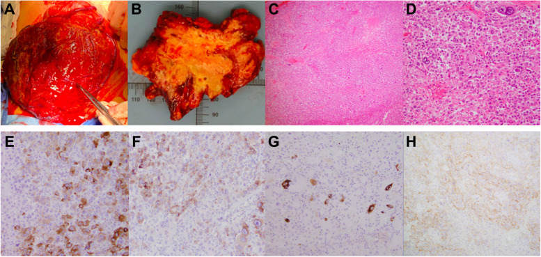
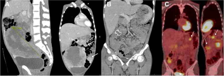
Case presentation: Here, we report a case of an aggressive, 23 cm mesenteric malignant PEComa with multiple peritoneal metastases in a young male patient. Pathological examination of the initial biopsy showed a malignant epithelioid neoplasm with high-grade morphology and atypical immunoprofile, which precluded a definitive diagnosis. Because of the patient's excessive transfusion requirements due to intra-tumoral hemorrhage, a palliative R2 resection was performed. Histopathological examination of the tumor revealed focal immunoreactivity for Melan-A, HMB-45, desmin, and CD117. Although a diagnosis of malignant PEComa was favored, other entities such as epithelioid gastrointestinal stromal tumor (GIST) or melanoma could not be definitively ruled out. Given the favored diagnosis, the patient was started on sirolimus, an mTOR inhibitor, rather than chemotherapy. Molecular analyses were performed and the tumor was found to harbor mutations in TP53 and TSC2, supporting a definitive diagnosis of malignant PEComa. The patient was then switched to nab-sirolimus, with initial stabilization of the disease.
Conclusions: This report details a multidisciplinary approach for the diagnosis and management of a highly aggressive, metastatic malignant PEComa in a young male patient. The basis for the treatment of malignant PEComas with the recently FDA-approved mTOR inhibitor, nab-sirolimus, is also reviewed. In summary, this case highlights the importance of molecular analysis, particularly TSC1/2 alterations, for both the definitive diagnosis of malignant PEComas and predicting their response to nab-sirolimus.
Keywords: Malignant PEComa; Nab-sirolimus; TP53; TSC2; mTOR.
© 2023. The Author(s).
Conflict of interest statement
The authors declare no competing interests.
Figures
Similar articles
-
Clinical activity of mTOR inhibition with sirolimus in malignant perivascular epithelioid cell tumors: targeting the pathogenic activation of mTORC1 in tumors.J Clin Oncol. 2010 Feb 10;28(5):835-40. doi: 10.1200/JCO.2009.25.2981. Epub 2010 Jan 4. J Clin Oncol. 2010. PMID: 20048174 Free PMC article.
-
Extrarenal perivascular epithelioid cell tumors (PEComas) respond to mTOR inhibition: clinical and molecular correlates.Int J Cancer. 2013 Apr 1;132(7):1711-7. doi: 10.1002/ijc.27800. Epub 2012 Sep 21. Int J Cancer. 2013. PMID: 22927055 Free PMC article.
-
Cutaneous perivascular epithelioid cell tumor (PEComa): case report and world literature review of clinical and molecular characteristics.Dermatol Online J. 2022 Jan 15;28(1). doi: 10.5070/D328157058. Dermatol Online J. 2022. PMID: 35499412
-
Treatment of Advanced Malignant Uterine Perivascular Epithelioid Cell Tumor with mTOR Inhibitors: Single-institution Experience and Review of the Literature.Anticancer Res. 2016 Nov;36(11):6161-6164. doi: 10.21873/anticanres.11208. Anticancer Res. 2016. PMID: 27793946 Review.
-
TFE3-associated perivascular epithelioid cell tumor with complete response to mTOR inhibitor therapy: report of first case and literature review.World J Surg Oncol. 2022 Mar 1;20(1):62. doi: 10.1186/s12957-021-02462-5. World J Surg Oncol. 2022. PMID: 35232443 Free PMC article. Review.
Cited by
-
Case report: Malignant epithelioid angiosarcoma in a Chinese female patient.Front Oncol. 2024 Aug 16;14:1398656. doi: 10.3389/fonc.2024.1398656. eCollection 2024. Front Oncol. 2024. PMID: 39220642 Free PMC article.
-
Case Report: Malignant perivascular epithelioid cell tumor with aggressive mediastinal invasion and pulmonary metastasis.Front Oncol. 2025 Aug 18;15:1551663. doi: 10.3389/fonc.2025.1551663. eCollection 2025. Front Oncol. 2025. PMID: 40900800 Free PMC article.
References
-
- Stacchiotti S, Frezza AM, Blay J-Y, Baldini EH, Bonvalot S, Bovée JVMG, et al. Ultra-rare sarcomas: A consensus paper from the Connective Tissue Oncology Society community of experts on the incidence threshold and the list of entities. Cancer. 2021;127:2934–2942. doi: 10.1002/cncr.33618. - DOI - PMC - PubMed
Publication types
MeSH terms
Substances
LinkOut - more resources
Full Text Sources
Medical
Research Materials
Miscellaneous
