Review
. 2023 Feb;16(2):347-356.
doi: 10.14202/vetworld.2023.347-356. Epub 2023 Feb 21.

A review on the evolution of methods for intestinal in vitro organ culture and its application in veterinary science

Affiliations
Review

A review on the evolution of methods for intestinal in vitro organ culture and its application in veterinary science

Barbara Ribeiro de Souza Cortez et al. Vet World. 2023 Feb.

Abstract

Different techniques have been reported in studies of intestinal in vitro organ culture (IVOC). A robust compilation of all available methods is lacking in the literature, making it difficult to choose a method that corresponds to the study's demands. In this review, readers can assess the most available methods, allowing them to evaluate which is more suitable for their purposes and requirements. A simplified view of culturing intestinal explants is presented, highlighting the approachability of IVOC. Relevant findings from diverse veterinarian studies, where explants played a major role, as well as the technique used in each, are described to illustrate its applications. Finally, the strengths and limitations of the innovative intestinal IVOC methods are discussed. This review provides a collection of methods for intestinal explant culture and their possible applications in veterinary research. In this way, it aims to broaden access to IVOC techniques and aid decision-making regarding the best suited for a study's purposes.

Keywords: enteropathogens; explants culture; intestinal pathogens; swine colon; ussing chamber.


Conflict of interest statement

The authors declare that they have no competing interests.

Figures
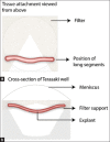
Figure-1
Figure-1
Schematic figure of explants under catenary culture [6]. (a) The tubular segments of the embryonic intestine have their edges fixed on a piece of paper filter cut in a “V” shape. (b) This setting is then placed inside a well of a Terasaki plate which is filled with culture media.
Figure-2
Figure-2
Diagram of Eastwood and Trier explant setting [2]. The apparatus was composed of a culture dish with an inner well, allowing the creation of a dead space filled with water-imbibed filter paper. A stainless steel grid was placed on top of the central well such that the culture medium underneath touched the bottom of the grid and provided nutrients through capillarity for the explant. The entire setting was placed inside an incubator with a 95% O2 and 5% CO2 atmosphere.
Figure-3
Figure-3
Diagram of explant setting [24]. The intestinal fragment striped from the serosal layer is attached with an agar block - the attachment matrix - which allows nutrients from the culture media around it to permeate and nourish the explant. Above the explant, an inoculation ring is attached with tissue glue then the desired stimulus can be placed and confined to the mucosal surface. The entire setting is set inside a hyperoxide chamber at 37°C.
Figure-4
Figure-4
Representation of porcine colon explant culture upgrade [1]. Each plate contains 6 explants independently cultured with polarized stimulus. The setting is composed of a 6-well culture plate, each with a layer of culture media underneath the cell strainer touching its bottom. One explant per well is placed with the mucosal side facing upwards and the serosal side facing downwards. Then an inoculation ring is attached with tissue glue, and the inoculum is placed inside. Several plates can be placed inside hyperoxide chambers and undergo culture at 37°C incubators.
Figure-5
Figure-5
Demonstration of segment model setup [29], (a) The intestinal segment is cleaned and placed on culture media. (b) The segment is cut open on its mesenteric border, placed (mucosa facing down) on a refrigerated platform, and cut with a biopsy punch. (c) The biopsied fragments are obtained and individually placed on 24-well culture plates containing culture media.
Figure-6
Figure-6
Representation of an Ussing chamber mounted with a fragment of the intestine [36]. The apical and basolateral sides have separated chambers that can receive different stimuli. A supply of oxygen and carbonic gas is constantly pumped into the media, which is recirculated inside each chamber. The device has an underlying electrical current used to measure trans-epithelial electrical resistance and detect ionic differences related to Na+/H+ and Cl-/HCO3- membrane exchangers.
Figure-7
Figure-7
Schematic drawing of an organoid [44]. The external layer is composed of all the different cell types of the intestinal mucosa, where the pale pink cells represent intestinal crypts and the blue cells represent the intestinal surface. The interior of the enteroid, in this case, has received a microbiota transplant, so its lumen contains a population of bacteria.
Figure-8
Figure-8
Schematic figure of Baydoun’s interphase microfluidic culture system [47]. Three explants are placed on top of the culture chamber, which is permeated by microchannels conducting the culture media from the inlets to the outlets and providing nutrients through the circular porous membrane. The individual culture apparatus is attached with polydimethylsiloxane, a silicon-like material that holds all elements in place.
Figure-9
Figure-9
Schematic image of an organ-on-a-chip model [48]. The image displays a culture of intestinal epithelium organized inside the central compartment of the chip. The surface and the basolateral side of the epithelium are separated, and each one receives a different substance (stimulus or culture media) in a constant flow.
Figure-10
Figure-10
Schematic representation of Yissachar’s setting [49]. (Left) A fragment of the intestine is plugged into an input and an output port responsible for allowing flow transit inside the lumen. The serosal side is also equipped with input and output plugs, which circulate the culture media at the intestine surroundings. (Right) Each set contains six above-described chambers, which are enbibed with a controlled O2/CO2 atmosphere.

Similar articles

References

    1. Costa M, Hill J, Dame M, Harding J.C.S. In vitro porcine colon culture. In:Methods in Molecular Biology. Humana Press Inc United States. (2018):185–195. - PubMed
    1. Randall K.J, Turton J, Foster J.R. Explant culture of gastrointestinal tissue:A review of methods and applications. Cell Biol. Toxicol. 2011;27(4):267–284. - PubMed
    1. Beck M, Thomson D. Some further research on the cultivation of tissues in vitro. J. R. Soc. Med. 1914;7:21–47.
    1. Fischer A. A pure strain of cartilage cells in vitro. J. Exp. Med. 1992;36(4):379–384. - PMC - PubMed
    1. Trowell O.A. A modified technique for organ culture in vitro. Exp. Cell. Res. 1954;6(1):246–248. - PubMed

LinkOut - more resources