. 2023 Apr 18;12(8):e028661.
doi: 10.1161/JAHA.122.028661. Epub 2023 Apr 12.

Ventricular Conduction Stability Noninvasively Identifies an Arrhythmic Substrate in Survivors of Idiopathic Ventricular Fibrillation

Affiliations

Ventricular Conduction Stability Noninvasively Identifies an Arrhythmic Substrate in Survivors of Idiopathic Ventricular Fibrillation

Ji-Jian Chow et al. J Am Heart Assoc. .

Abstract

Background Idiopathic ventricular fibrillation (VF) is a diagnosis of exclusion following normal cardiac investigations. We sought to determine if exercise-induced changes in electrical substrate could distinguish patient groups with various ventricular arrhythmic pathophysiological conditions and identify patients susceptible to VF. Methods and Results Computed tomography and exercise testing in patients wearing a 252-electrode vest were combined to determine ventricular conduction stability between rest and peak exercise, as previously described. Using ventricular conduction stability, conduction heterogeneity in idiopathic VF survivors (n=14) was compared with those surviving VF during acute ischemia with preserved ventricular function following full revascularization (n=10), patients with benign ventricular ectopy (n=11), and patients with normal hearts, no arrhythmic history, and negative Ajmaline challenge during Brugada family screening (Brugada syndrome relatives; n=11). Activation patterns in normal subjects (Brugada syndrome relatives) are preserved following exercise, with mean ventricular conduction stability of 99.2±0.9%. Increased heterogeneity of activation occurred in the idiopathic VF survivors (ventricular conduction stability: 96.9±2.3%) compared with the other groups combined (versus 98.8±1.6%; P=0.001). All groups demonstrated periodic variation in activation heterogeneity (frequency, 0.3-1 Hz), but magnitude was greater in idiopathic VF survivors than Brugada syndrome relatives or patients with ventricular ectopy (7.6±4.1%, 2.9±2.9%, and 2.8±1.2%, respectively). The cause of this periodicity is unknown and was not replicable by introducing exercise-induced noise at comparable frequencies. Conclusions In normal subjects, ventricular activation patterns change little with exercise. In contrast, patients with susceptibility to VF experience activation heterogeneity following exercise that requires further investigation as a testable manifestation of underlying myocardial abnormalities otherwise silent during routine testing.

Keywords: exercise testing; idiopathic ventricular fibrillation; myocardial ischemia; premature ventricular complex.


Figures
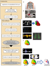
Figure 1
Figure 1. The V‐CoS score process.
A, The 252‐electrode ECG isensor vest is applied to the patient undergoing maximal Bruce protocol exercise. B, Recordings are made during 10 minutes of supine recovery, which is followed by noncontrast CT scan of chest. C, The CT scan is segmented into a 3D mesh. D, Cardiac cycles from the recording are selected for analysis. E, Body surface signals from the vest too noisy for analysis are identified and removed from the vest recording. F, Activation maps are drawn from the reconstructions. G, In the V‐CoS software, the QRS complex is identified. H, Reconstructed electrograms (compare with the body surface signals from E) are curated for excess artifact and removed from the analyzed map. The V‐CoS process compares the 2 activation maps and calculates the percentage score of electrograms with median‐relative LAT difference <10 ms. 3D indicates 3‐dimensional; CT, computed tomography; ECGi, electrocardiographic imaging; LAT, local activation time; and V‐CoS, ventricular conduction stability.
Figure 2
Figure 2. Methods of assigning a V‐CoS score to a patient.
A, The “single lowest value” method: following peak exercise, patient heart rate (blue trendline) and respiration rate (red trendline, correlating with chest movement) fall toward resting values. Cardiac cycles selected immediately after exercise and 3 stages of recovery undergo V‐CoS mapping (the 4 epicardial shells). The lowest calculated V‐CoS is defined as the patient score. In this method, cardiac cycles are selected at the user's discretion, and cardiac cycles are individually stripped of uninterpretable electrograms at user discretion. B, The V‐CoS matrix: this study uses a new method, where the mean of a 10‐by‐10 comparison matrix of consecutive cardiac cycles from the peak and rest phases is taken as the patient score. By standardizing the curation of electrograms between cardiac cycles and forcing the selection of consecutive beats, user‐induced variability is reduced, and temporal relationships are revealed. V‐CoS indicates ventricular conduction stability.
Figure 3
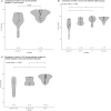
Figure 3. Mean V‐CoS scores compared between groups.
A, Comparison between survivors of idiopathic VF with a pooled control set of patients with ischemic VF with full recovery of left ventricular function and full revascularization, patients with benign ventricular ectopy, and unaffected BrS relatives. B, Comparison between survivors of idiopathic VF with control groups separated. C, Comparison between survivors of idiopathic VF experiencing either multiple or single episodes of ventricular arrhythmia and controls. Group comparisons were determined using the Kruskal‐Wallis test, and pairwise comparisons were determined using Dunn test with Benjamini‐Hochberg correction. For the numeric P values of all comparisons, please see Data S1. BrS indicates Brugada syndrome; IHD, ischemic heart disease; V‐CoS, ventricular conduction stability; VF, ventricular fibrillation; and VE, ventricular ectopy.
Figure 4
Figure 4. Eight example V‐CoS matrices: 2 from each condition group.
All patients demonstrate some variability in V‐CoS scores, but this seems to be greater in patients with idiopathic VF than those with recovered ischemic VF or the other controls. Beats immediately after peak exercise (consecutive beat x), pkex(x); beats from full recovery (consecutive beat ×), 10 minutes (×). BrS indicates Brugada syndrome; IHD, ischemic heart disease; V‐CoS, ventricular conduction stability; VE, ventricular ectopy; and VF, ventricular fibrillation.
Figure 5
Figure 5. Comparison of V‐CoS maps and edvlectrograms used to build the matrices.
Low V‐CoS scores from 2 matrices are selected, and the source epicardial activation maps are displayed. V‐CoS is the percentage of epicardial points with <10‐ms discordance in activation time. This discordance can be mapped: blue as advancement and red as delay of the test phase, with increasing color intensity representing the magnitude. A, Idiopathic VF survivor has clear differences between activation maps, resulting in greater discordance on the V‐CoS map and a low V‐CoS score. Example electrograms from regions “c” and “d” show that despite baseline noise, the software accurately finds the minimum dV/dt to define activation. The difference in activation times is stated as ∆AT. B, Control patient has much less discordance, and example electrograms from the most intensely colored regions “e” and “f” reveal the lower levels of discordance compared with the idiopathic VF survivor's map. dV/dt, change in voltage divided by change in time; V‐CoS indicates ventricular conduction stability; VE, ventricular ectopy; and VF, ventricular fibrillation.

References

    1. Haïssaguerre M, Duchateau J, Dubois R, Hocini M, Cheniti G, Sacher F, Lavergne T, Probst V, Surget E, Vigmond E, et al. Idiopathic ventricular fibrillation: role of purkinje system and microstructural myocardial abnormalities. JACC Clin Electrophysiol. 2020;6:591–608. doi: 10.1016/j.jacep.2020.03.010 - DOI - PMC - PubMed
    1. Franz MR, Schaefer J, Schöttler M, Seed WA, Noble MI. Electrical and mechanical restitution of the human heart at different rates of stimulation. Circ Res. 1983;53:815–822. doi: 10.1161/01.res.53.6.815 - DOI - PubMed
    1. Cluitmans MJM, Bear LR, Nguyên UC, van Rees B, Stoks J, Ter Bekke RMA, Mihl C, Heijman J, Lau KD, Vigmond E, et al. Noninvasive detection of spatiotemporal activation‐repolarization interactions that prime idiopathic ventricular fibrillation. Sci Transl Med. 2021;13:eabi9317. doi: 10.1126/scitranslmed.abi9317 - DOI - PubMed
    1. Leoni AL, Gavillet B, Rougier JS, Marionneau C, Probst V, Le Scouarnec S, Schott JJ, Demolombe S, Bruneval P, Huang CL, et al. Variable Na(v)1.5 protein expression from the wild‐type allele correlates with the penetrance of cardiac conduction disease in the Scn5a(+/−) mouse model. PLoS One. 2010;5:e9298. doi: 10.1371/journal.pone.0009298 - DOI - PMC - PubMed
    1. Brugada J, Pappone C, Berruezo A, Vicedomini G, Manguso F, Ciconte G, Giannelli L, Santinelli V. Brugada syndrome phenotype elimination by epicardial substrate ablation. Circ Arrhythm Electrophysiol. 2015;8:1373–1381. doi: 10.1161/CIRCEP.115.003220 - DOI - PubMed

Publication types

MeSH terms

Supplementary concepts

LinkOut - more resources