. 2023 Jun 1;210(11):1827-1836.
doi: 10.4049/jimmunol.2200694.

Identification and Characterization of Alveolar and Recruited Lung Macrophages during Acute Lung Inflammation

Affiliations

Identification and Characterization of Alveolar and Recruited Lung Macrophages during Acute Lung Inflammation

Wei Han et al. J Immunol. .

Abstract

To precisely identify mouse resident alveolar macrophages (AMs) and bone marrow (BM)-derived macrophages, we developed a technique to separately label AMs and BM-derived macrophages with a fluorescent lipophilic dye followed by FACS. We showed that this technique overcomes issues in cell identification related to dynamic shifts in cell surface markers that occurs during lung inflammation. We then used this approach to track macrophage subsets at different time points after intratracheal (i.t.) instillation of Escherichia coli LPS. By isolating BM-derived macrophages and AMs, we demonstrated that BM-derived macrophages were enriched in expression of genes in signal transduction and immune system activation pathways whereas resident AMs were enriched in cellular processes, such as lysosome/phagosome pathways, efferocytosis, and metabolic pathways related to fatty acids and peroxisomes. Taken together, these data indicate that more accurate identification of macrophage origin can result in improved understanding of differential phenotypes and functions between AMs and BM-derived macrophages in the lungs.


Figures

Figure 1.
Figure 1.
Analysis of leukocytes in the lungs by flow cytometry during LPS-induced lung inflammation/injury. A) Contour plots showing strategy for identifying major immune cell populations in the lungs, including CD45+ cells, neutrophils (Neu), and other myeloid cells at baseline (day 0) and 1, 3, 7, and 14 days following intratracheal (IT) injection of LPS (3μg/g). CD45+ cells were identified with CD45 BV650 Ab, neutrophils were defined by staining with Ly6G APC-Cy7 Ab, and other myeloid cells were defined as CD11b-APC and/or CD11c-PE-Cy7 positive cells. B) Quantification of CD45+ cells, neutrophils, and myeloid cells (excluding neutrophils) in the lungs at each time point. C) Gating strategy used for the identification of macrophage subsets in the lungs of untreated mice, including alveolar macrophages (AMs, SiglecF-PE positive), interstitial and bone marrow (BM)-derived macrophages (IM and BM-derived MΦ, F4/80-PE-Cy5 positive). D) Quantification of AMs and IM/BM-derived MΦ in the lungs at each time point. N=4 mice for each time point, Mean ± SEM.
Figure 2.
Figure 2.
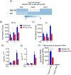
Identification of macrophages subsets in the lungs using fluorescent dyes. A) Schematic representation of intratracheal injection of PKH67 (green) dye and intra-tibial injection of PKH26 (red) dye to label lung resident cells and bone-marrow (BM)-derived cells along with the gating strategy to identify PKH67 and PKH26 positive cells by flow cytometry. B) Quantification of PKH67 and PKH26 positive cells in lungs. C) Gating strategy used to identify PKH67+ cells in lungs, including CD45+/− cells, alveolar macrophages (AMs), neutrophils (Neu), and other leukocytes. D) Quantification of PKH67+ cells in lungs. E) Gating strategy used to identify PKH26+ cells in lungs, including CD45+/− cells, monocytes/macrophages (Mono/Mac), neutrophils (Neu), and other leukocytes. F) Quantification of PKH26+ cells in lungs. N=5 mice per group, mean ± SEM.
Figure 3.
Figure 3.
Precision tracking of resident alveolar macrophages and bone marrow-derived macrophages during acute lung inflammation. A) Contour plots representing the gating strategy applied to the identification of PKH labeled resident alveolar macrophages (AM) (green) and bone marrow (BM)-derived macrophages (red). These plots are overlaid onto CD11b and CD11c gates to show expression of standard macrophage markers after IT LPS. The boxes outlined by dotted lines indicate the AM gate in untreated mice. B-C) Quantification of resident AMs and BM-derived macrophages in lungs based on PKH labeling. N=4–12 mice per time point, mean ± SEM, *=p<0.05 compared to untreated (day 0) controls.
Figure 4.
Figure 4.
Transcriptional analysis of resident alveolar macrophages and bone marrow-derived macrophages during acute inflammation. A) Volcano plots showing differentially expressed genes [log2(Fold-change) > 1 and adjusted p-value < 0.1] in resident AM versus BM-derived macrophages at day 1, 3 and 7 after IT LPS. B) Correlation analysis of differentially expressed genes (AMs enriched, blue; BM-derived Mφ enriched, red) between resident AMs and BM-derived macrophages at different time point after IT LPS (p < 0.00001 with Fisher’s exact test). N=3–5 samples per group.
Figure 5.
Figure 5.
Pathway enrichment analysis of differentially expressed genes shows increased inflammatory signaling in bone marrow-derived macrophage compared to resident alveolar macrophages. A) Number of enriched pathways in resident alveolar macrophages (AMs) (blue) and bone marrow (BM)-derived macrophages (red) at day 1, day 3 and day 7 after IT LPS). B) Selected KEGG pathways in specific categories enriched in AMs (blue) or BM-derived macrophages (red). Scale bar represents fold enrichment. N=3–5 samples per time point.
Figure 6.
Figure 6.
Differences in expression of NF-κB dependent genes in resident alveolar macrophages and bone marrow-derived macrophages after IT LPS. A) Heat map showing expression of transcripts regulated through the NF-κB pathway in resident alveolar macrophages (AMs) (blue) and bone marrow (BM)-derived macrophages (red) at day 1 after LPS treatment relative to AMs from untreated mice. Scale bar represents log2 fold-change compared to AMs from untreated mice (no LPS). B) Direct comparison of NF-κB target gene expression of AMs compared with BM-derived macrophages at day 1 after LPS treatment. C) RT-PCR and gene transcripts (normalized counts) for selected NF-κB dependent genes. N=4 samples per group. Mean ± SEM, *=p<0.05.
Figure 7.
Figure 7.
Enhanced efferocytosis in resident alveolar macrophages compared with bone marrow-derived macrophages. A) Enrichment of efferocytosis related gene expression in resident alveolar macrophages (AMs) compared with bone marrow (BM)-derived macrophages shown as log2 fold-change after LPS treatment. B-E) Expression levels of efferocytosis related genes MerTK and Axl by gene transcripts (normalized counts) and RT-PCR. N=4 samples for each time point. Mean ± SEM, *=p<0.05 compared to BM-derived macrophages at the same time point. F) Uptake of fluorescently labeled apoptotic neutrophils (efferocytosis) in resident AMs (blue) and BM-derived macrophages (red) expressed as relative fluorescence units (RFU). AMs and BM-derived macrophages were isolated from the lungs at day 7 after IT LPS. (N=3 and 2 replicates for this experiment, *=p<0.05).
Figure 8.
Figure 8.
Peroxisome pathway are up-regulation in resident alveolar macrophages compared with bone marrow-derived macrophages. A) Heatmaps show the expression profiles of three clusters of genes involved in the peroxisome pathway up regulated in alveolar macrophages (AM) compared to BM-derived macrophages. Data are shown as log2 fold-change of transcripts at day 1, 3, and 7 after IT LPS. B) STRING analysis showing network of differentially enriched genes in resident AMs compared to BM-derived macrophages. C-D) Up-regulation of PPARγ expression in resident AMs compared to BM-derived macrophages at day 1, 3, and 7 after IT LPS. N=4 per group per timepoint. Mean ± SEM, * p<0.05.

References

    1. Koay MA, Gao X, Washington MK, Parman KP, Sadikot RT, Blackwell TS, and Christman JW. 2002. Macrophages are necessary for maximal nuclear factor-kappaB activation in response to endotoxin. Am. J. Respir. Cell. Mol. Biol 26: 572–578. - PubMed
    1. Blackwell TS, Lancaster LH, Blackwell TR, Venkatakrishnan A, abd Christman JW. 1999. Differential NF-kappaB activation after intratracheal endotoxin. Am. J. Physiol 277: L823–830. - PubMed
    1. Xue J, Schmidt SV, Sander J, Draffehn A, Krebs W, Quester I, De Nardo D, Gohel TD, Emde M, Schmidleithner L, Ganesan H, Nino-Castro A, Mallmann MR, Labzin L, Theis H, Kraut M, Beyer M, Latz E, Freeman TC, Ulas T, and Schultze JL. 2014. Transcriptome-based network analysis reveals a spectrum model of human macrophage activation. Immunity 40: 274–288. - PMC - PubMed
    1. Boorsma CE, Draijer C, and Melgert BN. 2013. Macrophage heterogeneity in respiratory diseases. Mediat. Inflamm 2013: e769214. - PMC - PubMed
    1. Hashimoto D, Chow A, Noizat C, Teo P, Beasley MB, Leboeuf M, Becker CD, See P, Price J, Lucas D, Gteter M, Mortha A, Boyer SW, Forsberg EC, Tanaka M, Van Rooijen N, Carcia-Saste A, Stanley ER, Ginhoux F, Frenette PS, and Merad M. 2013. Tissue-resident macrophages self-maintain locally throughout adult life with minimal contribution from circulating monocytes. Immunity 38: 792–804. - PMC - PubMed

Publication types