. 2023 May 31;97(5):e0027023.
doi: 10.1128/jvi.00270-23. Epub 2023 Apr 12.

Transforming Growth Factor β Signaling Promotes HIV-1 Infection in Activated and Resting Memory CD4+ T Cells

Affiliations

Transforming Growth Factor β Signaling Promotes HIV-1 Infection in Activated and Resting Memory CD4+ T Cells

Lok Yan Yim et al. J Virol. .

Abstract

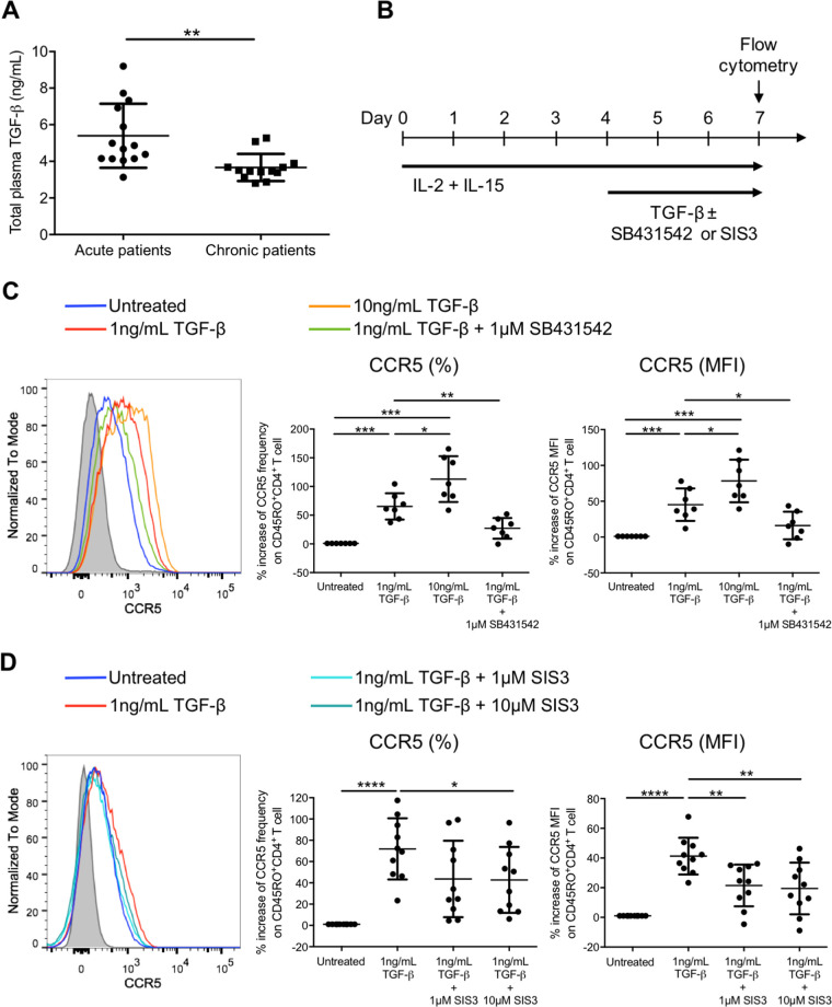
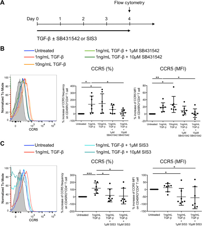
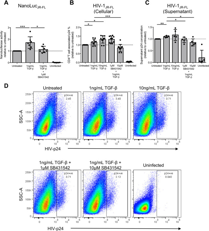
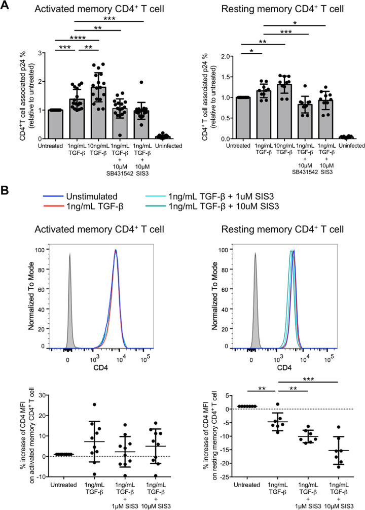
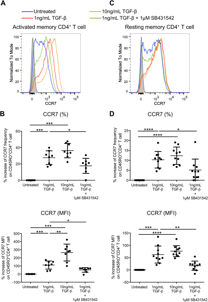
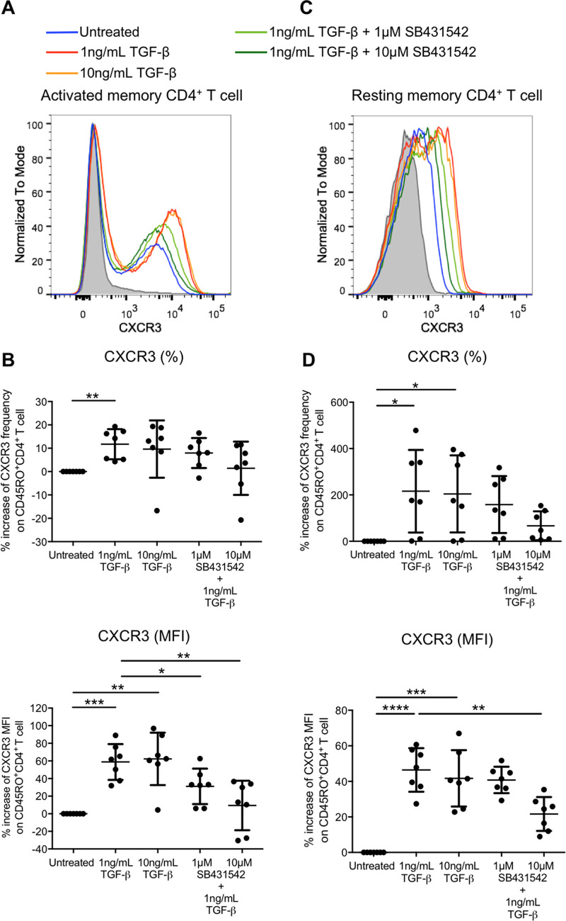
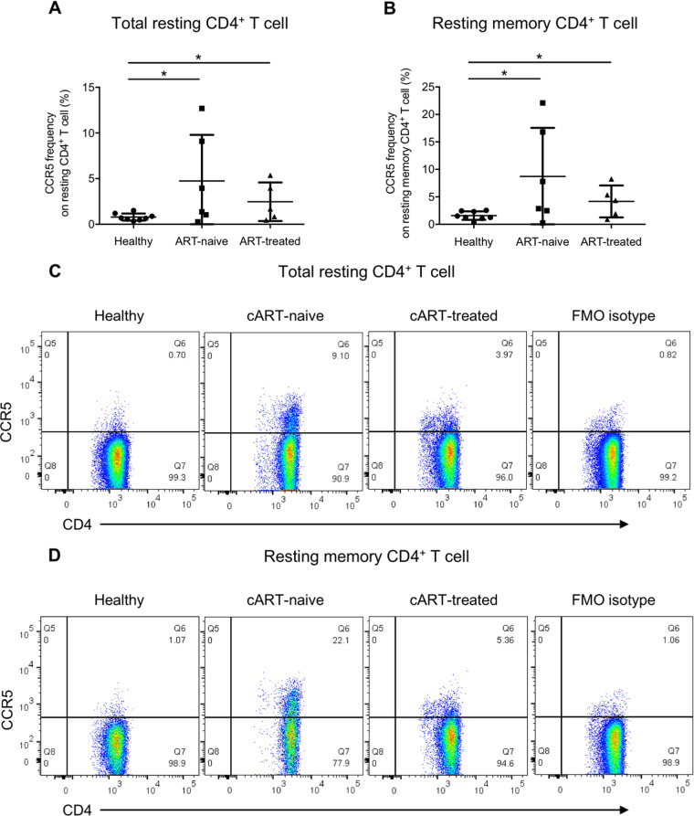
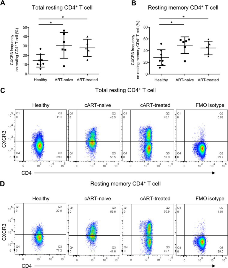
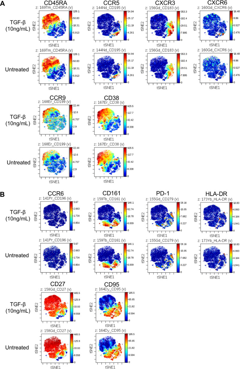
Understanding the facilitator of HIV-1 infection and subsequent latency establishment may aid the discovery of potential therapeutic targets. Here, we report the elevation of plasma transforming growth factor β (TGF-β) during acute HIV-1 infection among men who have sex with men (MSM). Using a serum-free in vitro system, we further delineated the role of TGF-β signaling in mediating HIV-1 infection of activated and resting memory CD4+ T cells. TGF-β could upregulate both the frequency and expression of the HIV-1 coreceptor CCR5, thereby augmenting CCR5-tropic viral infection of resting and activated memory CD4+ T cells via Smad3 activation. The production of live HIV-1JR-FL upon infection and reactivation was increased in TGF-β-treated resting memory CD4+ T cells without increasing CD4 expression or inducing T cell activation. The expression of CCR7, a central memory T cell marker that serves as a chemokine receptor to facilitate T cell trafficking into lymphoid organs, was also elevated on TGF-β-treated resting and activated memory CD4+ T cells. Moreover, the expression of CXCR3, a chemokine receptor recently reported to facilitate CCR5-tropic HIV-1 infection, was increased on resting and activated memory CD4+ T cells upon TGF-β treatment. These findings were coherent with the observation that ex vivo CCR5 and CXCR3 expression on total resting and resting memory CD4+ T cells in combination antiretroviral therapy (cART)-naive and cART-treated patients were higher than in healthy individuals. Overall, the study demonstrated that TGF-β upregulation induced by acute HIV-1 infection might promote latency reservoir establishment by increasing infected resting memory CD4+ T cells and lymphoid organ homing of infected central memory CD4+ T cells. Therefore, TGF-β blockade may serve as a potential supplementary regimen for HIV-1 functional cure by reducing viral latency. IMPORTANCE Incomplete eradication of HIV-1 latency reservoirs remains the major hurdle in achieving a complete HIV/AIDS cure. Dissecting the facilitator of latency reservoir establishment may aid the discovery of druggable targets for HIV-1 cure. This study showed that the T cell immunomodulatory cytokine TGF-β was upregulated during the acute phase of infection. Using an in vitro serum-free system, we specifically delineated that TGF-β promoted HIV-1 infection of both resting and activated memory CD4+ T cells via the induction of host CCR5 coreceptor. Moreover, TGF-β-upregulated CCR7 or CXCR3 might promote HIV-1 latent infection by facilitating lymphoid homing or IP-10-mediated viral entry and DNA integration, respectively. Infected resting and central memory CD4+ T cells are important latency reservoirs. Increased infection of these cells mediated by TGF-β will promote latency reservoir establishment during early infection. This study, therefore, highlighted the potential use of TGF-β blockade as a supplementary regimen with cART in acute patients to reduce viral latency.

Keywords: CCR5-tropic infection; TGF-β; activated memory CD4 T cells; human immunodeficiency virus; resting memory CD4 T cells.


Conflict of interest statement

The authors declare no conflict of interest.

Figures

FIG 1
FIG 1
TGF-β increases the frequency and expression level of the HIV-1 coreceptor CCR5 on activated memory CD4+ T cells via Smad3. (A) Plasma TGF-β levels in acute and chronic HIV-1 patients were compared. Plasma samples of patients were heat inactivated, and the level of TGF-β in plasma was determined by ELISA. (B) Schematic diagram showing in vitro activation and TGF-β treatment of purified CD45RO+ memory CD4+ T cells. Purified memory CD4+ T cells were activated by IL-2 (10 ng/mL) and IL-15 (20 ng/mL) for 4 days, followed by the addition of TGF-β (1 ng/mL or 10 ng/mL) for 3 days in the absence or presence of the TGF-β receptor kinase inhibitor SB431542 (1 μM) or the Smad3 inhibitor SIS3 (1 or 10 μM). The expression of CCR5 was subsequently evaluated using flow cytometry analysis. (C and D) Representative flow cytometry histogram and cumulative dot plots comparing the relative increase in frequency and expression (MFI) of CCR5 on purified activated memory CD4+ T cells compared with the untreated control in the presence of SB431542 (C) or SIS3 (D). For panels A, C, and D, each symbol represents an individual donor. The unpaired Student t test was used for statistical analysis (*, P ≤ 0.05; **, P ≤ 0.01; ***, P ≤ 0.001; ****, P ≤ 0.0001).
FIG 2
FIG 2
TGF-β increases the frequency and expression level of the HIV-1 coreceptor CCR5 on resting memory CD4+ T cells via Smad3. (A) Schematic diagram showing in vitro TGF-β treatment (1 or 10 ng/mL) of purified CD25 CD69 HLA-DR CD45RO+ resting memory CD4+ T cells in the absence or presence of the TGF-β receptor kinase inhibitor SB431542 (1 or 10 μM) or the Smad3 inhibitor SIS3 (1 or 10 μM) for 4 days, followed by flow cytometry analysis to evaluate CCR5 expression on resting memory CD4+ T cells after TGF-β treatment. (B and C) Representative flow cytometry histogram and cumulative dot plots comparing the relative increase in frequency and expression (MFI) of CCR5 on purified resting memory CD4+ T cells compared with the untreated control in the presence of SB431542 (B) or SIS3 (C). Each symbol represents an individual donor. The unpaired Student t test was used for statistical analysis (*, P ≤ 0.05; **, P ≤ 0.01).
FIG 3
FIG 3
TGF-β increases CCR5-tropic live HIV-1 infection and viral production of resting memory CD4+ T cells. Purified CD25 CD69 HLA-DR CD45RO+ resting memory CD4+ T cells were treated with TGF-β in the absence or presence of the TGF-β receptor kinase inhibitor SB431542, followed by infection of NanoLucJR-FL CCR5-tropic HIV-1 pseudovirus that carried a nanoluciferase reporter in the nef gene of the pseudovirus backbone. The nanoluciferase reading in the supernatant was measured 7 days after infection to evaluate infection of resting memory CD4+ T cells upon TGF-β treatment. (A) Cumulative dot plot showing nanoluciferase activity relative to that of the untreated control. (B) TGF-β treated purified resting memory CD4+ T cells were infected with live HIV-1JR-FL, and T cell-associated p24 was determined by flow cytometry analysis to evaluate HIV-1 infection. The cumulative dot plots show the changes of live HIV-1JR-FL infection on resting memory CD4+ T cells relative to the untreated control. (C) TGF-β-treated purified resting memory CD4+ T cells were infected with live HIV-1JR-FL. The cumulative dot plot shows the increase in HIV-1JR-FL production relative to the untreated control by evaluating the quantity of p24 in the culture supernatant using ELISA after reactivation by anti-CD3 and anti-CD28 stimulation from 2 independent experiments (n = 5). For panels A to C, each symbol represents an individual donor. The unpaired Student t test was used for statistical analysis (*, P ≤ 0.05; **, P ≤ 0.01; ***, P ≤ 0.001). (D) Representative flow cytometry plots showing the gating of T cell-associated p24 on resting memory CD4+ T cells for evaluating live HIV-1JR-FL infection by flow cytometry after TGF-β treatment.
FIG 4
FIG 4
TGF-β increases CCR5-tropic live HIV-1JR-FL infection of activated and resting memory CD4+ T cells via Smad3 phosphorylation. (A) Purified activated CD45RO+ activated memory CD4+ T cells (left) and purified CD25 CD69 HLA-DR CD45RO+ resting memory CD4+ T cells (right) were treated with TGF-β in the absence or presence of the TGF-β receptor kinase inhibitor SB431542 or Smad3 inhibitor SIS3 (10 μM). Cells were then infected with live HIV-1JR-FL and T cell-associated p24 was determined 3 days after infection by flow cytometry. (B) CD4 expression level on purified activated (left) and resting (right) memory CD4+ T cells was evaluated by flow cytometry analysis after TGF-β treatment as mentioned above. The top shows the representative flow cytometry histogram, and the bottom shows cumulative dot plots of relative CD4 expression (MFI) compared with the untreated control. For both panels, each symbol represents an individual donor. The unpaired Student t test was used for statistical analysis (*, P ≤ 0.05; **, P ≤ 0.01; ***, P ≤ 0.001; ****, P ≤ 0.0001).
FIG 5
FIG 5
TGF-β increases the frequency and expression level of the central memory marker CCR7 on activated and resting memory CD4+ T cells. Purified CD45RO+ activated memory (A and B) or purified CD25 CD69 HLA-DR CD45RO+ resting memory (C and D) CD4+ T cells were treated with TGF-β in the absence or presence of the TGF-β receptor kinase inhibitor SB431542. The expression of CCR7 was subsequently evaluated by flow cytometry analysis. Representative flow cytometry histograms of CCR7 expression on activated (A) and resting (C) memory CD4+ T cells are shown, and the cumulative dot plots show the increase of frequency and expression (MFI) of activated (B) and resting (D) memory CD4+ T cells relative to the untreated control. Each symbol represents an individual donor. The unpaired Student t test was used for statistical analysis (*, P ≤ 0.05; **, P ≤ 0.01; ****, P ≤ 0.0001).
FIG 6
FIG 6
TGF-β increases CXCR3 expression on activated and resting memory CD4+ T cells. Purified CD45RO+ CD4+ activated memory (A and B) or purified CD25 CD69 HLA-DR CD4+ cells gated on CD45RO+ resting memory (C and D) T cells were treated with TGF-β in the absence or presence of the TGF-β receptor kinase inhibitor SB431542. The expression of CXCR3 was subsequently evaluated by flow cytometry analysis. Representative flow cytometry histograms of CXCR3 expression on activated (A) and resting (C) memory CD4+ T cells are shown, and the cumulative dot plots show relative frequency and expression (MFI) of activated (B) and resting (D) memory CD4+ T cells compared to the untreated control. Each symbol represents an individual donor. The unpaired Student t test was used for statistical analysis (*, P ≤ 0.05; **, P ≤ 0.01; ****, P ≤ 0.0001).
FIG 7
FIG 7
CCR5 frequency on total resting and resting memory CD4+ T cells is elevated in cART-naive and cART-treated HIV-1 patients. Total PBMCs from cART-naive patients, cART-treated patients, and healthy donors were stained for CCR5 with the T cell and T cell activation markers CD3, CD4, CD69, CD25, and HLA-DR and the memory T cell marker CD45RO, followed by flow cytometry analysis. Ex vivo CCR5 frequency on total resting CD4+ T cells was evaluated by gating on CD3+ CD4+ CD25 CD69 HLA-DR T cells, and that on resting memory CD4+ T cells was evaluated by gating on CD3+ CD4+ CD25 CD69 HLA-DR CD45RO+ T cells. Cumulative dot plots show the ex vivo CCR5 frequencies on total resting (A) and resting memory (B) CD4+ T cells among cART-naive patients, cART-treated patients, and healthy donors. Representative flow cytometry plots of total resting and resting memory CD4+ T cells are also shown in panels C and D, respectively. The FMO isotype control was pooled from all samples for isotype staining. Each symbol represents an individual donor. The unpaired Student t test was used for statistical analysis (*, P ≤ 0.05).
FIG 8
FIG 8
CXCR3 frequency on total resting and resting memory CD4+ T cells is elevated in cART-naive and cART-treated HIV-1 patients. Total PBMCs from cART-naive patients, cART-treated patients, and healthy donors were stained for CXCR3 with the T cell and T cell activation markers CD3, CD4, CD69, CD25, and HLA-DR and the memory T cell marker CD45RO, followed by flow cytometry analysis. Ex vivo CXCR3 frequency on total resting CD4+ T cells was evaluated by gating on CD3+ CD4+ CD25 CD69 HLA-DR T cells, and that on resting memory CD4+ T cells was evaluated by gating on CD3+ CD4+ CD25 CD69 HLA-DR CD45RO+ T cells. Cumulative dot plots show ex vivo CXCR3 frequencies on total resting (A) and resting memory (B) CD4+ T cells among cART-naive patients, cART-treated patients, and healthy donors. Representative flow cytometry plots of total resting and resting memory CD4+ T cells are also shown in panels C and D, respectively. The FMO isotype control was pooled from all samples for isotype staining. Each symbol represents an individual donor. The unpaired Student t test was used for statistical analysis (*, P ≤ 0.05).
FIG 9
FIG 9
TGF-β does not induce pan-chemokine receptor upregulation on resting memory CD4+ T cells. Total resting CD4+ T cells were either treated with 10 ng/mL or TGF-β or left untreated for 4 days, followed by antibody staining and CyTOF analysis. Resting memory CD4+ T cells were identified as CD45RA cells on the top left of panel A. Targets with expression upregulation after TGF-β treatment are illustrated in panel A, except the CD45RA marker for distinguishing the naive or memory cells. Targets without expression changes or downregulated are illustrated in panel B. The experiment was conducted once as a preliminary experiment.

References

    1. Katlama C, Deeks SG, Autran B, Martinez-Picado J, van Lunzen J, Rouzioux C, Miller M, Vella S, Schmitz JE, Ahlers J, Richman DD, Sekaly RP. 2013. Barriers to a cure for HIV: new ways to target and eradicate HIV-1 reservoirs. Lancet 381:2109–2117. doi:10.1016/S0140-6736(13)60104-X. - DOI - PMC - PubMed
    1. Cohn LB, Chomont N, Deeks SG. 2020. The biology of the HIV-1 latent reservoir and implications for cure strategies. Cell Host Microbe 27:519–530. doi:10.1016/j.chom.2020.03.014. - DOI - PMC - PubMed
    1. Lee E, Bacchetti P, Milush J, Shao W, Boritz E, Douek D, Fromentin R, Liegler T, Hoh R, Deeks SG, Hecht FM, Chomont N, Palmer S. 2019. Memory CD4 + T-cells expressing HLA-DR contribute to HIV persistence during prolonged antiretroviral therapy. Front Microbiol 10:2214. doi:10.3389/fmicb.2019.02214. - DOI - PMC - PubMed
    1. Tran TA, de Goer de Herve MG, Hendel-Chavez H, Dembele B, Le Nevot E, Abbed K, Pallier C, Goujard C, Gasnault J, Delfraissy JF, Balazuc AM, Taoufik Y. 2008. Resting regulatory CD4 T cells: a site of HIV persistence in patients on long-term effective antiretroviral therapy. PLoS One 3:e3305. doi:10.1371/journal.pone.0003305. - DOI - PMC - PubMed
    1. Leite TF, Delatorre E, Cortes FH, Ferreira ACG, Cardoso SW, Grinsztejn B, de Andrade MM, Veloso VG, Morgado MG, Guimaraes ML. 2019. Reduction of HIV-1 reservoir size and diversity after 1 year of cART among Brazilian individuals starting treatment during early stages of acute infection. Front Microbiol 10:145. doi:10.3389/fmicb.2019.00145. - DOI - PMC - PubMed

Publication types

Substances