. 2023 Jun 15;11(3):e0063122.
doi: 10.1128/spectrum.00631-22. Epub 2023 Apr 12.

Formate Metabolism in Shigella flexneri and Its Effect on HeLa Cells at Different Stages during the Infectious Process

Affiliations

Formate Metabolism in Shigella flexneri and Its Effect on HeLa Cells at Different Stages during the Infectious Process

Ke-Chuan Wang et al. Microbiol Spectr. .

Abstract

Shigellosis caused by Shigella is one of the most important foodborne illnesses in global health, but little is known about the metabolic cross talk between this bacterial pathogen and its host cells during the different stages of the infection process. A detailed understanding of the metabolism can potentially lead to new drug targets remedying the pressing problem of antibiotic resistance. Here, we use stable isotope-resolved metabolomics as an unbiased and fast method to investigate how Shigella metabolizes 13C-glucose in three different environments: inside the host cells, adhering to the host cells, and alone in suspension. We find that especially formate metabolism by bacteria is sensitive to these different environments. The role of formate in pathogen metabolism is sparsely described in the literature compared to the roles of acetate and butyrate. However, its metabolic pathway is regarded as a potential drug target due to its production in microorganisms and its absence in humans. Our study provides new knowledge about the regulatory effect of formate. Bacterial metabolism of formate is pH dependent when studied alone in culture medium, whereas this effect is less pronounced when the bacteria adhere to the host cells. Once the bacteria are inside the host cells, we find that formate accumulation is reduced. Formate also affects the host cells resulting in a reduced infection rate. This was correlated to an increased immune response. Thus, intriguingly formate plays a double role in pathogenesis by increasing the virulence of Shigella and at the same time stimulating the immune response of the host. IMPORTANCE Bacterial infection is a pressing societal concern due to development of resistance toward known antibiotics. Central carbon metabolism has been suggested as a potential new target for drug development, but metabolic changes upon infection remain incompletely understood. Here, we used a cellular infection model to study how the bacterial pathogen Shigella adapts its metabolism depending on the environment starting from the extracellular medium until Shigella successfully invaded and proliferated inside host cells. The mixed-acid fermentation of Shigella was the major metabolic pathway during the infectious process, and the glucose-derived metabolite formate surprisingly played a divergent role in the pathogen and in the host cell. Our data show reduced infection rate when both host cells and bacteria were treated with formate, which correlated with an upregulated immune response in the host cells. The formate metabolism in Shigella thus potentially provides a route toward alternative treatment strategies for Shigella prevention.

Keywords: Shigella; formate; host-pathogen interactions; metabolism.


Conflict of interest statement

The authors declare no conflict of interest.

Figures
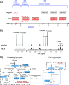
FIG 1
FIG 1
The Shigella infection model for the metabolic analysis by 1H NMR. (A) A three-step infection model. In the first part, Shigella is alone in suspension where it is cultured to mid-log for 3 h. HeLa is likewise separately cultured. In the second part, HeLa is infected with mid-log cultured Shigella (MOI, 100). In the third part, after an initial 1-h infection, HeLa cells are treated with 100 μg/mL gentamicin which kills the extracellular bacteria. (B) Part of 1H NMR spectrum obtained directly on the cell medium. The typical signal pattern produced by metabolites downstream of [U-13C] glucose 6-h after Shigella infection is highlighted. (C) The metabolic pathway of glucose for Shigella and HeLa cells showing how Shigella can utilize both aerobic and anaerobic metabolism, and HeLa cells mainly utilize aerobic metabolism.
FIG 2
FIG 2
Time course of extracellular 13C-labeled metabolites from HeLa cells infected with Shigella. The metabolism of [U-13C] glucose was followed in infected (infect) and noninfected HeLa cells (HeLa) (n = 2). Each sample was made according to the procedure of the continuous measurement of metabolism (see Fig. S1A in the supplemental material). Briefly, 100 μg/mL gentamicin was added at 1 h p.i. and was kept without medium replacement during the total 6-h incubation. The 13C-labeled metabolites are obtained from the symmetric 13C-satellites displaying the characteristic 1J(13C,1H) coupling of 130 Hz in the 1D 1H NMR spectra.
FIG 3
FIG 3
Comparison of the production of extracellular 13C-metabolites 1 h p.i. and up to 6 h p.i. from HeLa cells and Shigella-infected HeLa cells as measured by 1H NMR. Each sample was made according to the procedure of the continuous measurement of metabolism (see Fig. S1A in the supplemental material). Briefly, 100 μg/mL gentamicin was added at 1 h p.i. and was kept without medium replacement during the total 6-h incubation. Each sample was saved at 1 h p.i. and 6 h p.i. and was analyzed by 1H NMR. The statistical analysis was done by the comparison of the 13C-metabolites produced between 1 h p.i. and 6 h p.i. All data are represented as mean ± standard error of the mean (SEM), and an asterisk indicated a statistical difference (P ≤ 0.05); n = 4.
FIG 4
FIG 4
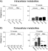
1H NMR analysis of 13C-metabolites produced from HeLa cells and Shigella-infected HeLa cells. The analyses of both extracellular and intracellular samples were performed according to the procedure of metabolism from intracellular bacteria (see Fig. S1B in the supplemental material). Briefly, after 1 h treatment with 100 μg/mL gentamicin, each medium was replaced with fresh medium containing 10 μg/mL of gentamicin for an additional 4-h incubation. At 4 h and 6 h p.i., the supernatants and samples of PCA extracted cell lysates were analyzed by 1H NMR. (A) The intracellular metabolites from the PCA extracted cells at 4 h and 6 h p.i. No 13C-formate could be detected (see Fig. S3 in the supplemental material for the NMR spectrum). (B) The extracellular supernatants collected at 4 h and 6 h p.i. showed no detection of 13C-formate. A significant production between 4 h and 6 h p.i. was determined for 13C-acetate in the infected group, whereas no significant production was measured for HeLa cells. Asterisks indicate the statistical difference (***, P ≤ 0.001) by ANOVA. Data are represented as mean ± SEM (n = 3).
FIG 5
FIG 5
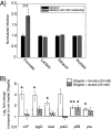
The role of metabolites from bacterial mixed-acid fermentation on Shigella virulence. (A) Shigella was treated with 20 mM metabolites formate, lactate, ethanol, and acetate for 3 h before infection (or not). The treatment hereafter continued in the additional 6-h adhesion and intracellular proliferation period. Each experiment was performed in triplicate. (B) Six selected genes related to virulence and glycolysis were analyzed with qRT-PCR. The nontreated group relative to the 20 mM formate-treated (open bars) group and the nontreated group relative to the 20 mM lactate-treated (slash bars) group were analyzed individually. The expression level of each mRNA was calculated according to the ΔΔCT method. Data was measured in triplicate. All data are represented as mean ± SEM, and asterisks indicated a statistical difference as follows: *, P ≤ 0.05; **, P ≤ 0.01; and ***, P ≤ 0.001 compared to the nontreated group.
FIG 6
FIG 6
Profiles of cell density, pH, and the consumption and production of metabolites during Shigella growth. Shigella was grown in TSB medium at 37°C with shaking at 200 rpm for 8 h. Cell density and pH were measured on a supernatant sample taken every hour, and metabolic profiles were measured on the same sample with 1H NMR.
FIG 7
FIG 7
Impact of pH on Shigella metabolism in the presence of HeLa cells. (A) pH dependence of 13C-formate and 13C-acetate production as function of host cell density. Extracellular metabolite production from [U-13C] glucose was measured with 1H NMR 1 h after addition of the same number of Shigella (5 × 107) to varying density of HeLa cells (0, 50, 90, and >90% corresponding to 0, 0.25, 0.5, or 1 million). For each cell concentration, the growth medium had been adjusted to either pH 6 or 7 prior to the addition of Shigella. Gray box indicates the conditions used for the infection data, and the number of Shigella used was also the same (MOI = 100 for 0.5 million cells). Signal-to-noise for 13C-formate and acetate are shown in Fig. S6 in the supplemental material. Each experiment was performed in triplicate and represented as mean ± SEM. (B) Difference of Shigella adherence on HeLa cells at pH 7 with or without 20 mM formate under the conditions marked with the gray box in Fig. 7A.
FIG 8
FIG 8
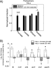
The role of metabolites from bacterial mixed-acid fermentation on HeLa defense. (A) HeLa was treated with 20 mM metabolites formate, lactate, ethanol, and acetate for 3 h before infection (or not). The treatment hereafter continued in the additional 6-h adhesion and proliferation period. Each experiment was performed in triplicate and represented as mean ± SEM. (B) Six selected genes related to immune response (il8, nod1, pi3k, and orai1) and glycolysis (glut1 and gapdh) were analyzed with qRT-PCR. The nontreated group relative to 20 mM formate-treated (open bars) group and the nontreated group relative to 20 mM lactate-treated (slash bars) group were analyzed individually. The expression level of each mRNA was calculated according to the ΔΔCT method. Data was measured in triplicate and represented as mean ± SEM. Asterisks indicated a statistical difference. *, P ≤ 0.05; ***, P ≤ 0.001.
FIG 9
FIG 9
The role of metabolites from bacterial mixed-acid fermentation on Shigella and HeLa. (A) HeLa and Shigella were treated with 20 mM metabolites acetate, formate, ethanol, and lactate for 3 h before infection (or not). The treatment hereafter continued in the additional 6-h adhesion and proliferation period. Each experiment was measured in triplicate. The results were shown as the mean ± SEM. Asterisks indicated a statistical difference. ***, P ≤ 0.001. (B) Summary of the effect of formate in the three different stages of the infection process (separate cell proliferation, adhesion, and intracellular proliferation). Indication of Shigella’s ability to produce formate and how both Shigella and HeLa cells react to pretreatment with 20 mM formate.

References

    1. Puzari M, Sharma M, Chetia P. 2018. Emergence of antibiotic resistant Shigella species: a matter of concern. J Infect Public Health 11:451–454. doi:10.1016/j.jiph.2017.09.025. - DOI - PubMed
    1. Miethke M, Pieroni M, Weber T, Bronstrup M, Hammann P, Halby L, Arimondo PB, Glaser P, Aigle B, Bode HB, Moreira R, Li Y, Luzhetskyy A, Medema MH, Pernodet JL, Stadler M, Tormo JR, Genilloud O, Truman AW, Weissman KJ, Takano E, Sabatini S, Stegmann E, Brotz-Oesterhelt H, Wohlleben W, Seemann M, Empting M, Hirsch AKH, Loretz B, Lehr CM, Titz A, Herrmann J, Jaeger T, Alt S, Hesterkamp T, Winterhalter M, Schiefer A, Pfarr K, Hoerauf A, Graz H, Graz M, Lindvall M, Ramurthy S, Karlen A, van Dongen M, Petkovic H, Keller A, Peyrane F, Donadio S, Fraisse L, et al. . 2021. Towards the sustainable discovery and development of new antibiotics. Nat Rev Chem 5:726–749. doi:10.1038/s41570-021-00313-1. - DOI - PMC - PubMed
    1. Hartman HB, Fell DA, Rossell S, Jensen PR, Woodward MJ, Thorndahl L, Jelsbak L, Olsen JE, Raghunathan A, Daefler S, Poolman MG. 2014. Identification of potential drug targets in Salmonella enterica sv. Typhimurium using metabolic modelling and experimental validation. Microbiology (Reading) 160:1252–1266. doi:10.1099/mic.0.076091-0. - DOI - PubMed
    1. Eisenreich W, Rudel T, Heesemann J, Goebel W. 2020. Persistence of intracellular bacterial pathogens-with a focus on the metabolic perspective. Front Cell Infect Microbiol 10:615450. doi:10.3389/fcimb.2020.615450. - DOI - PMC - PubMed
    1. Bongers S, Hellebrekers P, Leenen LPH, Koenderman L, Hietbrink F. 2019. Intracellular penetration and effects of antibiotics on Staphylococcus aureus inside human neutrophils: a comprehensive review. Antibiotics 8:54. doi:10.3390/antibiotics8020054. - DOI - PMC - PubMed

Publication types

LinkOut - more resources