Can gadolinium contrast agents be replaced with saline for direct MR arthrography of the hip? A pilot study with arthroscopic comparison
- PMID: 37042981
- PMCID: PMC10415454
- DOI: 10.1007/s00330-023-09586-0
Can gadolinium contrast agents be replaced with saline for direct MR arthrography of the hip? A pilot study with arthroscopic comparison
Erratum in
-
Correction: Can gadolinium contrast agents be replaced with saline for direct MR arthrography of the hip? A pilot study with arthroscopic comparison.Eur Radiol. 2023 Dec;33(12):9481. doi: 10.1007/s00330-023-09844-1. Eur Radiol. 2023. PMID: 37402005 Free PMC article. No abstract available.
Abstract
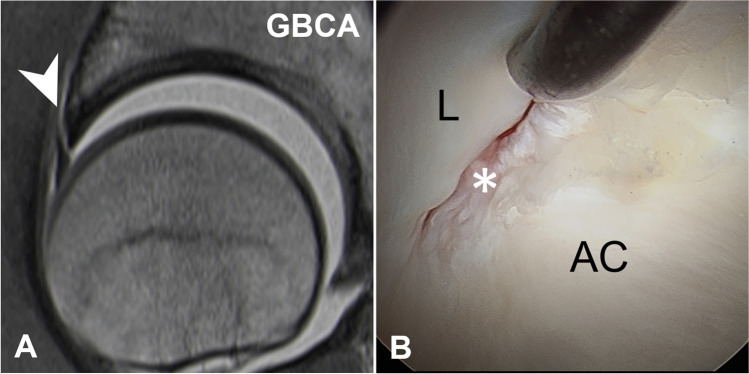
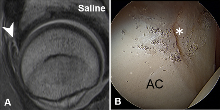
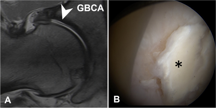
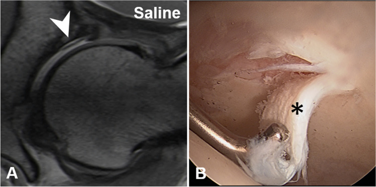
Objective: To compare image quality and diagnostic performance of preoperative direct hip magnetic resonance arthrography (MRA) performed with gadolinium contrast agent and saline solution.
Methods: IRB-approved retrospective study of 140 age and sex-matched symptomatic patients with femoroacetabular impingement, who either underwent intra-articular injection of 15-20 mL gadopentetate dimeglumine (GBCA), 2.0 mmol/L ("GBCA-MRA" group, n = 70), or 0.9% saline solution ("Saline-MRA" group, n = 70) for preoperative hip MRA and subsequent hip arthroscopy. 1.5 T hip MRA was performed including leg traction. Two readers assessed image quality using a 5-point Likert scale (1-5, excellent-poor), labrum and femoroacetabular cartilage lesions. Arthroscopic diagnosis was used to calculate diagnostic accuracy which was compared between groups with Fisher's exact tests. Image quality was compared with the Mann-Whitney U tests.
Results: Mean age was 33 years ± 9, 21% female patients. Image quality was excellent (GBCA-MRA mean range, 1.1-1.3 vs 1.1-1.2 points for Saline-MRA) and not different between groups (all p > 0.05) except for image contrast which was lower for Saline-MRA group (GBCA-MRA 1.1 ± 0.4 vs Saline-MRA 1.8 ± 0.5; p < 0.001). Accuracy was high for both groups for reader 1/reader 2 for labrum (GBCA-MRA 94%/ 96% versus Saline-MRA 96%/93%; p > 0.999/p = 0.904) and acetabular (GBCA-MRA 86%/ 83% versus Saline-MRA 89%/87%; p = 0.902/p = 0.901) and femoral cartilage lesions (GBCA-MRA 97%/ 99% versus Saline-MRA 97%/97%; both p > 0.999).
Conclusion: Diagnostic accuracy and image quality of Saline-MRA and GBCA-MRA is high in assessing chondrolabral lesions underlining the potential role of non-gadolinium-based hip MRA.
Key points: • Image quality of Saline-MRA and GBCA-MRA was excellent for labrum, acetabular and femoral cartilage, ligamentum teres, and the capsule (all p > 0.18). • The overall image contrast was lower for Saline-MRA (Saline-MRA 1.8 ± 0.5 vs. GBCA-MRA 1.1 ± 0.4; p < 0.001). • Diagnostic accuracy was high for Saline-MRA and GBCA-MRA for labrum (96% vs. 94%; p > 0.999), acetabular cartilage damage (89% vs. 86%; p = 0.902), femoral cartilage damage (97% vs. 97%; p > 0.999), and extensive cartilage damage (97% vs. 93%; p = 0.904).
Keywords: Arthrography; Arthroscopy; Contrast agent; Hip; MRI.
© 2023. The Author(s).
Conflict of interest statement
The authors of this manuscript declare no relationships with any companies, whose products or services may be related to the subject matter of the article.
Figures