. 2023 Jun 1;34(7):ar64.
doi: 10.1091/mbc.E22-09-0411. Epub 2023 Apr 12.

The RhoGEF protein Plekhg5 regulates medioapical and junctional actomyosin dynamics of apical constriction during Xenopus gastrulation

Affiliations

The RhoGEF protein Plekhg5 regulates medioapical and junctional actomyosin dynamics of apical constriction during Xenopus gastrulation

Austin Baldwin et al. Mol Biol Cell. .

Abstract

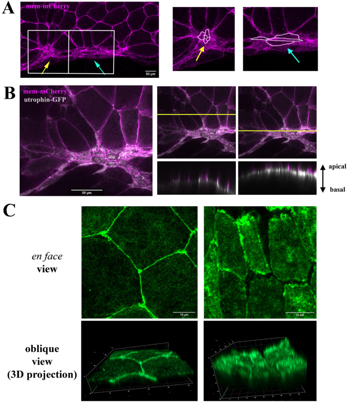
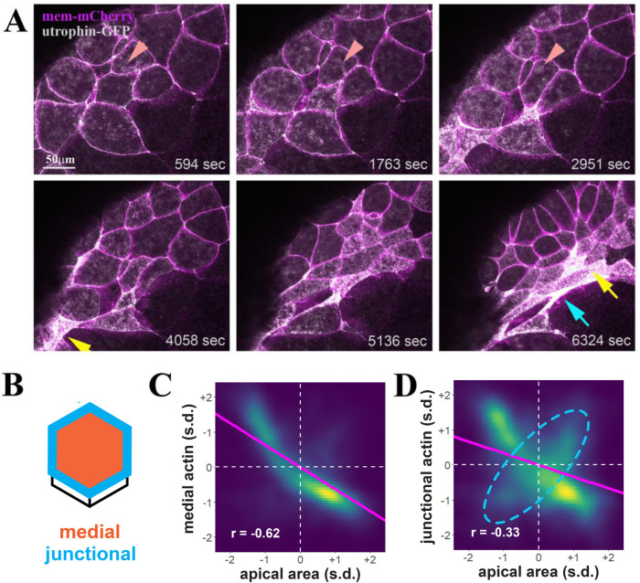
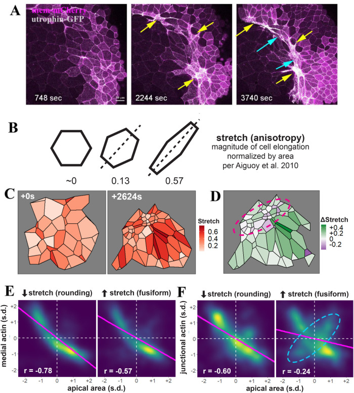
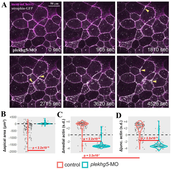
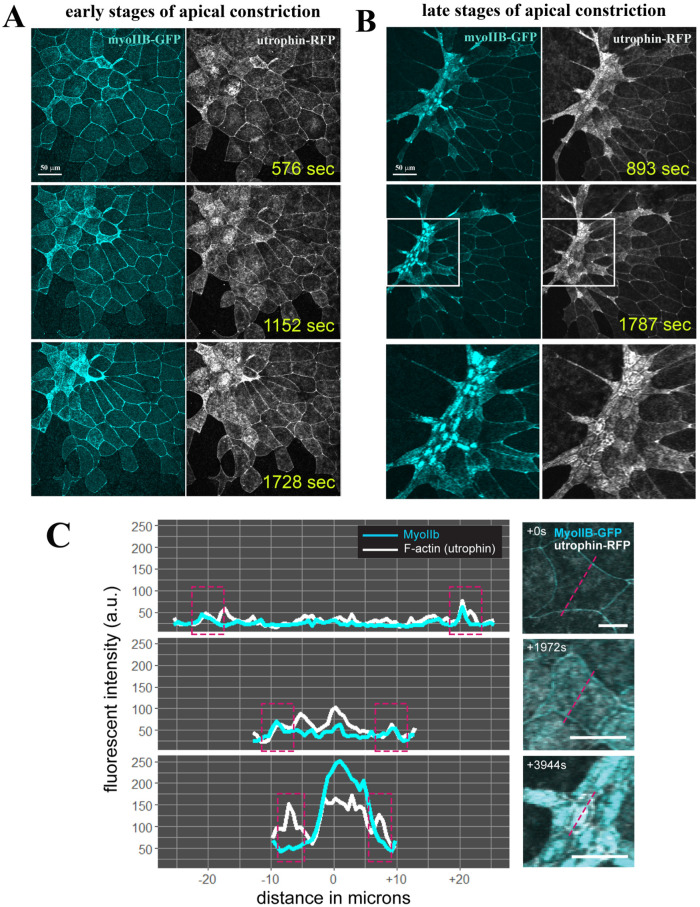
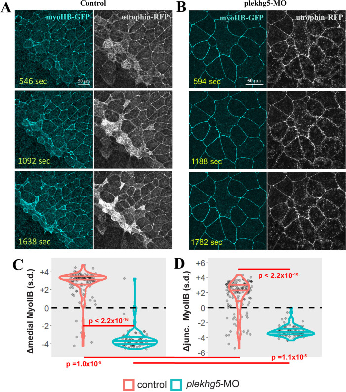
Apical constriction results in apical surface reduction in epithelial cells and is a widely used mechanism for epithelial morphogenesis. Both medioapical and junctional actomyosin remodeling are involved in apical constriction, but the deployment of medial versus junctional actomyosin and their genetic regulation in vertebrate embryonic development have not been fully described. In this study, we investigate actomyosin dynamics and their regulation by the RhoGEF protein Plekhg5 in Xenopus bottle cells. Using live imaging and quantitative image analysis, we show that bottle cells assume different shapes, with rounding bottle cells constricting earlier in small clusters followed by fusiform bottle cells forming between the clusters. Both medioapical and junctional actomyosin signals increase as surface area decreases, though correlation of apical constriction with medioapical actomyosin localization appears to be stronger. F-actin bundles perpendicular to the apical surface form in constricted cells, which may correspond to microvilli previously observed in the apical membrane. Knockdown of plekhg5 disrupts medioapical and junctional actomyosin activity and apical constriction but does not affect initial F-actin dynamics. Taking the results together, our study reveals distinct cell morphologies, uncovers actomyosin behaviors, and demonstrates the crucial role of a RhoGEF protein in controlling actomyosin dynamics during apical constriction of bottle cells in Xenopus gastrulation.


Figures

FIGURE 1:
FIGURE 1:
Distinct cell shapes, apical actin accumulation, and F-actin bundles in bottle cells. (A) Membrane mCherry–labeled bottle cells show two distinct morphologies, with round cells (yellow arrow) interspersed with fusiform cells (blue arrow). The panels on the right are the enlarged images of the boxed areas. (B) Coexpression of membrane mCherry with utrophin-GFP reveals accumulation of F-actin predominantly in the medial regions of apical cell cortex. The right two panels show the z-view of F-actin and mem-mCherry signals in regions without or with apical constriction. (C) En face images of phalloidin-stained fixed gastrula embryos show predominant cell junction localization of F-actin in not-yet-constricting cells (left top panel), whereas F-actin intensity is enhanced in the apical compartment of constricting cells (right top panel). Oblique views of 3D projections of the images reveal formation of dense F-actin bundles perpendicular to the apical surface in the constricting bottle cells (right bottom panel).
FIGURE 2:
FIGURE 2:
Dynamic F-actin remodeling accompanies apical surface reduction during bottle cell formation. (A) Selected frames from a time-lapse movie demonstrate dynamic F-actin organization before an overt decrease in apical cell area (top panels). The dynamic F-actin signal in a single cell is marked by the arrowheads. As apical constriction commences, groups of cells constrict first and take round shape (yellow arrow), with neighboring, later-constricting cells assuming fusiform (blue arrow). The clusters of round cells can appear as one large cell as cell constriction and F-actin signal increase often obscure cell boundaries (e.g., the cluster indicated by the yellow arrow at the 6324 s frame consists of five constricted cells seen in the time-lapse movie). (B) Schematics of quantification of medial (orange) and junctional (blue) F-actin intensity within single cells. (C, D) Density plots of apical cell area vs. medial (C) and junctional (D) F-actin. Magenta line indicates linear model. s.d. = standard deviations. n = 11,748 observations of 132 cells from four embryos. (C) A negative correlation of medial F-actin intensity and apical area is uncovered, with the correlation coefficient r value of –0.62. (D) Junctional F-actin intensity is also negatively correlated with the apical area, though less so with the r value of –0.33. This weaker correlation may be in part due to a subpopulation of cells with a positive correlation between junctional F-actin and the apical area (dashed cyan ellipsis).
FIGURE 3:
FIGURE 3:
Distinct cell shape changes accompany bottle cell formation. (A) Selected frames from a time-lapse movie reveal that apical constriction does not spread from a single point. Clusters of cells some distance apart can constrict around similar times. These cells tend to constrict early and form a round shape (yellow arrows), with the cells in between stretching in the circumferential direction to take the fusiform shape (blue arrows). The bottom yellow arrow in the 3740 s frame points to a constricted cluster of more than 10 cells. (B) Quantification of cell stretch by Tissue Analyzer, whereby the magnitude of cell elongation is normalized by cell area to generate a number ranging from 0 to 1, such that cells with the same shape but different areas will have the same stretch. (C) Example of heterogeneous cell constriction and stretch based on quantitative analysis of a time-lapse movie from the first and the last frames. (D) Mapping of changes of cell stretch onto the embryo images reveals that round, non-stretching cells are juxtaposed to the cells with increasing stretch in the blastopore lip (purple circle). The neighboring cells on the animal pole side of the blastopore lip also display distinct changes in stretch, with cells showing strong stretch preferentially abutting the round bottle cells. (E, F) Quantification of medial (E) or junctional (F) F-actin intensity reveals a stronger correlation with the round bottle cells than with the fusiform bottle cells. A subpopulation of fusiform cells with a positive correlation between junctional F-actin and apical area is labeled with a dashed cyan ellipsis. Magenta line indicates linear model. s.d. = standard deviations. n = 3171 observations for cells decreasing stretch (rounding) and 8709 observations for cells increasing stretch (fusiform) collected from four control embryos.
FIGURE 4:
FIGURE 4:
Knockdown of plekhg5 prevents apical accumulation of F-actin and reduction of apical cell size. (A) Selected frames from a time-lapse movie show that despite the initial F-actin dynamics and formation of F-actin puncta, the cells with plekhg5 knockdown fail to increase apical F-actin signal or reduce the apical surface. Instead, junctional F-actin flares can be seen frequently. The arrowheads point to examples of F-actin flares. (B) Quantification of apical cell area demonstrates that unlike cells in control embryos (Supplemental Movies 1 and 2), cells from plekhg5 knockdown embryos (Supplemental Movies 3 and 4) cannot reduce their apex efficiently. (C, D) While medial and junctional F-actin intensity increases during apical constriction of bottle cells in control embryos, the intensity of medial and junctional F-actin decreases in cells from plekhg5 knockdown embryos. Each dot is an individual cell. Horizontal lines within each violin delineate quartiles along each distribution. s.d. = standard deviations. P values were calculated via a KS test. Data were collected from four control and five plekhg5 knockdown embryos.
FIGURE 5:
FIGURE 5:
Coordinated enrichment of apical myosin IIB and F-actin during apical constriction of bottle cells. (A) Selected frames of a time-lapse movie reveal coordinated increase in MyoIIB and F-actin signals in the apical cortex of the bottle cells. The signal intensity of actomyosin inversely correlates with the apical cell area of the cells. The stretching of neighboring nonconstricting cells can also be seen. (B) During the late stage of apical constriction, the MyoIIB signal is down-regulated from cell junctions and is concentrated in the center of the bottle cells. F-actin signal can be seen in both the cell junctions and the medioapical region. Close-up view of the boxed regions is shown in the bottom panels. (C) Histogram of F-actin and MyoIIB signal intensity at the beginning, in the middle, and at the end of the apical constriction of the bottle cells reveals distinct patterns. While both medial and junctional F-actin and MyoIIB signals increase initially during bottle cell formation, junctional MyoIIB is reduced at the end of apical constriction while junctional F-actin remains strong. Approximate junctional region highlighted by dashed magenta boxes in left panels. Dashed magenta lines in right panels indicate quantified region of each cell.
FIGURE 6:
FIGURE 6:
Knockdown of plekhg5 prevents medial accumulation of MyoIIB. (A, B) Unlike cells in control embryos, in plekhg5 knockdown embryos, initial F-actin dynamics remains in the apical cortex but MyoIIB fails to show up in the medial domain. No actomyosin enrichment is observed with progression of time. (C, D) Quantification of medial and junctional MyoIIB intensity shows that unlike in control bottle cells where the intensity of MyoIIB increases, medial and junctional MyoIIB decrease in cells with plekhg5 knockdown. Each dot is an individual cell. Horizontal lines within each violin delineate quartiles along each distribution. s.d. = standard deviations. P values were calculated via a KS test. Data were collected from four control and five plekhg5 knockdown embryos.

Similar articles

Cited by

References

    1. Aigouy B, Cortes C, Liu S, Prud’Homme B (2020). EPySeg: a coding-free solution for automated segmentation of epithelia using deep learning. Development 147, dev194589. - PMC - PubMed
    1. Aigouy B, Farhadifar R, Staple DB, Sagner A, Roper JC, Julicher F, Eaton S (2010). Cell flow reorients the axis of planar polarity in the wing epithelium of Drosophila. Cell 142, 773–786. - PubMed
    1. Aigouy B, Umetsu D, Eaton S (2016). Segmentation and quantitative analysis of epithelial tissues. Methods Mol Biol 1478, 227–239. - PubMed
    1. An Y, Xue G, Shaobo Y, Mingxi D, Zhou X, Yu W, Ishibashi T, Zhang L, Yan Y (2017). Apical constriction is driven by a pulsatile apical myosin network in delaminating Drosophila neuroblasts. Development 144, 2153–2164. - PubMed
    1. Anderson DC, Gill JS, Cinalli RM, Nance J (2008). Polarization of the C. elegans embryo by RhoGAP-mediated exclusion of PAR-6 from cell contacts. Science 320, 1771–1774. - PMC - PubMed

Publication types

LinkOut - more resources