. 2023 Apr 25;42(4):112381.
doi: 10.1016/j.celrep.2023.112381. Epub 2023 Apr 11.

FXR1 regulates vascular smooth muscle cell cytoskeleton, VSMC contractility, and blood pressure by multiple mechanisms

Affiliations

FXR1 regulates vascular smooth muscle cell cytoskeleton, VSMC contractility, and blood pressure by multiple mechanisms

Amanda St Paul et al. Cell Rep. .

Abstract

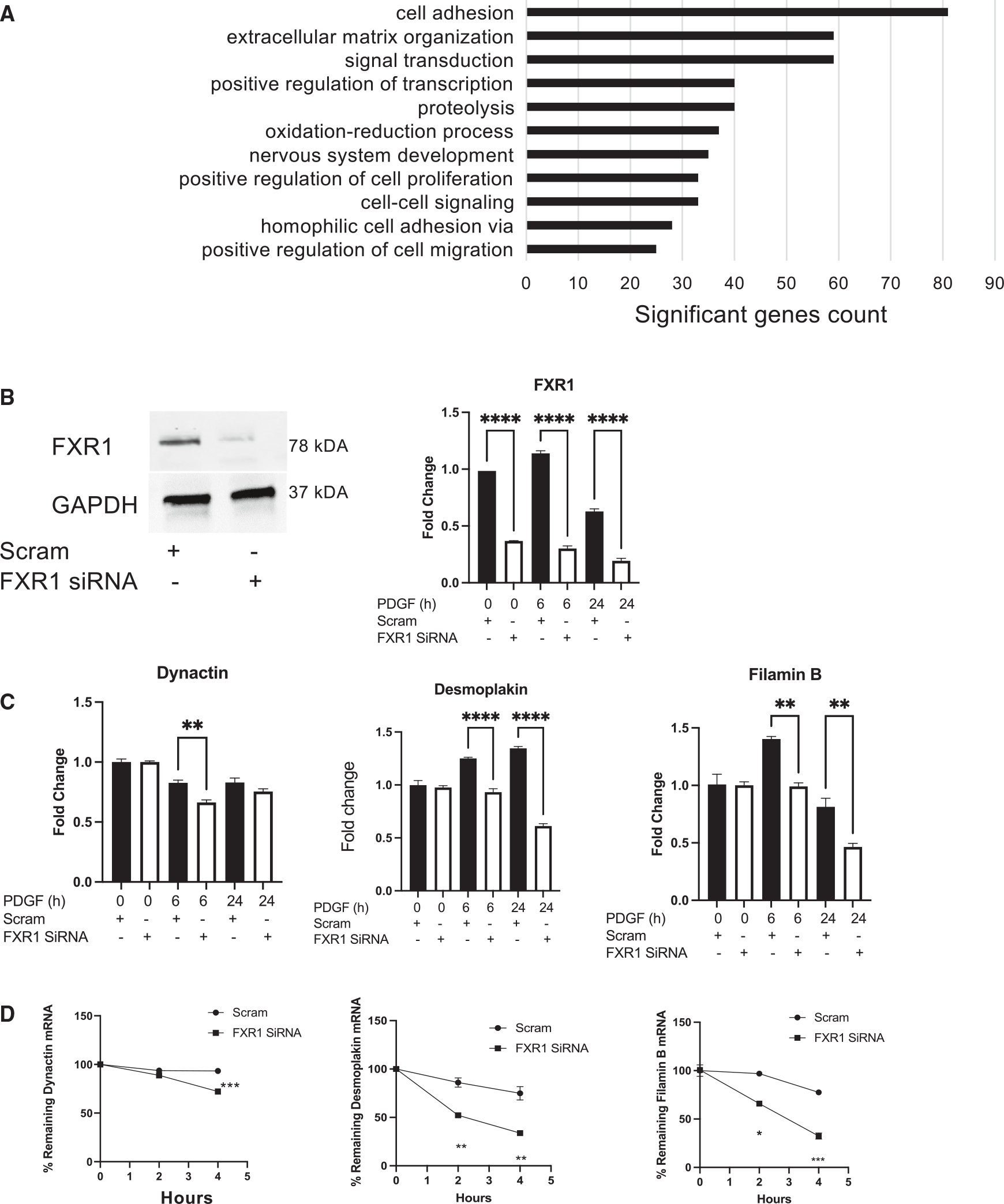
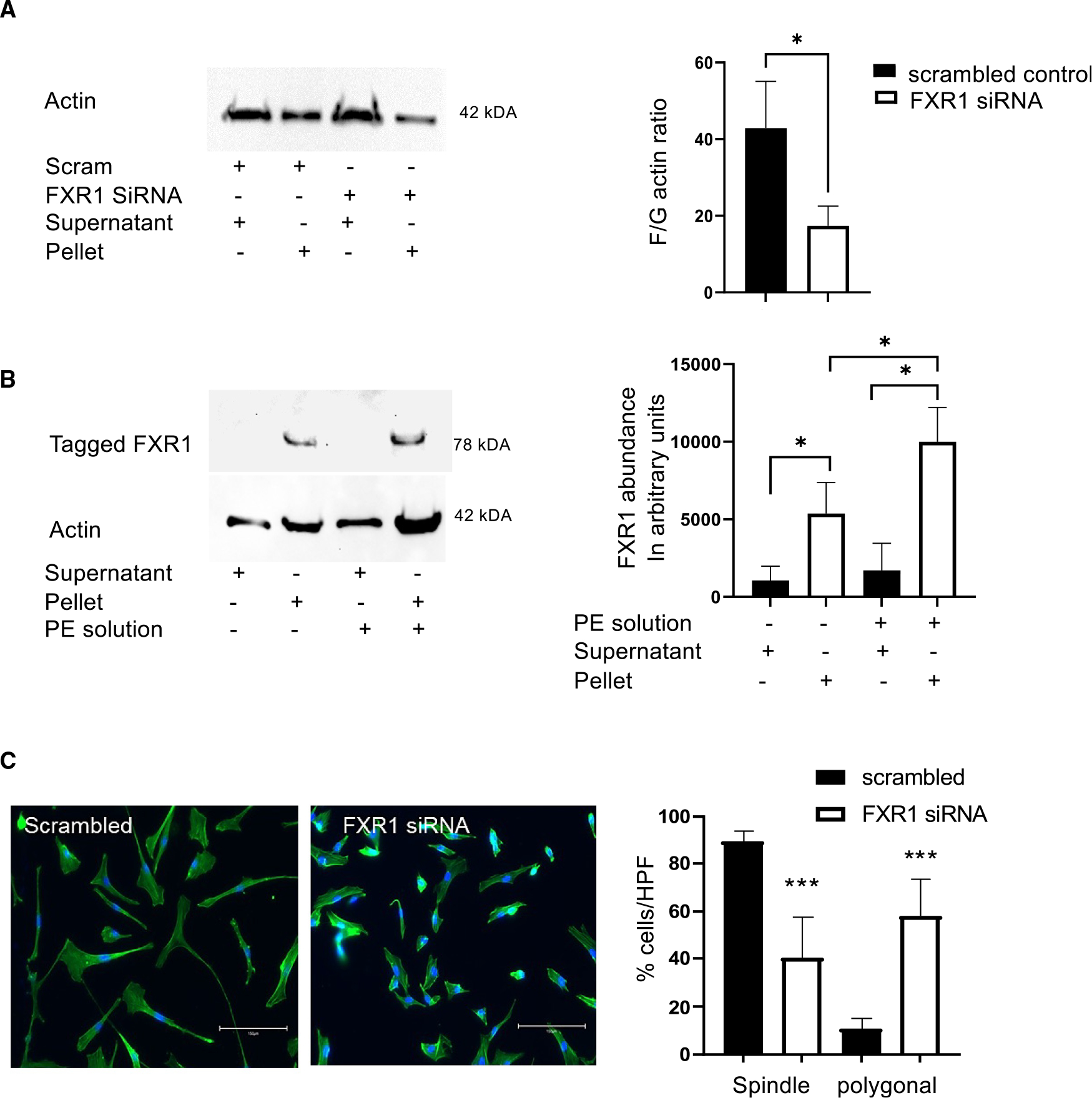
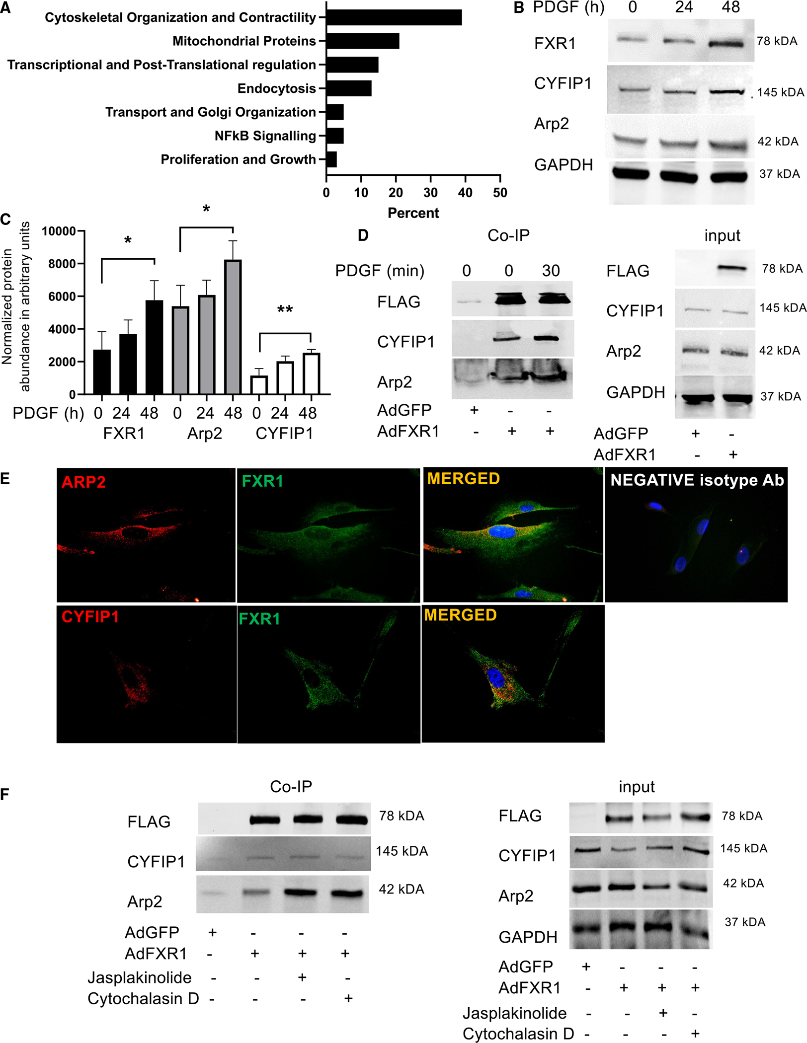
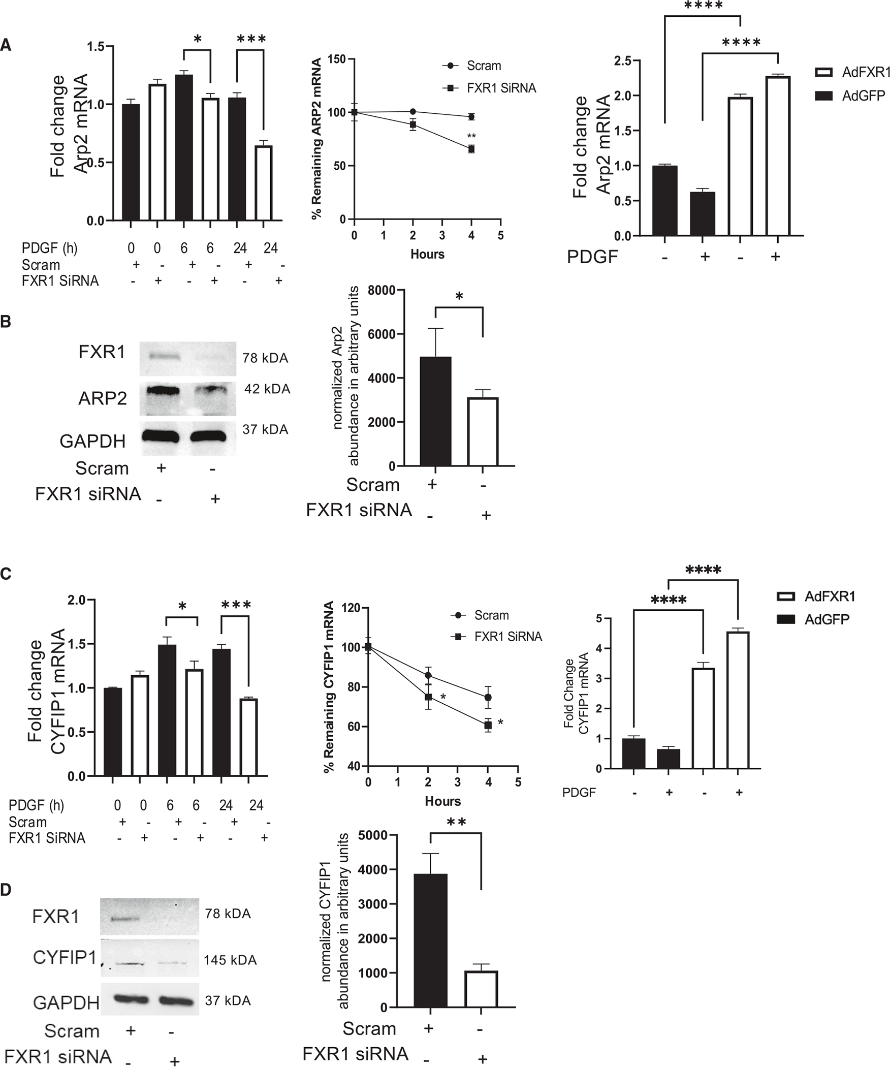
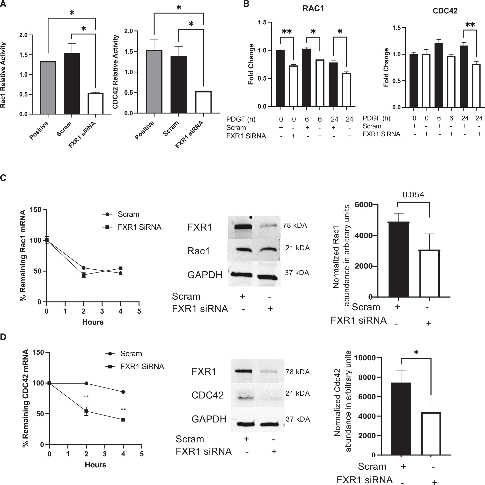
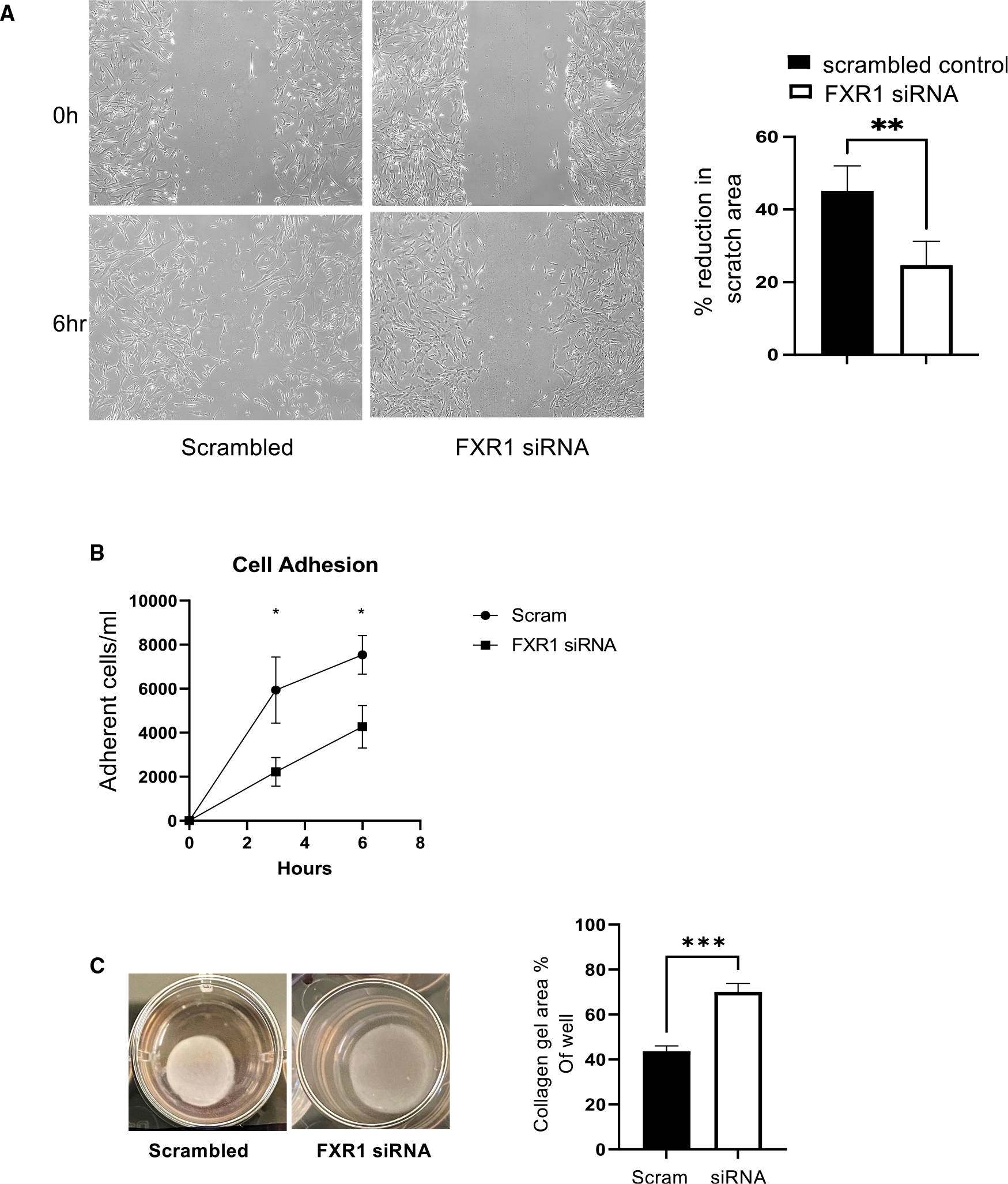
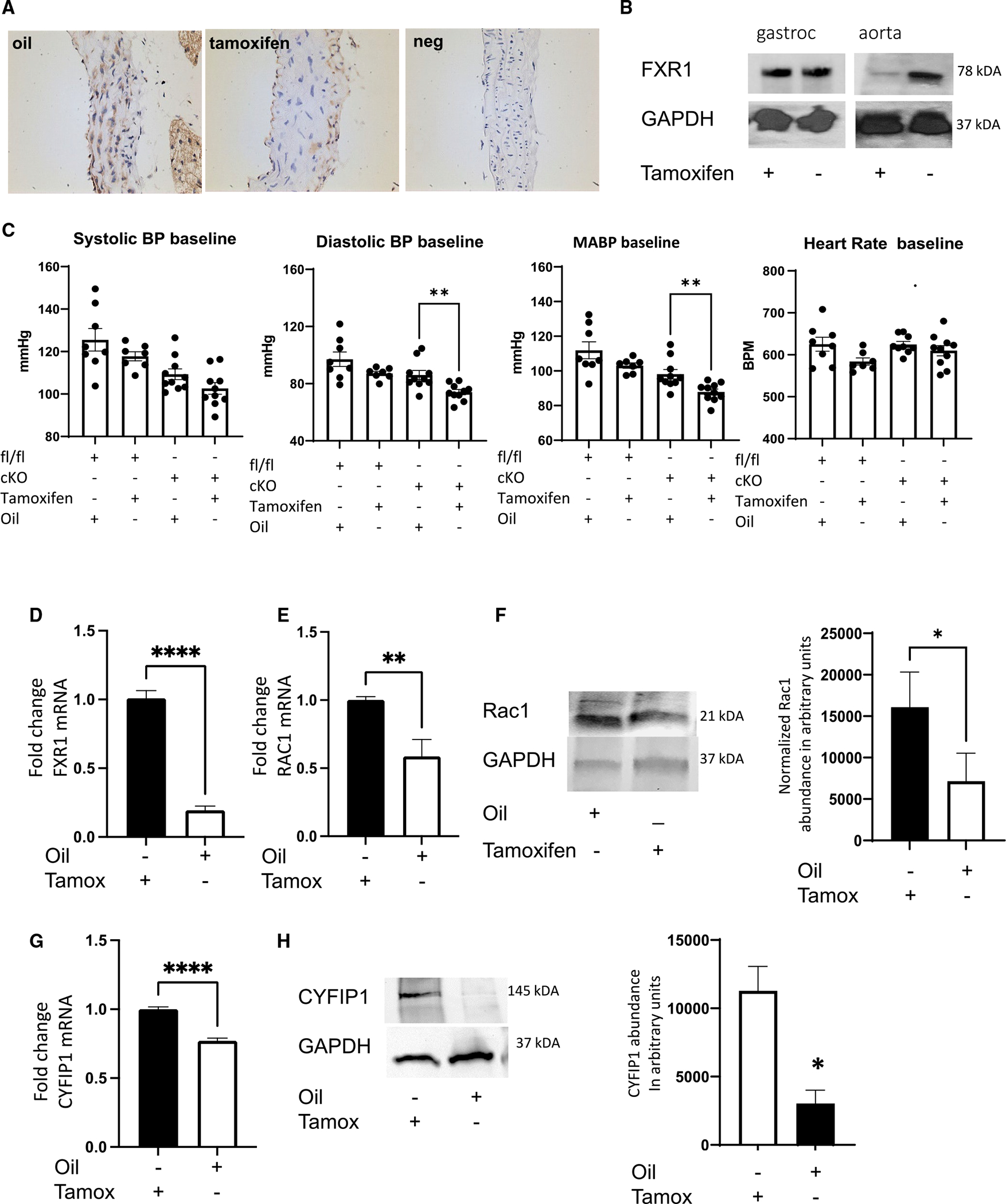
Appropriate cytoskeletal organization is essential for vascular smooth muscle cell (VSMC) conditions such as hypertension. This study identifies FXR1 as a key protein linking cytoskeletal dynamics with mRNA stability. RNA immunoprecipitation sequencing (RIP-seq) in human VSMCs identifies that FXR1 binds to mRNA associated with cytoskeletal dynamics, and FXR1 depletion decreases their mRNA stability. FXR1 binds and regulates actin polymerization. Mass spectrometry identifies that FXR1 interacts with cytoskeletal proteins, particularly Arp2, a protein crucial for VSMC contraction, and CYFIP1, a WASP family verprolin-homologous protein (WAVE) regulatory complex (WRC) protein that links mRNA processing with actin polymerization. Depletion of FXR1 decreases the cytoskeletal processes of adhesion, migration, contraction, and GTPase activation. Using telemetry, conditional FXR1SMC/SMC mice have decreased blood pressure and an abundance of cytoskeletal-associated transcripts. This indicates that FXR1 is a muscle-enhanced WRC modulatory protein that regulates VSMC cytoskeletal dynamics by regulation of cytoskeletal mRNA stability and actin polymerization and cytoskeletal protein-protein interactions, which can regulate blood pressure.

Keywords: CP: Cell biology; WRC proteins; blood pressure; cytoskeletal; mRNA stability; vascular smooth muscle cell.


Conflict of interest statement

Declaration of interests The authors have no interests to disclose.

Figures

Figure 1.
Figure 1.. RNA immunoprecipitation sequencing and effects of FXR1 deletion on cytoskeletal mRNA stability
(A) Gene Ontology analysis of mRNA transcripts identified by RNA immunoprecipitation sequencing (RIP-seq). Primary human VSMCs were transduced with FLAG-tagged AdFXR1 and immunoprecipitated with anti-FLAG antibody, and interacting transcripts were identified by RIP-seq. (B) qRT-PCR and representative western blot validating FXR1 knockdown with specific siRNA. (C) Depletion of FXR1 significantly reduces cytoskeletal mRNA abundance of selected cytoskeletal-associated transcripts. (D) Deletion of FXR1 results in reduced mRNA stability of selected cytoskeletal-associated transcripts. To determine mRNA stability, samples were stimulated with PDGF-AB for 16 h, actinomycin D added, RNA isolated at the indicated time points, and mRNA quantified by RT-PCR. *p < 0.05, **p < 0.01, ***p < 0.001, and ****p < 0.0001 from at least three experiments.
Figure 2.
Figure 2.. FXR1 regulates actin dynamics
(A) FXR1 regulates actin polymerization. Human VSMCs (hVSMCs) transfected with FXR1 siRNA or scrambled control were lysed in actin stabilization buffer followed by centrifugation to separate the F-actin from G-actin pool. Lysates were separated by SDS-PAGE and actin detected by western blot and quantified by ImageJ. The ratio of F-actin to G-actin was lower in hVSMCs in which FXR1 was depleted by siRNA compared with controls quantified from densitometry from three independent experiments. (B) FXR1 preferentially co-sediments with F-actin. hVSMCs transduced with FLAG-tagged AdFXR1 were lysed in actin stabilization buffer. Addition of polymerization-enhancing (PE) solution, 100× phalloidin, enhances actin polymerization, and significantly more FXR1 co-sediments with F-actin in phalloidin-treated samples compared with untreated samples. (C) hVSMCs depleted of FXR1 have an altered morphology, with significantly less VSMCs displaying the typical spindle shape indicative of primary cultured hVSMCs and significantly more VSMCs with a polygonal shape. Cells were stained with phalloidin, counterstained with DAPI, and counted from 4 high-powered fields (hpf) from three independent experiments. *p < 0.05 and ***p < 0.001 from at least three experiments.
Figure 3.
Figure 3.. FXR1 interacts with cytoskeletal and WRC proteins
(A) Gene Ontology analysis of proteins immunoprecipitated with FXR1 and identified by mass spectrometry (MS). hVSMCs were transduced with FLAG-tagged AdFXR1 and immunoprecipitated with anti-FLAG antibody, and immunoprecipitated proteins were identified by MS. (B) FXR1, CYFIP1, and Arp2 are PDGF-responsive proteins in hVSMCs. Representative western blot showing that FXR1, CYFIP1, and Arp2 expression is increased in PDGF-stimulated hVSMCs. hVSMCs were serum starved for 24 h and then stimulated for the indicated times with PDGF-AB. (C) Densiometric analysis of increased WAVE protein expression in PDGF-stimulated hVSMCs. FXR2 protein abundance is not increased in response to cytoskeleton-rearranging stimuli (Figure S3). (D) Validation of protein-protein interactions of CYFIP1 and Arp2 with FXR1. hVSMCs were transduced with AdFXR1 or AdGFP, and FXR1 was immunoprecipitated with anti-FLAG antibody. Some samples were stimulated with PDGF for 30 min. Immunoprecipitated proteins were then identified by western blot using antibody to the shown proteins. Blot shown is representative of at least 3 independent experiments performed. (E) In vivo co-localization of endogenous FXR1 and WAVE proteins. Immunocytochemistry of PDGF-stimulated (30 min) hVSMCs co-stained with antibody specific for FXR1, Arp2, and CYFIP1. FXR1 co-localized with these proteins primarily in the cytoplasm, magnification 600×. Negative isotype control is the same secondary antibody used for each primary antibody, merged. (F) FXR1 actin dynamics modulate FXR1 protein-protein interactions. hVSMCs were treated with the actin stabilizer jasplakinolide and the actin destabilizer cytochalasin-B prior to co-immunoprecipitation. *p < 0.05 and **p < 0.01 from at least three experiments.
Figure 4.
Figure 4.. FXR1 knockdown reduces WAVE mRNA abundance, mRNA stability, and protein abundance
hVSMCs were transfected with control or FXR1 siRNA, stimulated with PDGF-AB for the times noted, and total mRNA determined by qRT-PCR. To determine mRNA stability, samples were stimulated with PDGF-AB for 16 h, actinomycin D added, RNA isolated at the indicated time points, and mRNA quantified by RT-PCR. For RIP-PCR, transcripts were recognized and pulled down by FLAG-tagged FXR1 adenovirus, reverse transcribed, and amplified by PCR. For protein abundance, protein was isolated from lysates from hVSMCs transfected with control or FXR1 siRNA and detected by western blot. Densiometric analysis was determined from 3–5 independent experiments. (A) Arp2 RNA abundance, stability, and mRNA interaction by RIP-PCR. (B) Arp2 protein abundance. (C) CYFIP1 RNA abundance, stability, and mRNA interaction by RIP-PCR. (D) CYFIP1 protein abundance. Representative blots are shown of at least 3 independent experiments performed. *p < 0.05, **p < 0.01, ***p < 0.001, and ****p < 0.0001 from at least three experiments.
Figure 5.
Figure 5.. FXR1 depletion reduces GTPase activity
(A) siRNA depletion of FXR1 reduces Rac1 and CDC42 activity in PDGF-stimulated hVSMCs compared with controls. GTPase activity levels were measured by ELISA using a commercially available kit described in the STAR Methods. (B) siRNA depletion of FXR1 reduces Rac1 and CDC42 mRNA abundance in PDGF-stimulated hVSMCs compared with controls. Cultured hVSMCs were serum reduced for 24 h and then stimulated with 20 ng/mL PDGF-AB for the times indicated. (C) FXR1 depletion reduces Rac1 protein abundance. Cultured hVSMCs were serum reduced for 24 h and then stimulated with 20 ng/mL PDGF-AB for 24 h. Proteins were identified with specific antibody by chemiluminescence. (D) FXR1 reduces CDC42 mRNA stability and protein abundance. Cultured hVSMCs were treated as described in (C). Western blot shown is representative of at least three experiments. *p < 0.05, **p < 0.01, and ***p < 0.001 from at least three experiments.
Figure 6.
Figure 6.. FXR1 knockdown reduces cytoskeletal-dependent processes in hVSMCs
(A) FXR1 knockdown decreases hVSMC migration. hVSMCs were transfected with FXR1 siRNA and scrambled control and seeded on a glass chamber slide. Seventy-two hours post-transfection, images were captured at 6 h after a 2-mm scratch was made. The area of the scratch wound was quantified using ImageJ image analysis from three independent experiments. (B) FXR1 depletion reduces hVSMC adhesion. Equal numbers of hVSMCs were plated into a 24-well tray 72 h after transfection with FXR1 siRNA or scrambled control. Cells adhered to the plate were counted at 3 and 6 h. (C) FXR1 siRNA knockdown inhibits hVSMC contraction. hVSMCs were suspended into collagen gels as described in STAR Methods. Collagen gels were photographed after 72 h, and gel surface area was calculated by ImageJ in triplicate. **p < 0.001.
Figure 7.
Figure 7.. Conditional SMC-specific FXR1 knockout mice are hypotensive
(A) Conditional expression of FXR1. Immunohistochemistry of FXR1 protein in conditional FXR1SMC/SMC mice following 5 days of intraperitoneal (i.p.) tamoxifen injections (80 μg/kg/day) or corn oil. Figure shows FXR1 expression (red-brown staining) only in endothelium and adventitia of tamoxifen-injected mice. (B) Western blot from gastroc and aorta showing differing FXR1 expression following tamoxifen injections. (C) Conditional FXR1smc/smc mice have decreased diastolic and mean arterial blood pressure compared with controls. Telemetric measurements were taken for 26 h. **p < 0.001. WRC, GTPase, and cytoskeleton-associated gene expression are reduced in aorta from FXR1SMC/SMC. (D) RNA isolated from aorta from mice injected with oil or tamoxifen were subjected to qRT-PCR to validate FXR1 knockout. (E and F) Rac1 mRNA and protein are significantly decreased in aorta from tamoxifen-injected compared with control mice. (G and H) CYFIP1 mRNA and protein are significantly decreased in aorta from tamoxifen-injected compared with control mice. Western blot shown is representative of at least 3 experiments performed from aorta from at least 3 mice. *p < 0.05, **p < 0.01, and ***p < 0.001 from at least three aortas.

Similar articles

Cited by

References

    1. Blacher J, Evans A, Arveiler D, Amouyel P, Ferrières J, Bingham A, Yarnell J, Haas B, Montaye M, Ruidavets JB, et al. (2010). Residual cardiovascular risk in treated hypertension and hyperlipidaemia: the PRIME Study. J. Hum. Hypertens. 24, 19–26. 10.1038/jhh.2009.34. - DOI - PubMed
    1. Gunst SJ, and Zhang W (2008). Actin cytoskeletal dynamics in smooth muscle: a new paradigm for the regulation of smooth muscle contraction. Am. J. Physiol. Cell Physiol. 295, C576–C587. 10.1152/ajp-cell.00253.2008. - DOI - PMC - PubMed
    1. van Nieuw Amerongen GP, and van Hinsbergh VW (2001). Cytoskeletal effects of rho-like small guanine nucleotide-binding proteins in the vascular system. Arterioscler. Thromb. Vasc. Biol. 21, 300–311. 10.1161/01.atv.21.3.300. - DOI - PubMed
    1. Loirand G, and Pacaud P (2010). The role of Rho protein signaling in hypertension. Nat. Rev. Cardiol. 7, 637–647. 10.1038/nrcar-dio.2010.136. - DOI - PubMed
    1. Seko T, Ito M, Kureishi Y, Okamoto R, Moriki N, Onishi K, Isaka N, Hartshorne DJ, and Nakano T (2003). Activation of RhoA and inhibition of myosin phosphatase as important components in hypertension in vascular smooth muscle. Circ. Res. 92, 411–418. 10.1161/01.RES.0000059987.90200.44. - DOI - PubMed

Publication types