. 2023 Aug 1;146(8):3162-3171.
doi: 10.1093/brain/awad124.

Cation leak through the ATP1A3 pump causes spasticity and intellectual disability

Affiliations

Cation leak through the ATP1A3 pump causes spasticity and intellectual disability

Daniel G Calame et al. Brain. .

Abstract

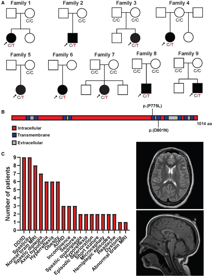
ATP1A3 encodes the α3 subunit of the sodium-potassium ATPase, one of two isoforms responsible for powering electrochemical gradients in neurons. Heterozygous pathogenic ATP1A3 variants produce several distinct neurological syndromes, yet the molecular basis for phenotypic variability is unclear. We report a novel recurrent variant, ATP1A3(NM_152296.5):c.2324C>T; p.(Pro775Leu), in nine individuals associated with the primary clinical features of progressive or non-progressive spasticity and developmental delay/intellectual disability. No patients fulfil diagnostic criteria for ATP1A3-associated syndromes, including alternating hemiplegia of childhood, rapid-onset dystonia-parkinsonism or cerebellar ataxia-areflexia-pes cavus-optic atrophy-sensorineural hearing loss (CAPOS), and none were suspected of having an ATP1A3-related disorder. Uniquely among known ATP1A3 variants, P775L causes leakage of sodium ions and protons into the cell, associated with impaired sodium binding/occlusion kinetics favouring states with fewer bound ions. These phenotypic and electrophysiologic studies demonstrate that ATP1A3:c.2324C>T; p.(Pro775Leu) results in mild ATP1A3-related phenotypes resembling complex hereditary spastic paraplegia or idiopathic spastic cerebral palsy. Cation leak provides a molecular explanation for this genotype-phenotype correlation, adding another mechanism to further explain phenotypic variability and highlighting the importance of biophysical properties beyond ion transport rate in ion transport diseases.

Keywords: ATP1A3; neurodevelopmental disorders; sodium-potassium ATPase; spastic paraparesis; spasticity.


Conflict of interest statement

The authors report no competing interests.

Figures

Figure 1
Figure 1
Clinical and molecular synopsis of ATP1A3:c.2324C>T; p.(Pro775Leu). (A) Pedigrees of families with ATP1A3:c.2324C>T; p.(Pro775Leu). Affected individuals are indicated by filled circles and squares and with arrows. Genotype at the locus is indicated below pedigree symbols if available; variant alleles are indicated in red. (B) Protein domain structure of ATP1A3. Intracellular domains are shown in red, transmembrane domains in blue, and extracellular domains in grey. (C) Phenotypic traits associated with ATP1A3:c.2324C>T; p.(Pro775Leu) are shown in order of frequency. Representative T2-weighted axial brain magnetic resonance imaging (top) and T1-weighted sagittal brain MRI (bottom) from proband in Family 1 showing normal supratentorial brain structure, cerebellum and brainstem.
Figure 2
Figure 2
P775L eliminates normal α3-Na+/K+-ATPase function and introduces inward current leak. (A) P775L shows loss-of-function in an ouabain complementation assay. HEK293 T cells were either mock-transfected (Mock) or transfected with ouabain-resistant ATP1A3 expression constructs with no additional variants (WT) or the D801N and P775L variants as shown. Two days after transfection, half of the cells were transferred to medium with or without 10 μM ouabain for another 2 days. The percentage of surviving cells in ouabain compared to the same sample without ouabain is shown. Each point represents one replicate with error bars at 95% CI. **P < 0.01 versus wild-type (WT). (B) P775L eliminates normal ion transport currents. Na+/K+-ATPase pump current was measured by two-electrode voltage clamp in Na+-loaded Xenopus oocytes expressing wild-type (WT), D801N or P775L variant human α3 Na+/K+-ATPase. Pump currents were elicited by perfusing external solution containing 110 mM Na+ and 5 mM K+. Currents specific to the exogenously expressed α3-Na+/K+-ATPase were isolated by subtracting measurements in 10 mM ouabain from measurements in 2 μM ouabain, and values at 0 mV (the holding potential used) are shown. n = 10 replicates for each construct; error bars = 95% CI; ***P < 0.0001 versus wild-type. (C) P775L introduces a negative leak current. Steady-state pump currents in 110 mM Na+ and 5 mM K+ were measured by ouabain subtraction of 100 ms pulses from 0 mV to the indicated voltages. n = 10 replicates per construct; error bars = 95% CI. (D) The P775L leak current does not depend on extracellular K+. Steady-state pump currents in 115 mM Na+ without K+ were measured by ouabain subtractions in the same oocytes from C after washout of ouabain for 9 min. Fifty millisecond pulses from 0 mV to the indicated voltages were used due to cell death during long pulses to negative potentials. n = 10, error bars = 95% CI. (E) P775L variant α3-Na+/K+-ATPases reach the cell membrane. In two-electrode voltage clamp measurements, off-transient currents representing Na+ movement out of its binding sites on α3-Na+/K+-ATPase were measured by ouabain subtraction in 115 mM Na+ after 50 ms pulses from 0 mV to the indicated voltages. These currents were fitted to monoexponential decay functions and integrated over time, yielding the charge moved Q at each voltage. n = 10, error bars = 95% CI.
Figure 3
Figure 3
P775L leak current primarily involves inward movement of Na+, with a secondary H+ component inhibited by extracellular Na+. Xenopus oocytes expressing P775L α3-Na+/K+-ATPase (ATP1A3 P775L; left) were compared to those expressing L104R α1-Na+/K+-ATPase (ATP1A1 L104R; right) in two-electrode voltage clamp current measurements with ouabain subtractions. Error bars representing the 95% CI are shown in all figures unless too small to be visible. (A) Removal of Na+ results in a negative shift of the reversal potential of P775L and L104R leak currents. Leak currents were measured by ouabain subtraction in 115 mM Na+, 115 mM trimethylammonium (TMA+) or 115 mM N-methyl-D-glucamine (NMDG+) external solution. Pairwise measurements were done in order to compare currents in the same oocyte, using either Na+ and TMA+ or Na+ and NMDG+; therefore, the plot shows pooled values for Na+. For P775L, n = 6 for Na+/NMDG+ and n = 8 for Na+/TMA+; for L104R, n = 8 for Na+/NMDG+ and n = 8 for Na+/TMA+. (B) Normalization of currents corrects for variability between oocytes. Current measurements from A were normalized by dividing all measurements in each oocyte by the magnitude of current in that oocyte at −80 mV in Na+. Reversal potentials ± standard deviation in mV were 0 ± 5 (Na+), −57 ± 8 (TMA+), and −40 ± 2 (NMDG+) for P775L and −6 ± 3 (Na+), −48 ± 3 (TMA+), and −25 ± 4 (NMDG+) for L104R. (C) Increasing the extracellular H+ concentration results in a positive shift of the reversal potential of P775L and L104R leak currents. Ouabain-subtraction currents are shown for pairwise measurements in the same oocyte at pH 7.6 versus 6.6 in external solution containing 110 mM Na+ and 5 mM K+ (top), 115 mM Na+ (middle) or 115 mM NMDG+ (bottom). Currents were normalized to the magnitude of current at pH 7.6 at −80 mV in the same oocyte, i.e. the NMDG+ curves were normalized to the value in NMDG+ at pH 7.6, not Na+ as in A. A magnified view is shown to the right of each plot to better illustrate the reversal potentials. Reversal potentials ± standard deviation in mV were −0.8 ± 8 (Na+/K+ pH 7.6), 15 ± 5 (Na+/K+ pH 6.6), −43 ± 7 (NMDG+ pH 7.6) and −14 ± 4 (NMDG+ pH 6.6) for P775L. They were −12 ± 3 (Na+/K+ pH 7.6), 22 ± 4 (Na+/K+ pH 6.6), −21 ± 2 (NMDG+ pH 7.6) and −15 ± 1 (NMDG+ pH 6.6) for L104R. n = 6 replicates for each combination of condition and variant. (D) Schematic of the ion flows indicated by the data from AC. P775L conducts primarily Na+ and some H+ leak in the presence of extracellular Na+, while L104R conducts a mixed Na+/H+ leak as previously demonstrated.
Figure 4
Figure 4
P775L disrupts the substrate ion binding sites, altering the kinetics of extracellular Na+ binding. (A) Simplified schematic of the Na+ binding/unbinding transitions studied. From right to left, three extracellular Na+ sequentially bind to and occlude at the three sites within the enzyme, respectively producing fast (kfast), medium (kmed) and slow (kslow) components of Na+ on-transient currents. Applying negative voltage pulses as in B and C drives the enzyme state toward the left such that the first ion to bind represents the fast component. After the end of the pulse, Na+ ions unbind from their binding sites, producing measurable off-transient currents. Therefore, the slow component represents the deocclusion and release of the first Na+ to unbind when considered in the normal direction of the enzymatic cycle. (B) Na+ transient currents are abnormal in P775L variant α3-Na+/K+-ATPases. Cut-open vaseline gap voltage clamp was done in Xenopus oocytes expressing P775L or wild-type (WT) ouabain-resistant α3-Na+/K+-ATPases. Every 40 s, 18 voltage pulses to −190 mV of various durations were applied, separated by 150 ms intervals. For clarity, five representative pulses with durations of 15, 1.5, 25, 2 and 10 ms are shown. (C) P775L reduces the contribution of the slow component to Na+ transient currents. The amounts of charge moved by the slow (Qs), medium (Qm) and fast (Qf) components were normalized to the total amount of charge at the longest (25 ms) prepulse and plotted as a function of time. Solid lines represent exponential functions with τs, τm and τf fixed to the average values obtained from the independent fittings of each experiment. Below each plot, the non-normalized charge Q moved by each component and the time constant τ calculated from the longest (25 ms) pulses are shown. n = 9 for wild-type (WT) and n = 5 for P775L.

References

    1. Clausen MV, Hilbers F, Poulsen H. The structure and function of the Na,K-ATPase isoforms in health and disease. Front Physiol. 2017;8:371. - PMC - PubMed
    1. Jiao S, Johnson K, Moreno C, Yano S, Holmgren M. Comparative description of the mRNA expression profile of Na(+)/K(+)-ATPase isoforms in adult mouse nervous system. J Comp Neurol. 2022;530:627–647. - PMC - PubMed
    1. de Carvalho Aguiar P, Sweadner KJ, Penniston JT, et al. . Mutations in the Na+/K+-ATPase alpha3 gene ATP1A3 are associated with rapid-onset dystonia parkinsonism. Neuron. 2004;43:169–175. - PubMed
    1. Rosewich H, Thiele H, Ohlenbusch A, et al. . Heterozygous de-novo mutations in ATP1A3 in patients with alternating hemiplegia of childhood: a whole-exome sequencing gene-identification study. Lancet Neurol. 2012;11:764–773. - PubMed
    1. Heinzen EL, Swoboda KJ, Hitomi Y, et al. . De novo mutations in ATP1A3 cause alternating hemiplegia of childhood. Nat Genet. 2012;44:1030–1034. - PMC - PubMed

Publication types