. 2023 Apr 14;9(15):eade5041.
doi: 10.1126/sciadv.ade5041. Epub 2023 Apr 12.

Milk-derived extracellular vesicles protect intestinal barrier integrity in the gut-liver axis

Affiliations

Milk-derived extracellular vesicles protect intestinal barrier integrity in the gut-liver axis

Lingjun Tong et al. Sci Adv. .

Abstract

Milk-derived extracellular vesicles (mEVs) have been proposed as a potential nanomedicine for intestinal disorders; however, their impact on intestinal barrier integrity in gut inflammation and associated metabolic diseases has not been explored yet. Here, mEVs derived from bovine and human breast milk exert similar protective effects on epithelial tight junction functionality in vitro, survive harsh gastrointestinal conditions ex vivo, and reach the colon in vivo. Oral administration of mEVs restores gut barrier integrity at multiple levels, including mucus, epithelial, and immune barriers, and prevents endotoxin translocation into the liver in chemical-induced experimental colitis and diet-induced nonalcoholic steatohepatitis (NASH), thereby alleviating gut disorders, their associated liver inflammation, and NASH. Oral administration of mEVs has potential in the treatment of gut inflammation and gut-liver axis-associated metabolic diseases via protection of intestinal barrier integrity.


Figures

Fig. 1.
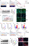
Fig. 1.. Characterization of bovine and human milk-derived extracellular vesicles (mEVs).
(A) Representative transmission electron microscope (TEM) images of mEVs derived from bovine (B-mEVs) and human (H-mEVs) milk. (B and C) Western blot analysis of extracellular vesicles (EVs) markers and non-EV proteins. (D) Size distribution of mEVs analyzed via nanoparticle tracking analysis (NTA). (E) Kyoto Encyclopedia of Genes and Genomes (KEGG) pathway analysis of B-mEV proteins and microRNAs associated with intestinal barrier function. (F) Venn diagrams of top 40 microRNAs in bovine and human mEVs. (G) Ranking of microRNAs in bovine mEVs corresponding to the top 20 microRNAs in human mEVs. Supt, supernatant.
Fig. 2.
Fig. 2.. Milk-derived extracellular vesicles (mEVs) alleviate dextran sulfate sodium (DSS)–induced disruption of epithelial tight junctions.
Caco-2 cells were pretreated with mEVs for 12 hours before exposure to DSS and TNF-α. (A) A schematic illustration of experimental procedures assessing epithelial permeability in the D_T treatment–induced epithelial injury model. (B) Transepithelial electrical resistance (TEER) determined hourly by the Millicell ERS-2 Voltohmmeter. (C) Western blot analysis of tight junction protein Occludin upon D_T treatment at 20 hours. (D) The immunofluorescence assessment of tight junction proteins ZO-1 and Occludin in Caco-2 cells at 20 hours. (E) A schematic illustration of the Transwell coculture system for intestinal epithelial cells and macrophages. (F and G) mRNA levels of tight junction proteins upon D_T treatment at 4 hours. (H and I) mRNA expression of inflammatory cytokines in bone marrow–derived macrophages (BMDM). (J) The illustration of TEER assays of miR-148a treatment (20 nM). miR-148a packed in LNPs (miR-148a-LNPs) was used for simulating mEVs to obtain data. (K to N) Scrambled miR (Scr-miR) packed in LNPs was used as control. (K) TEER was determined hourly. (L and M) Western blot analysis and quantification of Occludin expression in Caco-2 cells. (N) Immunofluorescence assessment of ZO-1 expression in Caco-2 cells. Gapdh was used as the reference gene. ns, not significant; *P < 0.05, **P < 0.01, and ***P < 0.001; one-way ANOVA. Three independent experiments were performed in triplicate. D_T, DSS + TNF-α; B-mEVs, bovine mEVs; H-mEVs, human milk EVs.
Fig. 3.
Fig. 3.. Milk-derived extracellular vesicles (mEVs) survive gastrointestinal environment and reach colon immune cells.
(A) Morphology and size distribution of bovine mEVs (B-mEVs) and liposomes before and after going through the ex vivo digestive system. Three independent experiments were performed in triplicate. (B) Representative IVIS images of healthy mice (top) and dissected colons (bottom) at 12 hours after oral administration of mEVs. (C) Fluorescence microscopy imaging of colon cryosections showing B-mEVs in red (Cy5-labeled), DC2.4 dendritic cells (DCs) strained with CD11c in green (top), and macrophages stained with CD68 in green (bottom). Scale bars, 100 μm. Colons were dissected from healthy mice as in (B). (D to G) Fluorescence-activated cell sorting (FACS) analysis showing cellular uptake of B-mEVs by colon immune cells in healthy and DSS-induced colon impaired mice (n = 3 mice per group). Control, healthy mice received free dye orally; Cy5-B-mEVs, healthy mice received Cy5-labeled B-mEVs orally; DSS-Cy5-B-mEVs, DSS-induced colon impaired mice received Cy5-labeled B-mEVs orally. *P < 0.05 and ****P < 0.0001, one-way ANOVA.
Fig. 4.
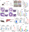
Fig. 4.. Oral administration of bovine milk-derived extracellular vesicles (B-mEVs) protects the colon from dextran sulfate sodium (DSS)–induced chronic impairment in colitis.
(A) Scheme of the DSS-induced chronic colitis model and administration of B-mEVs. Mice were euthanized on day 33. Body weight (B), DAI (C), and colon length (D and E) were monitored to assess colitis severity (n = 6 mice per group). (F) Representative images of colon sections stained with H&E to assess histological score (histopathological evaluations were conducted to examine intestinal damage and inflammatory infiltration). Scale bars, 100 μm. (G) Masson’s trichrome (MT) staining and quantitative analysis of fibrotic area (blue) to assess fibrosis. Scale bars, 100 μm. (H to K) Serum levels of proinflammatory cytokines assessed using ELISA (n = 4 to 5 mice per group). L, low dose, 0.6 mg/kg per day; H, high dose, 3 mg/kg per day. *P < 0.05, **P < 0.01, and ***P < 0.001, one-way ANOVA.
Fig. 5.
Fig. 5.. Oral administration of bovine milk-derived extracellular vesicles (B-mEVs) alleviates immunological barrier dysfunction in dextran sulfate sodium (DSS)–induced chronic colitis.
(A) Staining and quantification of CX3CR1hi resident macrophages in the colon (n = 3 to 4). (B) IL-10 protein levels in the colon determined by ELISA and normalized to tissue weight (n = 5). (C) Staining and quantification of CD4+ T cells in the colon (n = 3). (D) Flow cytometry analysis of CD4+CD25+FoxP3+ Treg cells in the colonic lamina propria (LP) and spleen. (E) Quantitative analysis of FoxP3 protein in the colon by immunofluorescence intensity. (F) Flow cytometry analysis of CD4+CD25+FoxP3+ Treg cells in the spleen. (G) Spleen weight. (H and I) Proportion of TH2 cells in the colonic LP and spleens determined by flow cytometry. N = 3 to 4 mice per group. Scale bars, 100 μm. L, 0.6 mg/kg per day; H, 3 mg/kg per day. *P < 0.05, **P < 0.01, and ***P < 0.001, one-way ANOVA.
Fig. 6.
Fig. 6.. Oral administration of bovine milk-derived extracellular vesicles (B-mEVs) efficiently ameliorates physical barrier dysfunction in dextran sulfate sodium (DSS)–induced chronic colitis.
(A) Western blot analysis of key proteins in the adenosine 5′-monophosphate (AMP)–activated protein kinase (AMPK) and GLP2 signaling pathways that regulate expression of tight junction proteins. (B to D) Quantification of protein expression levels of p-AMPK, AMPK, GLP-2, and IGF-1, normalized to GAPDH. (E) Quantitative analysis of ZO-1 protein expression in the colon by immunofluorescence intensity. (F and G) Representative images of Occludin and JAM-1 proteins in the colon. Scale bars, 200 μm. (H and I) Quantitative analysis of Occludin and JAM-1 protein expression in the colon by immunofluorescence intensity. (J) Intestinal permeability assessed by fluorescein isothiocyanate (FITC)–dextran test. N = 3 to 4 mice per group. L, 0.6 mg/kg per day; H, 3 mg/kg per day. Data are means ± SD; *P < 0.05, **P < 0.01, and ***P < 0.001, one-way ANOVA.
Fig. 7.
Fig. 7.. Oral administration of bovine milk-derived extracellular vesicles (B-mEVs) efficiently alleviates mucosal barrier dysfunction in dextran sulfate sodium (DSS)–induced chronic colitis.
(A) Left: Representative images of colon sections with Alcian blue staining for assessment of mucosal damage. Right: Enlarged view of colon sections, magnification ×400. M, mucin; GC, goblet cells; ME, muscularis externa; L, lumen; MDF, mucin-depleted foci. (B and C) Staining and quantification of MUC2 (red) in colon tissue with fluorescence microscopy imaging. Scale bars, 100 μm. N = 3 to 4 mice per group. (D) Apical junctional lines and complexes (red arrow) in colonic villus epithelium shown by Alcian blue staining. L, 0.6 mg/kg per day; H, 3 mg/kg per day. ***P < 0.001, one-way ANOVA.
Fig. 8.
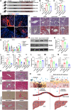
Fig. 8.. Bovine milk-derived extracellular vesicles (B-mEVs), but not EV-depleted supernatant, alleviate dextran sulfate sodium (DSS)–induced intestinal barrier injury and liver inflammation in acute colitis.
(A) Scheme of acute colitis induction (3.5% DSS daily for 7 days) and administration of B-mEVs (0.6 mg/kg per day). Mice were euthanized for further investigation on day 11. (B and C) Colon length was determined to assess severity of colonic impairment (n = 4 to 6). (D) Representative images of H&E-stained colon sections. Enlarged view of the red square demarcated regions is shown on the right. (E) mRNA expression levels of tight junction proteins, Zo-1 and Occludin, in the colon. Gapdh was used as the reference gene. (F) Illustration of liver inflammation in DSS-induced acute colitis. (G and H) Amount of CD45+ immune cells and CD45+Ly6G+ neutrophils in the liver determined by flow cytometry. (I) mRNA expression levels of cytokines in the liver. Different treatments were indicated as in (E) (n = 3 to 4 mice per group). *P < 0.05, ***P < 0.001, and ****P < 0.0001, one-way ANOVA.
Fig. 9.
Fig. 9.. Bovine milk-derived extracellular vesicles (B-mEVs) repair intestinal barrier integrity and alleviate liver pathologies in methionine-choline–deficient (MCD) diet–induced nonalcoholic steatohepatitis (NASH) mice.
(A) Scheme of the experimental design. Mice were fed milk-derived extracellular vesicles (mEVs) (1.2 mg/kg) every other day. (B) Intestinal barrier integrity assessed by oral administration of fluorescein isothiocyanate (FITC)–dextran. (C) Plasma levels of nitric oxide. (D) Immunofluorescence staining of tight junction protein ZO-1 in the colon. White arrows indicate the presence of ZO-1 in epithelial mucosa. (E) mRNA expression of tight junction proteins and inflammatory cytokine Tnf-α. Gapdh was used as the reference gene. (F) Liver histology analyzed by H&E and Oil Red O (ORO) staining. (G to I) Western blot analysis of key inflammatory signaling molecules iNOS and iRAK4 in the liver. Protein levels were normalized to GAPDH, n = 3 mice per group. (J) mRNA expression of inflammatory cytokines Tnf-α and Il-1β in the liver, n = 4 mice per group. (K) Masson’s trichrome (MT) staining and PicroSirius red (PRS) staining of collagen fibers in liver sections. Green arrows indicate the fibrosis areas. (L to N) Levels of endotoxin detected in the feces, liver, and plasma (n = 3 to 4 mice per group). (O) Schematic illustration showing that B-mEVs alleviate NASH via reducing endotoxin translocation from the gut to the liver. Blue dots indicate endotoxin. Supernatant, EV-depleted supernatants. *P < 0.05, **P < 0.01, ***P < 0.001, and ****P < 0.0001, one-way ANOVA.
Fig. 10.
Fig. 10.. Schematic illustration of potential mechanisms via which milk-derived extracellular vesicles (mEVs) protect gut barrier integrity in the gut-liver axis.
mEVs protect gut barrier integrity by reinforcing intestinal epithelial tight junctions, suppressing inflammation, and inhibiting translocation of endotoxin from the gut into the bloodstream and the liver, thereby ameliorating inflammatory bowel disease and nonalcoholic steatohepatitis (NASH).

References

    1. J. R. Turner, Intestinal mucosal barrier function in health and disease. Nat. Rev. Immunol. 9, 799–809 (2009). - PubMed
    1. L. Miele, V. Valenza, G. La Torre, M. Montalto, G. Cammarota, R. Ricci, R. Masciana, A. Forgione, M. L. Gabrieli, G. Perotti, F. M. Vecchio, G. Rapaccini, G. Gasbarrini, C. P. Day, A. Grieco, Increased intestinal permeability and tight junction alterations in nonalcoholic fatty liver disease. Hepatology 49, 1877–1887 (2009). - PubMed
    1. M. E. Johansson, M. Phillipson, J. Petersson, A. Velcich, L. Holm, G. C. Hansson, The inner of the two Muc2 mucin-dependent mucus layers in colon is devoid of bacteria. Proc. Natl. Acad. Sci. U.S.A. 105, 15064–15069 (2008). - PMC - PubMed
    1. C. K. Heazlewood, M. C. Cook, R. Eri, G. R. Price, S. B. Tauro, D. Taupin, D. J. Thornton, C. W. Png, T. L. Crockford, R. J. Cornall, R. Adams, M. Kato, K. A. Nelms, N. A. Hong, T. H. Florin, C. C. Goodnow, M. A. McGuckin, Aberrant mucin assembly in mice causes endoplasmic reticulum stress and spontaneous inflammation resembling ulcerative colitis. PLOS Med. 5, e54 (2008). - PMC - PubMed
    1. J. M. Allaire, S. M. Crowley, H. T. Law, S. Y. Chang, H. J. Ko, B. A. Vallance, The intestinal epithelium: Central coordinator of mucosal immunity. Trends Immunol. 39, 677–696 (2018). - PubMed