Review
. 2023 Apr 11;56(4):704-722.
doi: 10.1016/j.immuni.2023.03.014.

The ins and outs of innate and adaptive type 2 immunity

Affiliations
Review

The ins and outs of innate and adaptive type 2 immunity

Ari B Molofsky et al. Immunity. .

Abstract

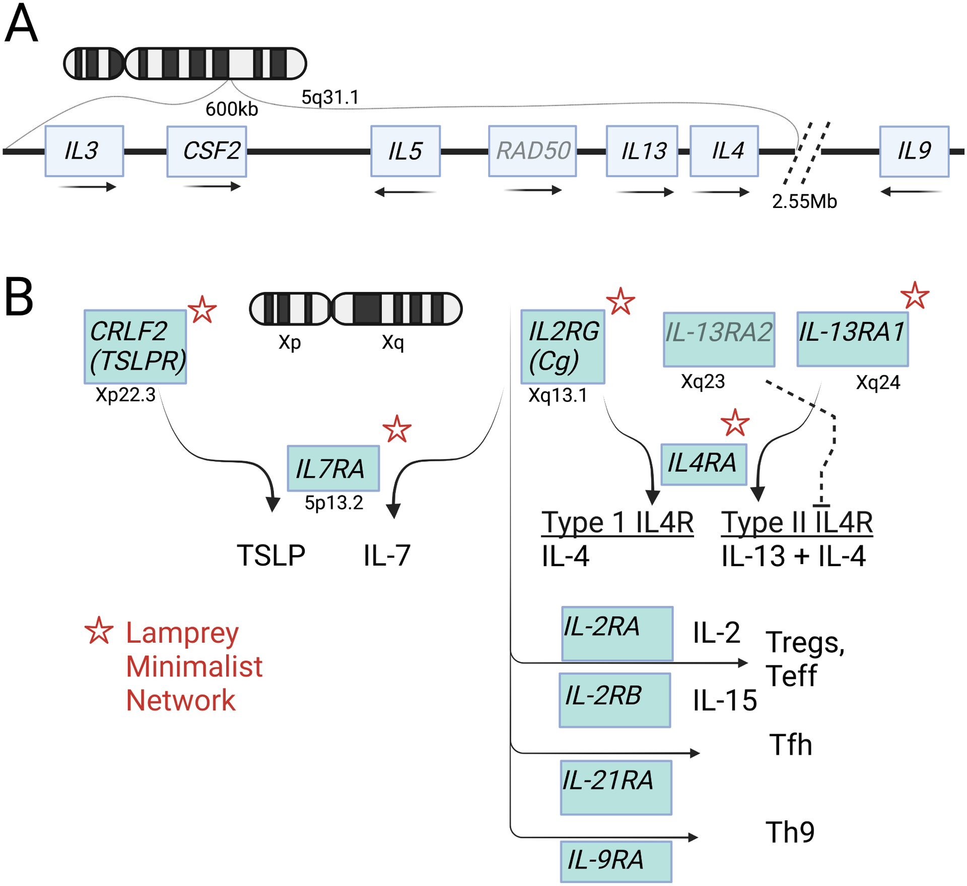
Type 2 immunity is orchestrated by a canonical group of cytokines primarily produced by innate lymphoid cells, group 2, and their adaptive counterparts, CD4+ helper type 2 cells, and elaborated by myeloid cells and antibodies that accumulate in response. Here, we review the cytokine and cellular circuits that mediate type 2 immunity. Building from insights in cytokine evolution, we propose that innate type 2 immunity evolved to monitor the status of microbe-rich epithelial barriers (outside) and sterile parenchymal borders (inside) to meet the functional demands of local tissue, and, when necessary, to relay information to the adaptive immune system to reinforce demarcating borders to sustain these efforts. Allergic pathology likely results from deviations in local sustaining units caused by alterations imposed by environmental effects during postnatal developmental windows and exacerbated by mutations that increase vulnerabilities. This framework positions T2 immunity as central to sustaining tissue repair and regeneration and provides a context toward understanding allergic disease.


Conflict of interest statement

Declaration of interests R.M.L. is a member of the Scientific Advisory Board for Genentech and a participant on the Editorial Advisory Board at Immunity.

Figures

Figure 1.
Figure 1.. The cytokines and core components of type 2 immunity.
A. Genomic organization of the core type 2 cytokines on human chromosome 5q31.1. Transcription orientation indicated by arrows. B. Human cytokine signaling receptors on the X chromosome. Asterisks indicate homologs found in the lamprey cytokine receptor network. Further diversification of subsets of CD4 lymphocytes in jawed vertebrates is accompanied by appearance of additional partners for the common γ chain.
Figure 2.
Figure 2.. Layered ontogeny underlies the distributed network of type 2 immune lymphocytes.
Although tissue resident ILC2s and Th2s derive from upstream precursors, we figuratively represent their origins from populations designated ILC2s or Th2s for illustrative purposes. Tissue ILC2s are comprised of populations originating from primitive hematopoiesis originating in yolk sac and liver, and bone marrow definitive HSCs. Additional heterogeneity may reflect derivation of some ILC2s from thymic pre-T cell precursors (dotted red borders). Adaptive Th2s arise from definitive adult bone marrow HSCs after thymic education and export as naive T cell precursors. Neonatal adaptive T cells can also arise from fetal HSCs, comprising a small population of tissue cells with rapid effector function (dotted blue borders). The triangles at top denote the different layering of peripheral tissues at the time of birth in mouse and human.
Figure 3.
Figure 3.. The ins- and-outs of tissue surveillance by type 2 immune lymphocytes – an organizational model.
The Center grouping depicts core shared components of ILC2s in a stromal niche that likely applies to resident Th2s in the adapted state. Tissue function is translated by many cell types into signaling pathways that are integrated and thresholded by resident alarmin signals, transmitted in part by stromal niche fibroblasts to ILC2s which release type 2 cytokines that feedback on niche stromal cells and regulate myeloid cell activation and recruitment. With sufficient perturbation, ILC2s proliferate, increase cytokine outputs and migrate from the niche to interact with resident cells and tissues. Outer Barriers (upper left) are externally-oriented epidermis in skin and mucosa, where ILC2s can be activated by barrier-specific alarmins, like IL-25 in small intestine or TSLP/IL-18 in skin. Outer Barrier tissues are supported by Outer Borders (lower left) consisting of subepithelial fascial planes that support resident ILC2s that when activated can facilitate niche enlargement to house recruited Th2 cells and myeloid cells that increase local collagen deposition and reinforce the physical border internal to the external barrier. Inner Borders (upper right) represent serosal mesothelial linings surrounding internal organs, which also support ILC2/stromal niches and use type 2 cytokines to enlarge the niche and enhance structural support. Internal Boundaries (lower right) depict organization of ILC2s in proximity to vascular and ductal structures that allow monitoring of regional homeostasis in tissues.
Figure 4.
Figure 4.. Vulnerabilities to type 2 pathology.
A. In the basal state, injury induces alarmin-mediated ILC2/Th2 outputs that inhibit normal stem cell transitions while the epithelium regenerates by engaging immediate type 3 responses and later type 1 immunity to enforce training. With repair, type 2 cytokines activate myeloid cells (AAMs, eosinophils) to mediate regulatory control and expand the type 2 immune cell niche using structural reinforcement as necessary. Systemic spread of type 2 lymphocytes contributes to metabolic homeostasis needed to support the altered tissue state and local function is sustained or even improved, depending on the context of the inciting event. B. Vulnerabilities occur when mutations interact with environmental exposures during critical developmental windows that affect local tissue functions or output of type 2 cytokines. The tempo of repair becomes altered, resulting in an excess of tissue AAMs, eosinophils and alarmin+ stroma that support an enlarged niche with potential for development of tertiary lymphoid structures promoting local Tfh and Th2 differentiation. The misalignment of repair and regeneration with immune signals, particularly as affected by underlying mutations, can lead to aberrant epithelial and tissue responses manifest as allergic pathology and loss of function.

References

    1. Palm NW, Rosenstein RK, Medzhitov R. 2012. Allergic host defenses. Nature 484:465–472. - PMC - PubMed
    1. Fahy JV. 2015. Type 2 inflammation in asthma – present in most, absent in many. Nat Rev Immunol 15:57–65. - PMC - PubMed
    1. Hammad H, Lambrecht BN. 2021. The basic immunology of asthma. Cell 184:1469–1485. - PubMed
    1. Zhu Z, Lee PH, Chaffin MD, Chung W, Loh P-R, Lu Q, Christiani, Liang L. 2018. A genome-wide cross trait analysis from UK Biobank highlights the shared genetic architecture of asthma and allergic diseases. Nat Genet 50:857–864. - PMC - PubMed
    1. Sharma M, Leung D, Momenilandi M, Jones LCW, Pacillo L, James AE, Murrell JR, Delafontaine S, Maimaris J, Vaseghi-Shanjani M, Del Bel KL, Lu HY, Chua GT, Di Cesare S, Fornes O, Liu Z, De Matteo G, Fu MP, Amodio D, et al. 2023. Human germline heterozygous gain-of-function STAT6 variants cause severe allergic disease. J Exp Med 220:20221437. - PMC - PubMed

Publication types