. 2023 Apr 11;18(4):1030-1047.
doi: 10.1016/j.stemcr.2023.03.006.

Scalable generation of sensory neurons from human pluripotent stem cells

Affiliations

Scalable generation of sensory neurons from human pluripotent stem cells

Tao Deng et al. Stem Cell Reports. .

Abstract

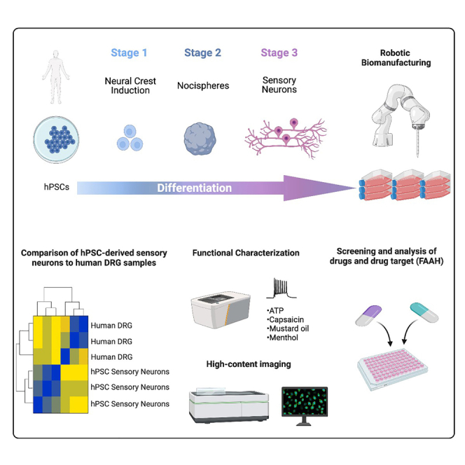
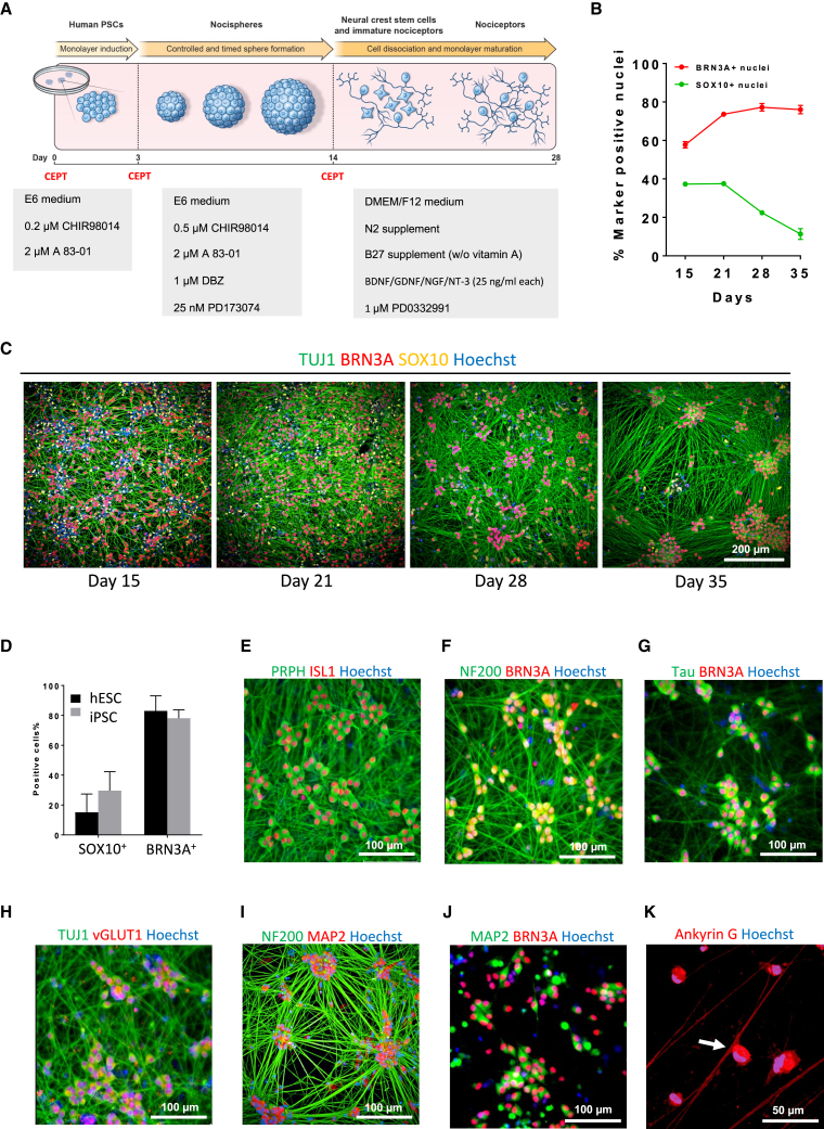
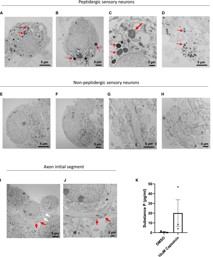
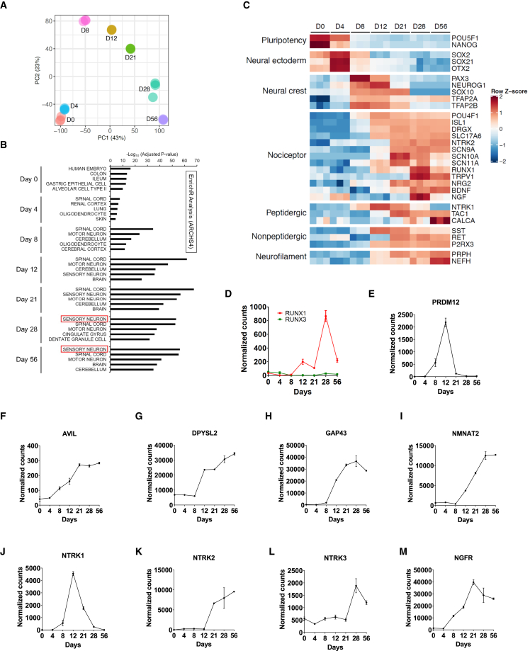
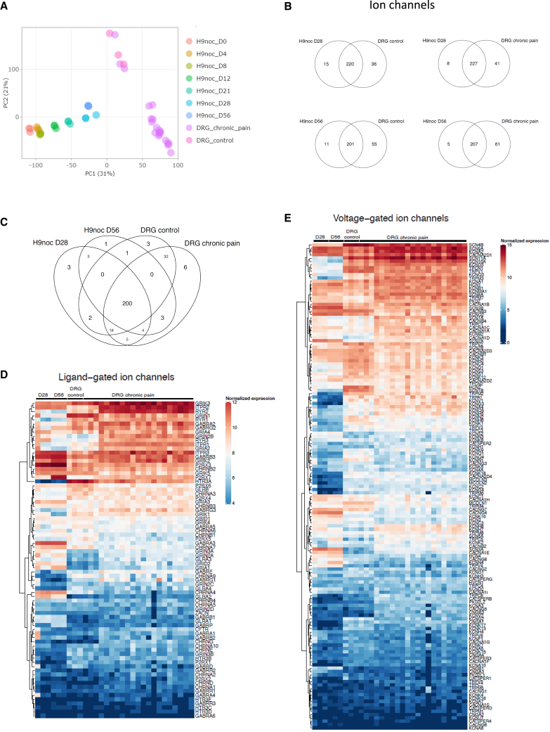
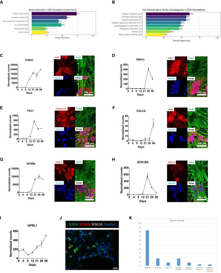
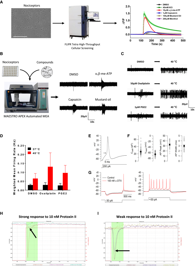
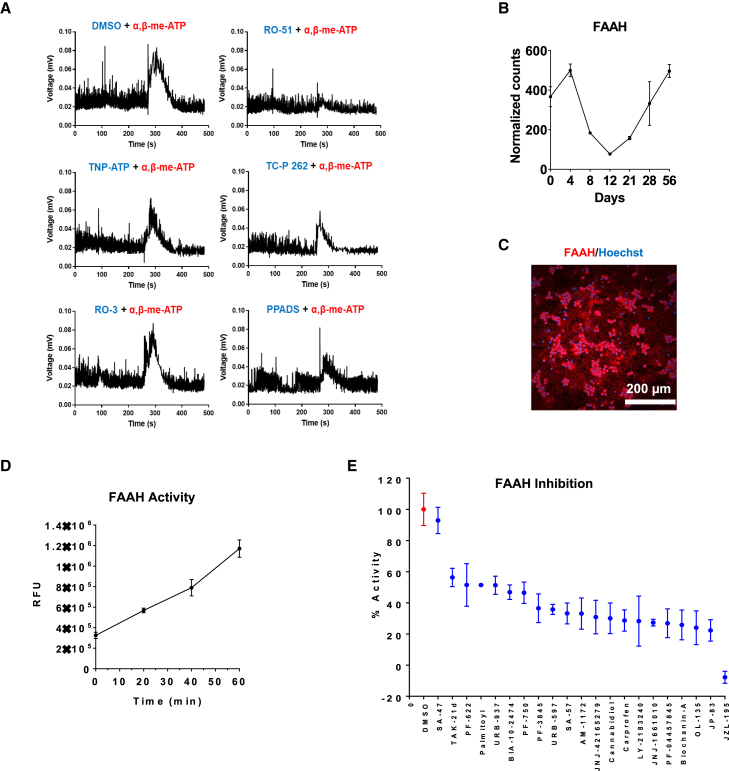
Development of new non-addictive analgesics requires advanced strategies to differentiate human pluripotent stem cells (hPSCs) into relevant cell types. Following principles of developmental biology and translational applicability, here we developed an efficient stepwise differentiation method for peptidergic and non-peptidergic nociceptors. By modulating specific cell signaling pathways, hPSCs were first converted into SOX10+ neural crest, followed by differentiation into sensory neurons. Detailed characterization, including ultrastructural analysis, confirmed that the hPSC-derived nociceptors displayed cellular and molecular features comparable to native dorsal root ganglion (DRG) neurons, and expressed high-threshold primary sensory neuron markers, transcription factors, neuropeptides, and over 150 ion channels and receptors relevant for pain research and axonal growth/regeneration studies (e.g., TRPV1, NAV1.7, NAV1.8, TAC1, CALCA, GAP43, DPYSL2, NMNAT2). Moreover, after confirming robust functional activities and differential response to noxious stimuli and specific drugs, a robotic cell culture system was employed to produce large quantities of human sensory neurons, which can be used to develop nociceptor-selective analgesics.

Keywords: CEPT cocktail; analgesics; cell differentiation; drug testing; iPS cells; neural crest; nociceptor; opioid crisis; pain; sensory neuron.


Conflict of interest statement

Conflict of interests T.D., A.S., and I.S. are co-inventors on a US Department of Health and Human Services patent covering the nociceptor differentiation method and its utilization.

Figures

None
Graphical abstract
Figure 1
Figure 1
Directed differentiation of hPSC into pseudo-unipolar nociceptors (A) Schematic overview of nociceptor differentiation method. (B) Quantitative analysis of cells expressing SOX10 and BRN3A during cell differentiation. Note that most cells express the pan-sensory marker BRN3A at days 28 and 35 (n = 3, mean ± SD). Data shown are from three different wells of one representative experiment (WA09) from multiple repeats. (C) Representative images of differentiating cells (WA09) at different timepoints immunolabeled for neural crest marker SOX10 and neuronal markers TUJ1 and BRN3A. (D) Quantification of cells expressing SOX10 or BRN3A derived from hESCs (WA09) and iPSCs (LiPSC-GR1.1) showing consistency of the differentiation method (n = 3, mean ± SD). Data shown are from three different wells of one representative experiment from multiple repeats. (E–G) Representative images of nociceptor cultures (WA09) at day 28 expressing the axonal markers PRPH (peripherin), NF200, and transcription factors ISL1 and BRN3A. (H) Sensory neurons derived from hESCs (WA09) expressing vGLUT1 consistent with their glutamatergic phenotype. See also Figure S3C. (I and J) Immunostaining for NF200, MAP2, and BRN3A at day 28 (WA09). Note that MAP2 immunoreactivity is confined to cell bodies. (K) Immunostaining for Ankyrin G and confocal microscopy showing axon splitting of nociceptors (WA09) (see also Figures S3F–S5H for confocal z stack images).
Figure 2
Figure 2
Ultrastructural analysis of peptidergic and non-peptidergic sensory neurons derived from hESCs (WA09) (A–D) Electron microscopic images showing peptidergic nociceptors. Prominent large dense core vesicles (LDCVs; red arrows) are present in cell bodies (A–C) and in axonal profiles sectioned in different planes (D). (E and F) Cell bodies of non-peptidergic nociceptor soma are devoid of LDCVs. (G and H) Typical axonal profiles of non-peptidergic neurons lacking LDCVs. (I) Overview image showing nociceptor cell bodies, axonal structures, and LDCVs. White arrows indicate the axon initial segment, which emanates from the cell soma. Red arrows point to more distal portions of the same axon. (J) Higher magnification of the axon initial segment shown in (I). (K) ELISA for substance P detection in the supernatant of day 28 nociceptors after DMSO and 10 μM capsaicin treatment. Data shown are from three different wells of one representative experiment using iPSC-derived nociceptors (NCRM5 cell line).
Figure 3
Figure 3
Transcriptomic analysis of the differentiation of hPSCs (WA09) into neural crest and nociceptors (A) PCA plot of RNA-seq experiment showing distinct molecular signatures at each timepoint. (B) Gene enrichment analysis using ENRICHR and ARCHS4 database identify “sensory neuron” as the top category at days 28 and 56. (C) Heatmap analysis showing stepwise differentiation of pluripotent cells into neural crest and nociceptors (n = 3). Data shown are from three different wells of one representative experiment. (D–M) Time-course expression profile of different genes with importance in sensory neuron development.
Figure 4
Figure 4
Molecular comparison of hPSC-derived nociceptors (WA09/H9) and human DRG samples (A) PCA of differentiating cells harvested at different timepoints and DRG samples (normal and chronic pain patient). (B and C) Venn diagrams comparing expressed ion channels in nociceptors (days 28 and 56) and DRG samples. (D and E) Heatmap comparison of ligand- and voltage-gated ion channels expressed by in vitro-generated nociceptors and DRGs (n = 3). See Figure S5 for detailed analysis of other gene families. Data shown are from three different wells of one representative experiment.
Figure 5
Figure 5
Analysis of gene families and specific targets expressed by hPSC-derived nociceptors (WA09) (A and B) Overview of genes and gene families expressed by hPSC-derived nociceptors (RNA-seq). (C–I) Pain research-relevant targets expressed by nociceptors, as confirmed at the transcript and protein level (RNA-seq and immunocytochemistry) (n = 3, mean ± SD). Data shown are from three different wells of one representative experiment. (J and K) Quantitative in situ hybridization (RNAscope) and analysis of sodium channel expression and co-expression of Nav 1.7, 1.8, and 1.9 in nociceptors.
Figure 6
Figure 6
Functional characterization of hPSC-derived nociceptors (WA09) (A) Calcium flux analysis after stimulation of nociceptors with KCL, ATP, capsaicin, mustard oil, and menthol (n = 8, mean ± SD). Data shown are from eight different wells of one representative experiment. (B) Automated MEA showing that nociceptors are activated by specific ligands. (C and D) MEA experiment showing that nociceptors are stimulated by a temperature increase from 37 to 40°C and are presensitized after treatment with oxaliplatin or PGE2 (n = 3, mean ± SD). Data shown are from three different wells of one representative experiment. (E) Typical action potential in nociceptors evoked by current injection. Arrow indicates the threshold voltage (−35 mV). (F) Collected results for passive and active electrical properties of the neurons. Open circles show measurements from individual cells and closed circles show mean ± SEM (n = 53 for cell capacitance and resting potential; n = 25 for action potential peak and width). (G) Enhancement of excitability by the Kv1-inhibitor α-dendrotoxin (DTX). (H) Automated single-cell patch-clamp (Sophion Qube) recording showing effect of 10 nM Protoxin-II and 1 μM TTX (in the continuing presence of Protoxin-II) on sodium current in an hPSC-derived nociceptor. (I) Collected results for percentage inhibition of sodium current by Protoxin-II alone and by Protoxin-II plus TTX (217 neurons analyzed).
Figure 7
Figure 7
Drug testing using hPSC-derived nociceptors (WA09) (A) MEA experiment comparing the potency of various P2RX3 inhibitors in human nociceptor cultures. (B) Gene expression of FAAH during cell differentiation into nociceptors (RNA-seq) (n = 3, mean ± SD). Data shown are from three different wells of one representative experiment. (C) Immunocytochemical analysis of FAAH expression in nociceptors (day 28). (D) Real-time fluorescent assay monitoring FAAH activity in nociceptors (n = 8, mean ± SD). Data shown are from eight different wells of one representative experiment. (E) FAAH inhibition assay testing the relative efficacy of 21 known inhibitors in day-28 nociceptor cultures (n = 8, mean ± SD). Data shown are from eight different wells of one representative experiment.

References

    1. Aguet F., Brown A.A., Castel S.E., Davis J.R., He Y., Jo B., Mohammadi P., Park Y.S., Parsana P., Segrè A.V., et al. Genetic effects on gene expression across human tissues. Nature. 2017;550:204–213. - PMC - PubMed
    1. Anastassiadis T., Deacon S.W., Devarajan K., Ma H., Peterson J.R. Comprehensive assay of kinase catalytic activity reveals features of kinase inhibitor selectivity. Nat. Biotechnol. 2011;29:1039–1045. - PMC - PubMed
    1. Black B.J., Atmaramani R., Kumaraju R., Plagens S., Romero-Ortega M., Dussor G., Price T.J., Campbell Z.T., Pancrazio J.J. Adult mouse sensory neurons on microelectrode arrays exhibit increased spontaneous and stimulus-evoked activity in the presence of interleukin-6. J. Neurophysiol. 2018;120:1374–1385. - PMC - PubMed
    1. Bartesaghi L., Wang Y., Fontanet P., Wanderoy S., Berger F., Wu H., Akkuratova N., Bouçanova F., Médard J.J., Petitpré C., et al. PRDM12 is required for initiation of the nociceptive neuron lineage during neurogenesis. Cell Rep. 2019;26:3484–3492.e4. - PMC - PubMed
    1. Basbaum A.I., Bautista D.M., Scherrer G., Julius D. Cellular and molecular mechanisms of pain. Cell. 2009;139:267–284. - PMC - PubMed

Publication types